. 2010 Nov 11:10:284.
doi: 10.1186/1471-2180-10-284.

Rhizobium leguminosarum bv. trifolii rosR is required for interaction with clover, biofilm formation and adaptation to the environment

Affiliations

Rhizobium leguminosarum bv. trifolii rosR is required for interaction with clover, biofilm formation and adaptation to the environment

Monika Janczarek et al. BMC Microbiol. .

Abstract

Background: Rhizobium leguminosarum bv. trifolii is a symbiotic nitrogen-fixing bacterium that elicits nodules on roots of host plants Trifolium spp. Bacterial surface polysaccharides are crucial for establishment of a successful symbiosis with legumes that form indeterminate-type nodules, such as Trifolium, Pisum, Vicia, and Medicago spp. and aid the bacterium in withstanding osmotic and other environmental stresses. Recently, the R. leguminosarum bv. trifolii RosR regulatory protein which controls exopolysaccharide production has been identified and characterized.

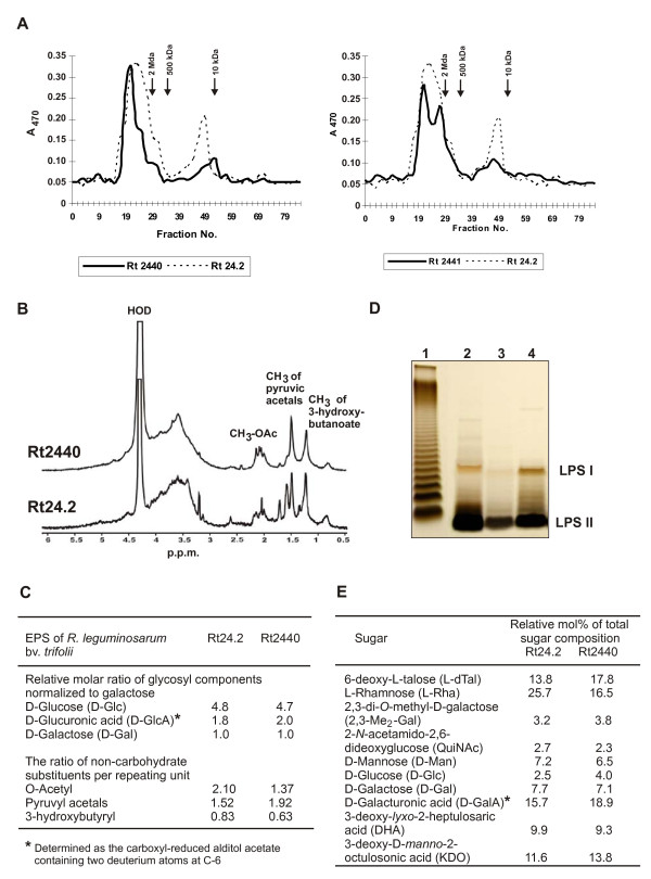
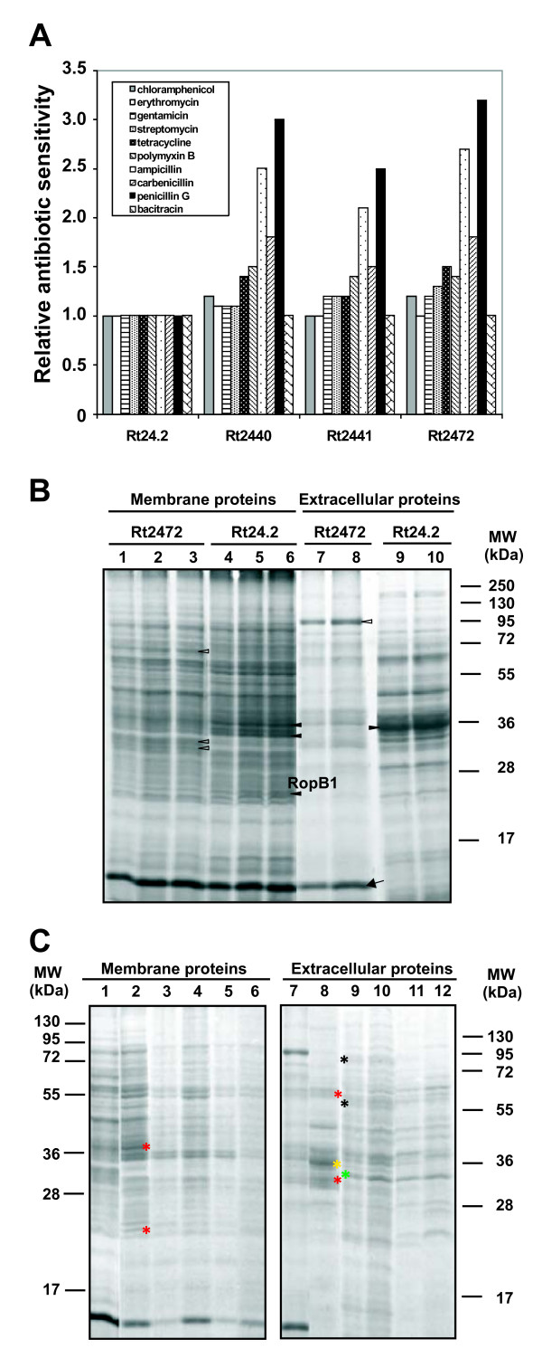
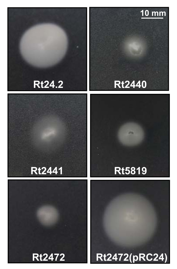
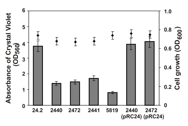
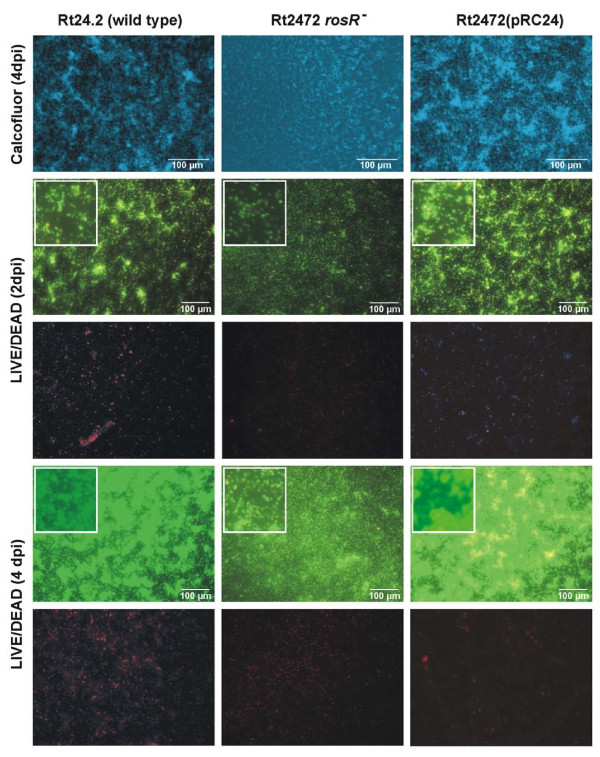
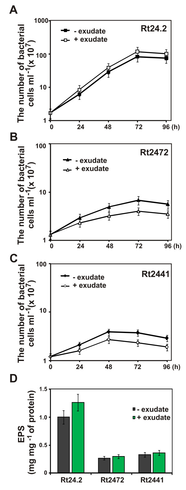
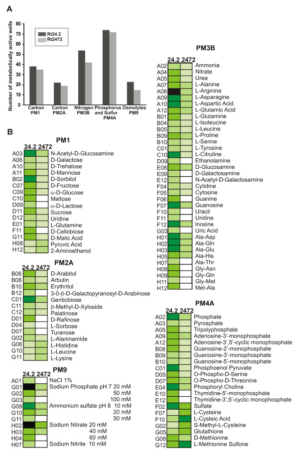
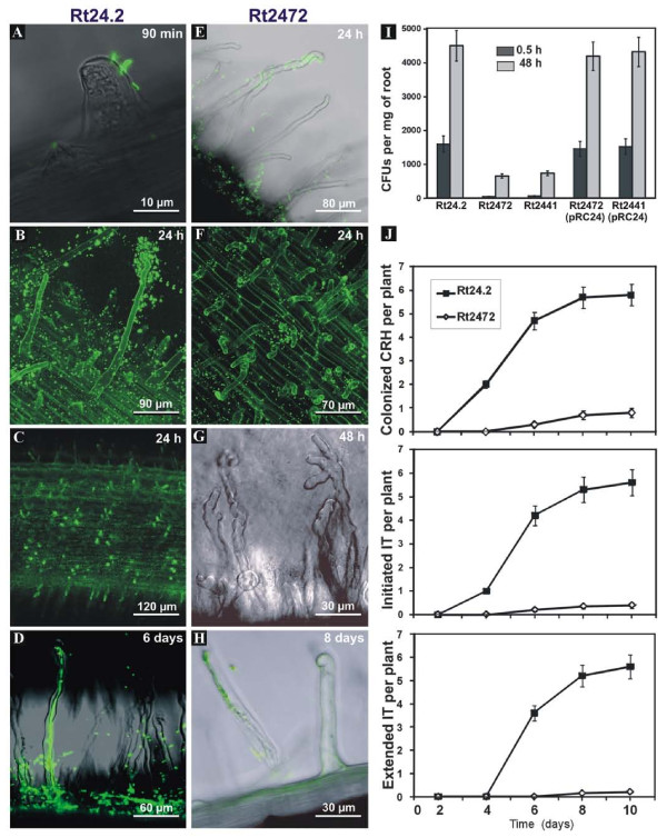
Results: In this work, we extend our earlier studies to the characterization of rosR mutants which exhibit pleiotropic phenotypes. The mutants produce three times less exopolysaccharide than the wild type, and the low-molecular-weight fraction in that polymer is greatly reduced. Mutation in rosR also results in quantitative alterations in the polysaccharide constituent of lipopolysaccharide. The rosR mutants are more sensitive to surface-active detergents, antibiotics of the beta-lactam group and some osmolytes, indicating changes in the bacterial membranes. In addition, the rosR mutants exhibit significant decrease in motility and form a biofilm on plastic surfaces, which differs significantly in depth, architecture, and bacterial viability from that of the wild type. The most striking effect of rosR mutation is the considerably decreased attachment and colonization of root hairs, indicating that the mutation affects the first stage of the invasion process. Infection threads initiate at a drastically reduced rate and frequently abort before they reach the base of root hairs. Although these mutants form nodules on clover, they are unable to fix nitrogen and are outcompeted by the wild type in mixed inoculations, demonstrating that functional rosR is important for competitive nodulation.

Conclusions: This report demonstrates the significant role RosR regulatory protein plays in bacterial stress adaptation and in the symbiotic relationship between clover and R. leguminosarum bv. trifolii 24.2.


Figures

Figure 1
Figure 1
Physical map of R. leguminosarum bv. trifolii rosR gene and genomic organization of rosR mutants. Physical and genetic map of pB31 plasmid carrying the rosR gene of Rhizobium leguminosarum bv. trifolii 24.2 (A). (B) The genomic organisation of the Rt2440, Rt2441, and Rt2472 mutants. The heavy line indicates the vector part in the Rt2441 integration mutant. B- BamHI, H- HindIII, S- SalI, P- PstI, N- NotI. P1 and P2 are promoter sequences of the rosR gene, and the RosR-box sequence is the target site recognized and bound by RosR protein.
Figure 2
Figure 2
The effect of additional copies of different regulatory rosR sequences on the EPS production by R. leguminosarum. Data shown are the means of three replicates ± SD.
Figure 3
Figure 3
Gel filtration chromatography of exopolysaccharides (EPS) produced by the R. leguminosarum bv. trifolii 24.2 wild type and the rosR mutants (Rt2440 and Rt2441). (A) EPS was fractionated on a Bio-Gel A-5m column, as described in the Methods. The retention times of molecular mass markers: dextran blue (2 MDa), dextran T250 (250 kDa), and dextran T10 (10 kDa) are indicated by arrows. (B) A 500 MHz 1H-NMR spectrometry analysis of the R. leguminosarum wild type and the rosR mutant (Rt2440). (C) The glycosyl components and non-carbohydrate substituents of EPS from the wild type and the mutant Rt2440. (D) Silver-stained Tricine SDS-PAGE profiles of LPS from the wild type and the rosR mutants. LPSs (2 μg) were loaded in 2 μl sample buffer. Lanes: 1- Salmonella enterica sv. Typhimurium (Sigma), 2- wild type Rt24.2, 3- Rt2440, 4- Rt2441. LPS I, high-molecular-weight LPS; LPS II, low-molecular-weight LPS. (E) The glycosyl composition of polysaccharides lacking lipid A released from LPS by mild acid hydrolysis from the wild type and the rosR mutant (Rt2440).
Figure 4
Figure 4
Sensitivity to antibiotics and profiles of membrane and extracellular proteins of R. leguminosarum bv. trifolii rosR mutants. Relative sensitivity of the R. leguminosarum bv. trifolii rosR mutants to antibiotics, determined by measuring the diameter of growth-inhibition zones (A). The values for the Rt24.2 wild type which were used for result normalization were as follows: chloramphenicol 16.9 ± 1.5 mm, erythromycin 24.0 ± 1.5 mm, gentamicin 22.8 ± 1.8 mm, streptomycin 23.5 ± 2.0 mm, tetracycline 45.2 ± 2.2 mm, polymyxin B 5.5 ± 1.0 mm, ampicillin 9.0 ± 1.0 mm, carbenicillin 24.5 ± 2.5 mm, penicillin G 3.5 ± 0.5 mm, bacitracin 14 ± 2.0 mm. Data shown are means of three replicates. (B) Profiles of membrane and extracellular proteins of the Rt24.2 wild type and Rt2472 rosR mutant grown in TY medium. The migration positions of molecular mass markers are shown. Lanes: 1, 2, 3 - Rt2472 membrane protein fraction: 3 μg, 6 μg, and 9 μg, respectively. Lanes: 4, 5, 6 - Rt24.2 wild type membrane protein fraction: 3 μg, 6 μg, and 9 μg, respectively. Lanes: 7, 8 - Rt2472 extracellular protein fraction isolated from 10 ml and 15 ml culture supernatants, respectively. Lanes: 9, 10 - Rt24.2 extracellular protein fraction isolated from 10 ml and 15 culture supernatants, respectively. The symbols indicate prominent proteins which vary apparently in the amount between the rosR mutant and the wild type: white triangles - proteins up-regulated in Rt2472 mutant, black triangles - proteins of increased amounts in Rt24.2 wild type, arrow - a protein unique to Rt2472 extracellular protein fraction. (C) Membrane and extracellular protein profiles of the wild type and the rosR mutant grown in TY and M1 medium with or without 5 μM exudates. Lane: 1- membrane proteins of Rt2472 grown in TY; 2- membrane proteins of Rt24.2 grown in TY; 3- membrane proteins of Rt24.2 grown in M1; 4 - membrane proteins of Rt24.2 grown in M1 with 5 μM exudates; 5- membrane proteins of Rt2472 grown in M1; 6 - membrane proteins of Rt2472 grown in M1 with 5 μM exudates. In the case of lanes 1 to 6, 5 μg of proteins were used. Lanes 7 and 8 - extracellular proteins isolated from TY supernatant of Rt2472 and Rt24.2 cultures, respectively; Lanes 9 and 10 - Rt24.2 extracellular proteins isolated from M1 and M1 with 5 μM exudates supernatants, respectively; Lanes 11 and 12 - Rt2472 extracellular proteins isolated from M1 and M1 with 5 μM exudates supernatants, respectively. In the case of lines 7 to 12, proteins from 10 ml culture supernatant were used. The asterisks indicate prominent proteins which vary apparently in the amount between TY and M1 media for the wild type and the rosR mutant: red asterisks - proteins unique to Rt24.2 and Rt2472 strains growing in TY medium, yellow asterisk - a protein unique to the extracellular protein fraction of Rt24.2 isolated from TY supernatant, green asterisk - a protein uniquely present in extracellular protein fractions of Rt24.2 and Rt2472 isolated from M1 supernatants, black asterisks - proteins present exclusively in the extracellular protein fraction of Rt24.2 isolated from M1 supernatant.
Figure 5
Figure 5
Motility of R. leguminosarum bv. trifolii 24.2 wild type and its derivatives after 3-day incubation at 28°C on 0.3% M1 agar plates.
Figure 6
Figure 6
Quantification of biofilm formation (bars) and bacterial growth (rombs) of R. leguminosarum bv. trifolii 24.2 wild type and its derivatives measured after 48 h. Data shown are the means of three replicates ± SD.
Figure 7
Figure 7
Developing stages of biofilm formation in R. leguminosarum bv. trifolii wild type 24.2, rosR mutant Rt2472 and Rt2472(pRC24) strains observed after 2 and 4 days. The rosR mutant Rt2472 did not form typical biofilm after 4 days and was restored to the wild type phenotype after introduction of the rosR gene cloned on pRC24 plasmid. Top panel shows 4 dpi biofilms stained with Calcofluor, and the remaining panels show horizontal projected images from 2 and 4 dpi biofilms, with live (Syto-9, green fluorescence) and dead (propidium iodide, red fluorescence) cells. The insets show details of individual stages of biofilm formation.
Figure 8
Figure 8
The effect of clover root exudates on the growth of Rt24.2 wild type (A), and Rt2472 (B) and Rt2441 (C) rosR mutants. (D) The effect of clover root exudates on the EPS production by the wild type and the rosR mutants. Data shown are the means of three replicates ± SD.
Figure 9
Figure 9
A quantitative and qualitative comparison of the carbon, nitrogen, phosphorus, and sulfur sources metabolized by the rosR mutant and the wild type strain. (A) The number of metabolized compounds by the rosR mutant Rt2472. (B) Metabolic differences between the wild type Rt24.2 and Rt2472 mutant in PMs. The following color code for the level of utilization of metabolic sources is used: OD600 <0.1, very light green; OD600 between 0.1 and 0.2, light green; OD600 between 0.2 and 0.3, medium green; OD600 between 0.3 and 0.4, dark green; OD600 > 0.4, black; unutilized metabolites are denoted by white boxes. Data shown are the means of two replicate experiments.
Figure 10
Figure 10
Root attachment and infection of clover roots by the rosR mutant and the wild type. Fluorescence microscopy analyses of clover root colonization and invasion by GFP-expressing cells of R. leguminosarum bv. trifolii wild type (A-D) and the rosR mutant (Rt2472) (E-H). The Rt24.2 cells attached very fast and effectively to root hairs (A-B), and formed caps on the top of root hairs (C). (D) Curled root hairs with an extended infection thread filled with the wild type cells. The infection thread started from the Shepherd's crook of the curled root hair and reached the base of root hair. The ability of root attachment and root cap formation of the rosR mutant was substantially decreased (E-F). Only individual cells of the Rt2472 rosR mutant attached to root hairs (E) and root caps were formed sporadically (F). Several root hairs showed abnormal deformation (G). The root hair colonized by the rosR mutant, which had developed an aborted infection thread (H). (I) Attachment to clover roots 0.5 h and 48 h post inoculation with the wild type, and the Rt2472 and Rt2441 rosR mutants, and their derivatives complemented with pRC24. For each strain, ten roots were examined. Data shown are the means of two replicates ± SD. (J) Kinetics of curled root hair (CRH) formation, infection thread (IT) initiation and extension on clover plants inoculated with the wild type and the rosR mutant (Rt2472). For each strain, 25 plants were used. Data shown are the means of two experiments.

Similar articles

Cited by

References

    1. Fraysse N, Couderc F, Poinsot V. Surface polysaccharide involvement in establishing the rhizobium - legume symbiosis. Eur J Biochem. 2003;270:1365–1380. doi: 10.1046/j.1432-1033.2003.03492.x. - DOI - PubMed
    1. Gage DJ. Infection and invasion of roots by symbiotic, nitrogen-fixing rhizobia during nodulation of temperate legumes. Microbiol Mol Biol Rev. 2004;68:280–300. doi: 10.1128/MMBR.68.2.280-300.2004. - DOI - PMC - PubMed
    1. Mathis R, Van Gijsegem F, De Rycke R, D'Haeze W, Van Maelsaeke E, Anthonio E, Van Montagu M, Holsters M, Vereecke D. Lipopolysaccharides as a communication signal for progression of legume endosymbiosis. Proc Natl Acad Sci USA. 2005;102:2655–2660. doi: 10.1073/pnas.0409816102. - DOI - PMC - PubMed
    1. Jones KM, Kobayashi H, Davies BW, Taga ME, Walker GC. How rhizobial symbionts invade plants: the Sinorhizobium - Medicago model. Nat Rev Microbiol. 2007;5:619–633. doi: 10.1038/nrmicro1705. - DOI - PMC - PubMed
    1. Becker A, Pühler A. In: Rhizobiaceae. Molecular Biology of Plant-Associated Bacteria. Spaink HP, Kondorosi A, Hooykaas PJJ, editor. Kluwer Dordrecht: Academic Press; 1998. Production of exopolysaccharides; pp. 97–118.

Publication types

LinkOut - more resources