

A live attenuated Listeria monocytogenes vaccine vector expressing SIV Gag is safe and immunogenic in macaques and can be administered repeatedly
- PMID: 21070847
- PMCID: PMC3039419
- DOI: 10.1016/j.vaccine.2010.10.072
A live attenuated Listeria monocytogenes vaccine vector expressing SIV Gag is safe and immunogenic in macaques and can be administered repeatedly
Abstract
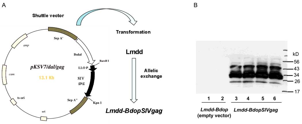
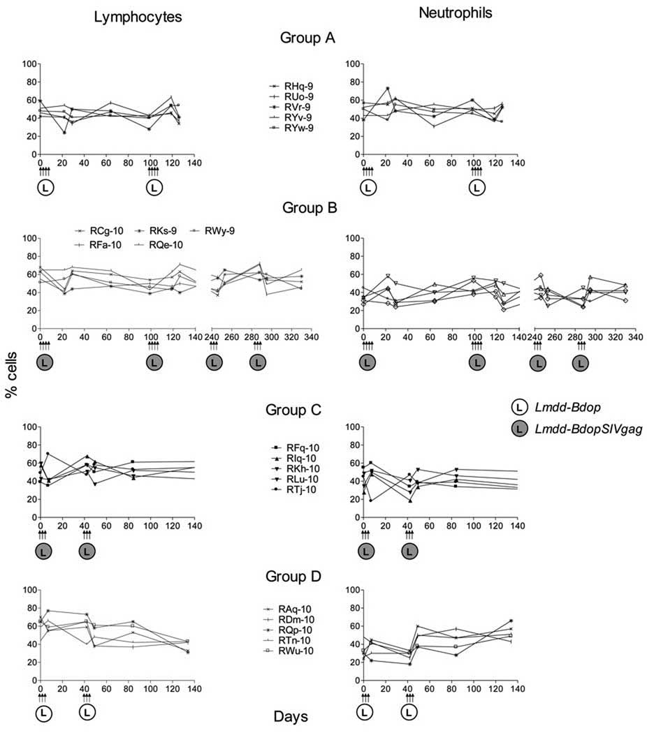
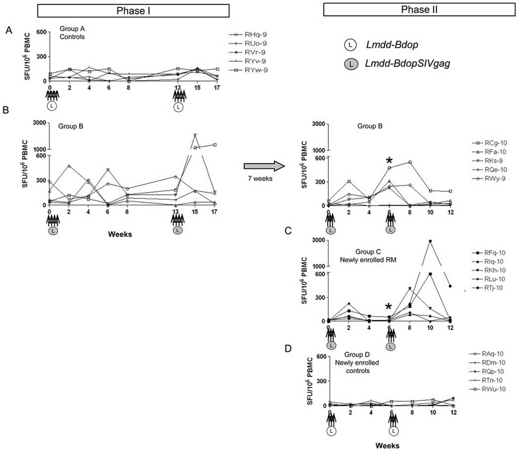
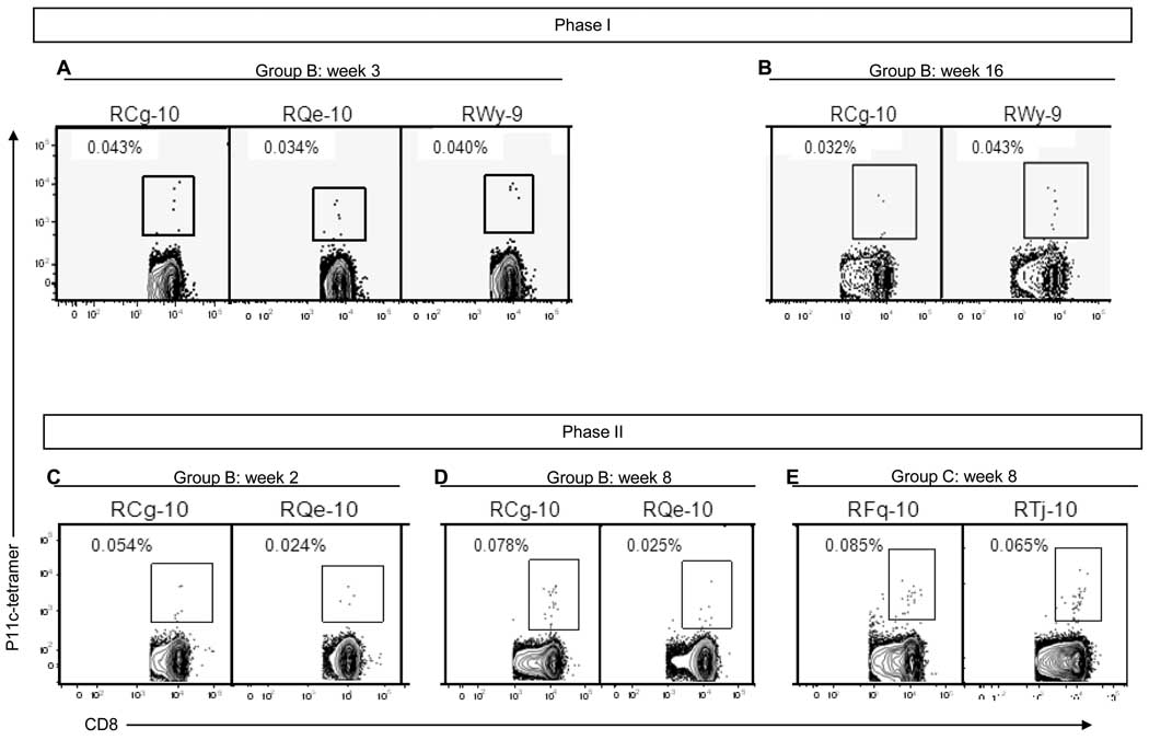
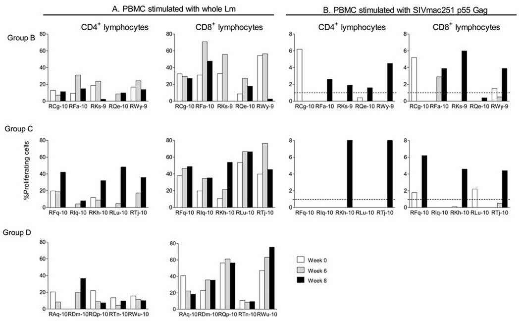
Listeria monocytogenes (Lm) is known to induce strong cellular immune responses. We constructed a live-attenuated Lm vector, Lmdd-BdopSIVgag, which encodes SIVmac239 gag. Intragastric (i.g.) administration of 3 × 10(12) bacteria to rhesus macaques was safe and induced anti-Gag cellular but no humoral immune responses. Boosting of Gag-specific cellular responses was observed after i.g. administration of Lmdd-BdopSIVgag to previously vaccinated RM despite preexisting anti-Lm immunity shown by lymphoproliferative responses. Surprisingly, anti-Lm cellular responses were also detected in non-vaccinated controls, which may reflect the fact that Lm is a ubiquitous bacterium. The novel, live-attenuated Lmdd-BdopSIVgag may be an attractive platform for oral vaccine delivery.
Copyright © 2010 Elsevier Ltd. All rights reserved.
Figures
Similar articles
-
Prime-boost vaccination with heterologous live vectors encoding SIV gag and multimeric HIV-1 gp160 protein: efficacy against repeated mucosal R5 clade C SHIV challenges.Vaccine. 2011 Aug 5;29(34):5611-22. doi: 10.1016/j.vaccine.2011.06.017. Epub 2011 Jul 14. Vaccine. 2011. PMID: 21693155 Free PMC article.
-
Live attenuated Listeria monocytogenes expressing HIV Gag: immunogenicity in rhesus monkeys.Vaccine. 2007 Oct 16;25(42):7470-9. doi: 10.1016/j.vaccine.2007.08.013. Epub 2007 Aug 27. Vaccine. 2007. PMID: 17854955 Free PMC article.
-
Recombinant rubella vectors elicit SIV Gag-specific T cell responses with cytotoxic potential in rhesus macaques.Vaccine. 2015 Apr 27;33(18):2167-74. doi: 10.1016/j.vaccine.2015.02.067. Epub 2015 Mar 21. Vaccine. 2015. PMID: 25802183 Free PMC article.
-
Robust vaccine-elicited cellular immune responses in breast milk following systemic simian immunodeficiency virus DNA prime and live virus vector boost vaccination of lactating rhesus monkeys.J Immunol. 2010 Dec 1;185(11):7097-106. doi: 10.4049/jimmunol.1002751. Epub 2010 Nov 1. J Immunol. 2010. PMID: 21041730 Free PMC article.
-
An attenuated Listeria monocytogenes vector primes more potent simian immunodeficiency virus-specific mucosal immunity than DNA vaccines in mice.J Virol. 2013 Apr;87(8):4751-5. doi: 10.1128/JVI.03085-12. Epub 2013 Feb 6. J Virol. 2013. PMID: 23388715 Free PMC article.
Cited by
-
Bacteria and cells as alternative nano-carriers for biomedical applications.Expert Opin Drug Deliv. 2022 Jan;19(1):103-118. doi: 10.1080/17425247.2022.2029844. Epub 2022 Jan 25. Expert Opin Drug Deliv. 2022. PMID: 35076351 Free PMC article. Review.
-
The attenuated hepatocellular carcinoma-specific Listeria vaccine Lmdd-MPFG prevents tumor occurrence through immune regulation of dendritic cells.Oncotarget. 2015 Apr 20;6(11):8822-38. doi: 10.18632/oncotarget.3558. Oncotarget. 2015. PMID: 25826093 Free PMC article.
-
Dendritic cell-based immunity and vaccination against hepatitis C virus infection.Immunology. 2012 Aug;136(4):385-96. doi: 10.1111/j.1365-2567.2012.03590.x. Immunology. 2012. PMID: 22486354 Free PMC article. Review.
-
A Listeria ivanovii balanced-lethal system may be a promising antigen carrier for vaccine construction.Microb Biotechnol. 2022 Nov;15(11):2831-2844. doi: 10.1111/1751-7915.14137. Epub 2022 Sep 7. Microb Biotechnol. 2022. PMID: 36069650 Free PMC article.
-
Prime-boost vaccination with heterologous live vectors encoding SIV gag and multimeric HIV-1 gp160 protein: efficacy against repeated mucosal R5 clade C SHIV challenges.Vaccine. 2011 Aug 5;29(34):5611-22. doi: 10.1016/j.vaccine.2011.06.017. Epub 2011 Jul 14. Vaccine. 2011. PMID: 21693155 Free PMC article.
References
-
- Baba TW, Liska V, Hofmann-Lehmann R, Vlasak J, Xu W, Ayehunie S, et al. Human neutralizing monoclonal antibodies of the IgG1 subtype protect against mucosal simian-human immunodeficiency virus infection. Nat Med. 2000 Feb;6(2):200–206. - PubMed
-
- Mascola JR, Stiegler G, VanCott TC, Katinger H, Carpenter CB, Hanson CE, et al. Protection of macaques against vaginal transmission of a pathogenic HIV-1/SIV chimeric virus by passive infusion of neutralizing antibodies. Nat Med. 2000 Feb;6(2):207–210. - PubMed
Publication types
MeSH terms
Substances
Grants and funding
LinkOut - more resources
Full Text Sources
