. 2011 Jan 1;50(1):179-95.
doi: 10.1016/j.freeradbiomed.2010.11.002. Epub 2010 Nov 9.

Fatty acid amide hydrolase is a key regulator of endocannabinoid-induced myocardial tissue injury

Affiliations

Fatty acid amide hydrolase is a key regulator of endocannabinoid-induced myocardial tissue injury

Partha Mukhopadhyay et al. Free Radic Biol Med. .

Abstract

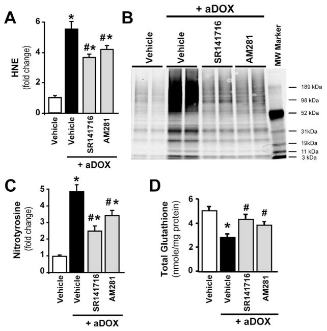
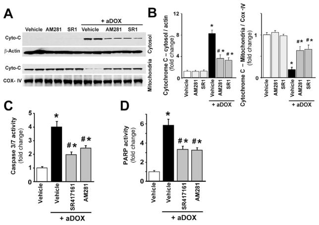
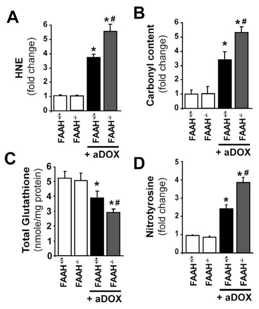
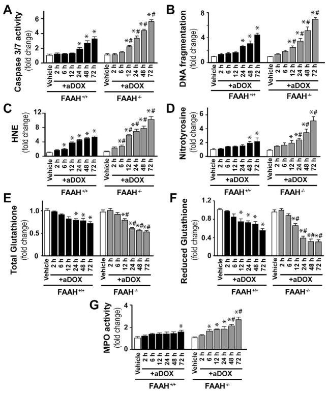
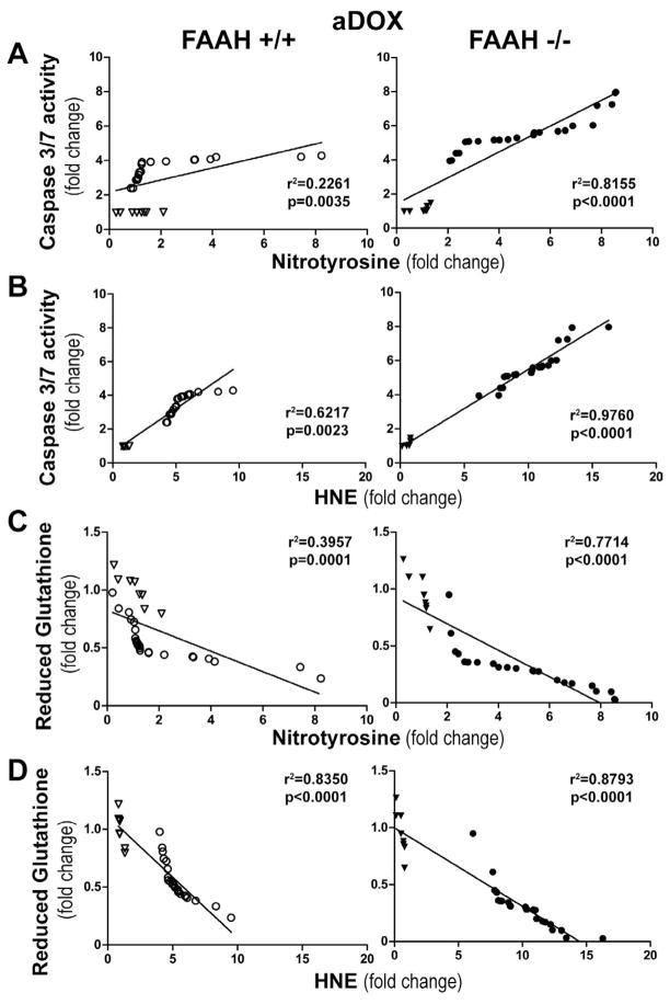
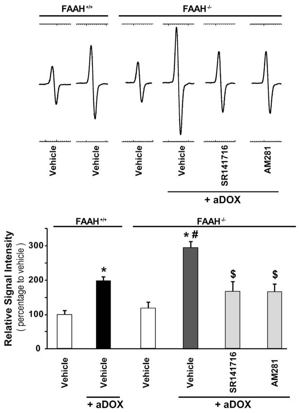
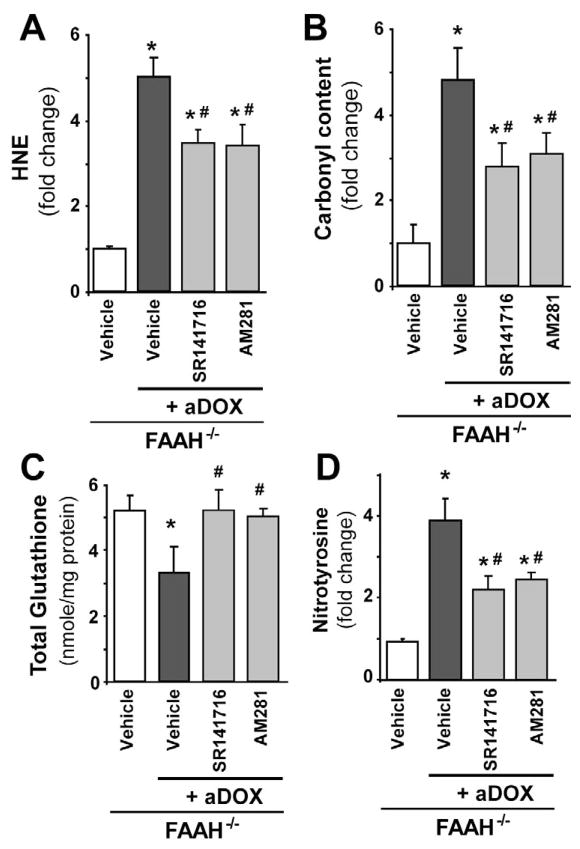
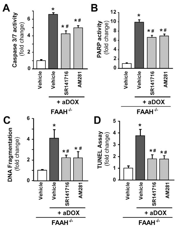
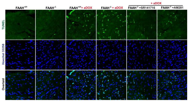
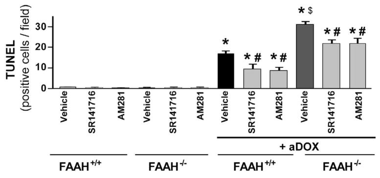
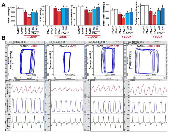
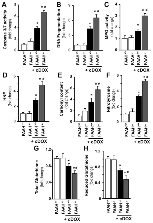
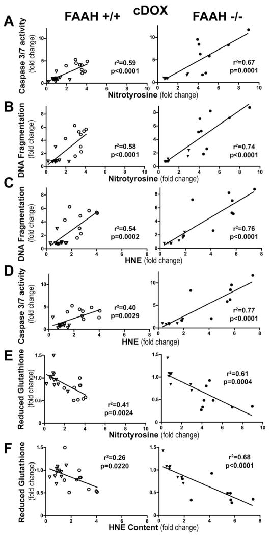
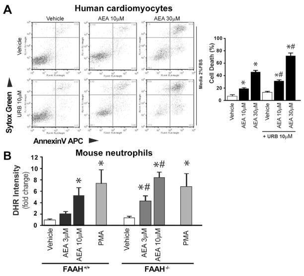
Previous studies have suggested that increased levels of endocannabinoids in various cardiovascular disorders (e.g., various forms of shock, cardiomyopathies, atherosclerosis) through the activation of CB(1) cannabinoid receptors may promote cardiovascular dysfunction and tissue injury. We have investigated the role of the main endocannabinoid anandamide-metabolizing enzyme (fatty acid amide hydrolase; FAAH) in myocardial injury induced by an important chemotherapeutic drug, doxorubicin (DOX; known for its cardiotoxicity mediated by increased reactive oxygen and nitrogen species generation), using well-established acute and chronic cardiomyopathy models in mice. The DOX-induced myocardial oxidative/nitrative stress (increased 4-hydroxynonenal, protein carbonyl, and nitrotyrosine levels and decreased glutathione content) correlated with multiple cell death markers, which were enhanced in FAAH knockout mice exhibiting significantly increased DOX-induced mortality and cardiac dysfunction compared to their wild type. The effects of DOX in FAAH knockouts were attenuated by CB(1) receptor antagonists. Furthermore, anandamide induced enhanced cell death in human cardiomyocytes pretreated with FAAH inhibitor and enhanced sensitivity to ROS generation in inflammatory cells of FAAH knockouts. These results suggest that in pathological conditions associated with acute oxidative/nitrative stress FAAH plays a key role in controlling the tissue injury that is, at least in part, mediated by the activation of CB(1) receptors by endocannabinoids.


Conflict of interest statement

Disclosures: No conflict of interest to disclose.

Figures

Figure 1
Figure 1. Cannabinoid CB1 receptor inhibition attenuates acute DOX-induced increased myocardial oxidative and nitrative stress and impaired antioxidant defense 5-day following DOX administration
Panel A: DOX administration enhanced myocardial HNE levels (marker of oxidative stress), which were attenuated by CB1 antagonists AM281 and SR141716. Panel B: Immunoprecipitation for myocardial nitrotyrosine (NT; marker of nitrative stress) shows DOX-induced increased NT formation, which was attenuated by CB1 antagonists. Panel C: Experiments from quantitative NT ELISA demonstrate DOX-induced increased myocardial NT formation, which was attenuated by CB1 antagonists. Panel D: Measurements of myocardial glutathione content showed DOX-induced decreases, which was attenuated by CB1 antagonists. Results are mean±SEM of 4–7 experiments/group for panels A, C and D. *P<0.05 vs. vehicle; #P<0.05 vs. DOX treated mice.
Figure 2
Figure 2. Cannabinoid CB1 receptor inhibition attenuates acute DOX-induced increased myocardial cytochrome C release and cell death 5-day following DOX administration
Panel A: representative immunoblot from myocardium shows enhanced release of cytochrome C (Cyto-C) into the cytosol after DOX administration, which was attenuated with CB1 receptor antagonists AM281 and SR141716. Panel B: quantification of cytochrome C release (in the cytosol it is normalized to β-actin, in the mitochondria to COX-IV). Panels C and D: DOX administration enhanced myocardial Caspase 3/7 (Panel C) and PARP (Panel D) activities (markers of cell death), which were attenuated by CB1 antagonists AM281 and SR141716. Results are mean±SEM of 6–8 experiments/group. *P<0.05 vs. vehicle; #P<0.05 vs. DOX treated mice.
Figure 3
Figure 3. Genetic deletion of FAAH is associated with enhanced acute DOX-induced myocardial cell death 3-day following DOX administration and decreased survival thereafter
Panel A: FAAH−/− mice treated with DOX show markedly reduced survival, compared to their wild type (FAAH+/+) littermates (n=11 and 13/group). P<0.05 vs. DOX-treated mice by Kaplan-Meier survival analysis. Panels B–D shows markedly increased myocardial cell death (both apoptotic and PARP-dependent) in FAAH−/− mice treated with DOX compared to their wild type (FAAH+/+) littermates. Results are mean±SEM of 6–8 experiments/group. *P<0.05 vs. vehicle in FAAH+/+ or FAAH−/− mice; #P<0.05 vs. DOX in FAAH+/+ treated mice.
Figure 4
Figure 4. Genetic deletion of FAAH is associated with enhanced acute DOX-induced myocardial oxidative and nitrative stress 3-day following DOX administration
Panels A–D show that FAAH−/− mice treated with DOX had significantly increased oxidative and nitrative stress compared to their wild type (FAAH+/+) littermates. Results are mean±SEM of 5–8 experiments/group. *P<0.05 vs. vehicle in FAAH+/+ or FAAH−/− mice; #P<0.05 vs. DOX in FAAH+/+ treated mice.
Figure 5
Figure 5. Genetic deletion of FAAH is associated with time-dependently enhanced DOX-induced cell death, oxidative and nitrative stress
Panel A–G show time-dependent increase of DOX-induced myocardial cell death, oxidative and nitrative stress, neutrophil infiltration (MPO activity) and decrease in reduced as well as total glutathione contents. These changes were aggravated in the absence of FAAH. Results are mean±SEM of 16–36 experiments/group. *P<0.05 vs. vehicle in FAAH+/+ or FAAH−/− mice; #P<0.05 vs. DOX in FAAH+/+ treated mice.
Figure 6
Figure 6. Myocardial oxidative and nitrative stress markers show direct positive correlation with cell death marker and negative correlation with reduced glutathione content 3 days following the administration of an acute single dose of DOX in FAAH+/+ and FAAH−/− mice
Panel A–B show direct correlation of nitrotyrosine and HNE protein adducts with caspase 3/7 activity in FAAH+/+ and FAAH−/− mice. Panel C–D show inverse correlation of reduced glutathione with nitrotyrosine and HNE protein adducts in FAAH+/+ and FAAH−/− mice. Results are mean±SEM of 8–24 experiments/group. Hollow triangles and circles denote data points from FAAH+/+ mice without and with DOX (left), respectively, while filled triangles and circles indicate data points from FAAH−/− mice without and with DOX (right), respectively.
Figure 7
Figure 7. The enhanced acute DOX-induced myocardial ROS generation in FAAH−/− mice is attenuated by CB1 antagonists
Upper representative EPR traces show markedly increased DOX-induced myocardial ROS generation in FAAH−/− mice compared to FAAH+/+ mice 3 days following DOX administration, which is attenuated by CB1 antagonists SR141716 and AM281. Below panel shows quantifications. Results are mean±SEM of 4–5 experiments/group. *P<0.05 vs. vehicle in FAAH+/+ or FAAH−/− mice; #P<0.05 vs. DOX in FAAH+/+ treated mice; $<0.05 vs. DOX in FAAH−/− treated mice.
Figure 8
Figure 8. The enhanced acute DOX-induced myocardial oxidative/nitrative stress in FAAH−/− mice is attenuated by CB1 antagonists
Panels A–D show markedly increased DOX-induced myocardial oxidative (HNE, protein carbonyl) and nitrative (nitrotyrosine) stress in FAAH−/− mice compared to FAAH+/+ littermates 3 days following DOX administration, which is attenuated by CB1 antagonists SR141716 and AM281. Results are mean±SEM of 5–7 experiments/group. *P<0.05 vs. vehicle in FAAH−/− mice; #P<0.05 vs. DOX in FAAH−/− mice.
Figure 9
Figure 9. The enhanced acute DOX-induced myocardial cell death in FAAH−/− mice is attenuated by CB1 antagonists
Panels A–D show markedly increased DOX-induced myocardial cell death in FAAH−/− mice compared to FAAH+/+ littermates 3 days following DOX administration, which is attenuated by CB1 antagonists SR141716 and AM281. Results are mean±SEM of 5–8 experiments/group. *P<0.05 vs. vehicle in FAAH−/− mice; #P<0.05 vs. DOX in FAAH−/− mice.
Figure 10
Figure 10. The enhanced acute DOX-induced myocardial apoptosis in FAAH−/− mice is attenuated by CB1 antagonists
Panels A: the upper row of images (green) shows representative TUNEL staining 3 days following DOX administration, the middle row (dark blue) is the nuclear staining, while the bottom one is the colocalization of both (light blue/white nuclei represents TUNEL positive cells). Panel B: quantification of myocardial TUNEL staining. Results are mean±SEM of 10–12 frames/group from 3–4 different hears. *P<0.05 vs. vehicle in FAAH−/− or FAAH+/+ mice; #P<0.05 vs. DOX in FAAH+/+ or FAAH−/− mice; $P<0.05 vs. DOX in FAAH+/+ mice.
Figure 11
Figure 11. FAAH−/− mice develop more severe acute DOX-induced cardiac dysfunction
Panel A shows decrease in markers of left ventricular contractile function (maximum first derivative of ventricular pressure with respect to time (+dP/dt), maximal left ventricular pressure (LVP), ejection fraction, stroke work and cardiac output) in FAAH+/+ and FAAH−/− mice 3 days following acute DOX treatment (aDOX). The DOX-induced myocardial dysfunction was significantly more pronounced in FAAH−/− mice compared to their wild-type littermates (Figure 11A–B) and was improved by CB1 antagonists SR141716 (SR1) or AM281 (AM) treatment (Figure 11A–B). Results are mean±SEM of 5–7 experiments/group. *P<0.05 vs. vehicle in FAAH+/+ or FAAH−/− mice; #P<0.05 vs. DOX in FAAH+/+ mice; $P<0.05 vs. DOX in FAAH−/− mice. Panel B shows representative pressure-volume (P–V) loops (upper row, blue), volume signal (second row, red), left ventricular (LV) pressure signal (third row, blue) and derivative of the LV pressure signal (bottom row, green) in FAAH+/+ and FAAH−/− mice 3 days following acute DOX administration, and in FAAH−/− mice treated with DOX with CB1 antagonists. Please note that the rightward shift of the P–V loops, decrease in stroke volume and in the LV pressure signal amplitude and maximum first derivative of LV pressure with respect to time (+dP/dt) indicates depressed myocardial contractile performance.
Figure 12
Figure 12. Enhanced chronic DOX-induced myocardial cell death, inflammation, and oxidative and nitrative stress in FAAH−/− mice
FAAH−/− mice treated with multiple low doses of DOX had markedly increased myocardial cell death (Panel A–B), neutrophil infiltration (Panel C; MPO activity) and significantly increased oxidative and nitrative stress (Panel D–H) compared to their wild type (FAAH+/+) littermates 30 days following the first DOX administration. Results are mean±SEM of 8–12 experiments/group. *P<0.05 vs. vehicle in FAAH+/+ or FAAH−/− mice; #P<0.05 vs. DOX in FAAH+/+ treated mice.
Figure 13
Figure 13. Myocardial oxidative and nitrative stress markers show direct correlation with cell death and reduced glutathione content after chronic DOX administration
Panels A–D show direct correlation of myocardial nitrotyrosine and HNE with DNA fragmentation and caspase 3/7 activity in FAAH+/+ and FAAH−/− mice. Panels E–F show inverse correlation of reduced myocardial glutathione content with nitrotyrosine and HNE protein adduct content in FAAH+/+ (open) and FAAH−/− mice (solid) after vehicle (triangle) and DOX (circle) treatment. Results are mean±SEM of 8 experiments/group.
Figure 14
Figure 14. Anandamide enhances cell death in human cardiomyocytes treated with FAAH inhibitor, and increased ROS production in FAAH knockout neutrophils
Panel A shows that anandamide (AEA) enhances concentration-dependent cell death in human primary cardiomyocytes, which is more pronounced in the presence of FAAH inhibitor URB527. Panel B shows the dose concentration-dependent effect of anandamide (AEA) on respiratory burst in neutrophil granulocytes isolated from FAAH+/+ and FAAH−/− mice. Phorbol Ester (PA) was used as positive control.

Similar articles

Cited by

References

    1. Howlett AC, Barth F, Bonner TI, Cabral G, Casellas P, Devane WA, Felder CC, Herkenham M, Mackie K, Martin BR, Mechoulam R, Pertwee RG. International Union of Pharmacology. XXVII. Classification of cannabinoid receptors. Pharmacol Rev. 2002;54:161–202. - PubMed
    1. Pacher P, Batkai S, Kunos G. The endocannabinoid system as an emerging target of pharmacotherapy. Pharmacol Rev. 2006;58:389–462. - PMC - PubMed
    1. Pacher P, Steffens S. The emerging role of the endocannabinoid system in cardiovascular disease. Semin Immunopathol. 2009;31:63–77. - PMC - PubMed
    1. Pacher P, Mukhopadhyay P, Mohanraj R, Godlewski G, Batkai S, Kunos G. Modulation of the endocannabinoid system in cardiovascular disease: therapeutic potential and limitations. Hypertension. 2008;52:601–607. - PMC - PubMed
    1. Batkai S, Pacher P. Endocannabinoids and cardiac contractile function: pathophysiological implications. Pharmacol Res. 2009;60:99–106. - PMC - PubMed

Publication types

MeSH terms