

Innate immune activation by the viral PAMP poly I:C potentiates pulmonary graft-versus-host disease after allogeneic hematopoietic cell transplant
- PMID: 21070856
- PMCID: PMC3015000
- DOI: 10.1016/j.trim.2010.11.004
Innate immune activation by the viral PAMP poly I:C potentiates pulmonary graft-versus-host disease after allogeneic hematopoietic cell transplant
Abstract
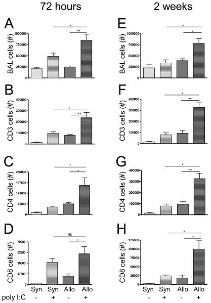
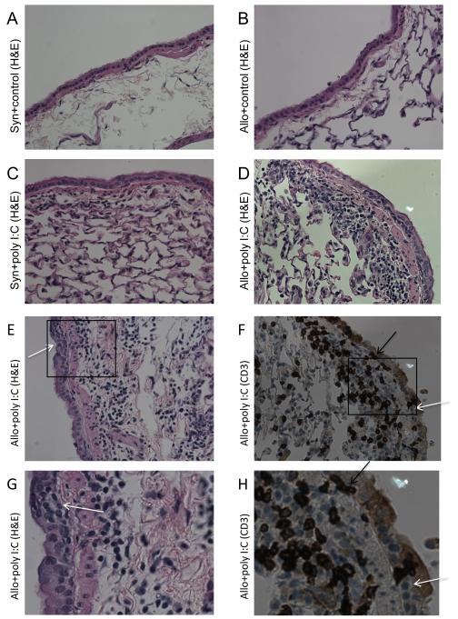
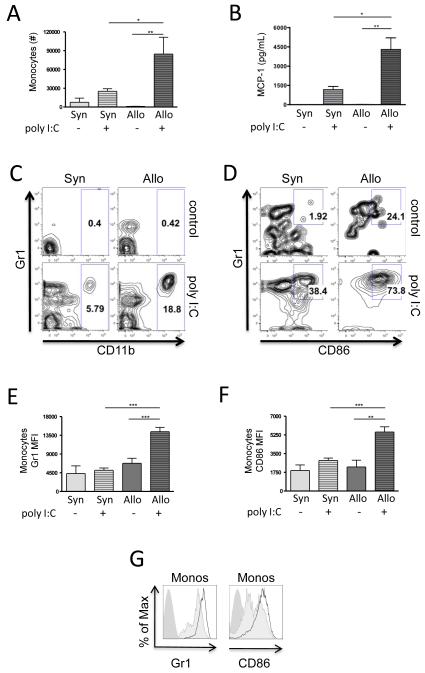
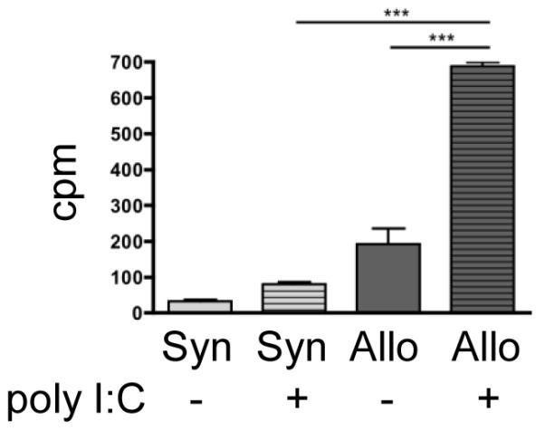
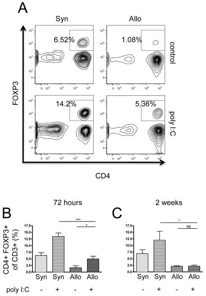
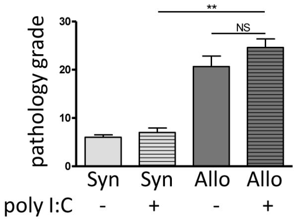
Respiratory viral infections cause significant morbidity and increase the risk for chronic pulmonary graft-versus-host disease (GVHD) after hematopoietic cell transplantation (HCT). Our overall hypothesis is that local innate immune activation potentiates adaptive alloimmunity. In this study, we hypothesized that a viral pathogen-associated molecular pattern (PAMP) alone can potentiate pulmonary GVHD after allogeneic HCT. We, therefore, examined the effect of pulmonary exposure to polyinosinic:polycytidylic acid (poly I:C), a viral mimetic that activates innate immunity, in an established murine HCT model. Poly I:C-induced a marked pulmonary T cell response in allogeneic HCT mice as compared to syngeneic HCT, with increased CD4+ cells in the lung fluid and tissue. This lymphocytic inflammation persisted at 2 weeks post poly I:C exposure in allogeneic mice and was associated with CD3+ cell infiltration into the bronchiolar epithelium and features of epithelial injury. In vitro, poly I:C enhanced allospecific proliferation in a mixed lymphocyte reaction. In vivo, poly I:C exposure was associated with an early increase in pulmonary monocyte recruitment and activation as well as a decrease in CD4+FOXP3+ regulatory T cells in allogeneic mice as compared to syngeneic. In contrast, intrapulmonary poly I:C did not alter the extent of systemic GVHD in either syngeneic or allogeneic mice. Collectively, our results suggest that local activation of pulmonary innate immunity by a viral molecular pattern represents a novel pathway that contributes to pulmonary GVHD after allogeneic HCT, through a mechanism that includes increased recruitment and maturation of intrapulmonary monocytes.
Copyright © 2010 Elsevier B.V. All rights reserved.
Figures
Similar articles
-
Protective role of T-bet and Th1 cytokines in pulmonary graft-versus-host disease and peribronchiolar fibrosis.Am J Respir Cell Mol Biol. 2012 Feb;46(2):249-56. doi: 10.1165/rcmb.2011-0131OC. Epub 2011 Sep 29. Am J Respir Cell Mol Biol. 2012. PMID: 21960548 Free PMC article.
-
Innate immune activation potentiates alloimmune lung disease independent of chemokine (C-X-C motif) receptor 3.J Heart Lung Transplant. 2011 Jun;30(6):717-25. doi: 10.1016/j.healun.2011.01.711. Epub 2011 Mar 27. J Heart Lung Transplant. 2011. PMID: 21444213 Free PMC article.
-
Allogeneic splenocyte transfer and lipopolysaccharide inhalations induce differential T cell expansion and lung injury: a novel model of pulmonary graft-versus-host disease.PLoS One. 2014 May 20;9(5):e97951. doi: 10.1371/journal.pone.0097951. eCollection 2014. PLoS One. 2014. PMID: 24844383 Free PMC article.
-
Alloreactivity as therapeutic principle in the treatment of hematologic malignancies. Studies of clinical and immunologic aspects of allogeneic hematopoietic cell transplantation with nonmyeloablative conditioning.Dan Med Bull. 2007 May;54(2):112-39. Dan Med Bull. 2007. PMID: 17521527 Review.
-
Hematopoietic stem cell graft manipulation as a mechanism of immunotherapy.Int Immunopharmacol. 2003 Aug;3(8):1121-43. doi: 10.1016/S1567-5769(03)00014-6. Int Immunopharmacol. 2003. PMID: 12860168 Review.
Cited by
-
Novel management of vaginal chronic graft-versus-host disease causing haematometra and haematocolpos.BMJ Case Rep. 2018 Apr 28;2018:bcr2017222720. doi: 10.1136/bcr-2017-222720. BMJ Case Rep. 2018. PMID: 29705733 Free PMC article.
-
Protective role of T-bet and Th1 cytokines in pulmonary graft-versus-host disease and peribronchiolar fibrosis.Am J Respir Cell Mol Biol. 2012 Feb;46(2):249-56. doi: 10.1165/rcmb.2011-0131OC. Epub 2011 Sep 29. Am J Respir Cell Mol Biol. 2012. PMID: 21960548 Free PMC article.
-
Nonalloreactive T cells prevent donor lymphocyte infusion-induced graft-versus-host disease by controlling microbial stimuli.J Immunol. 2012 Dec 15;189(12):5572-81. doi: 10.4049/jimmunol.1200045. Epub 2012 Nov 7. J Immunol. 2012. PMID: 23136200 Free PMC article.
-
The Microbiome, Systemic Immune Function, and Allotransplantation.Clin Microbiol Rev. 2016 Jan;29(1):191-9. doi: 10.1128/CMR.00063-15. Clin Microbiol Rev. 2016. PMID: 26656674 Free PMC article. Review.
-
Induction of endogenous retroelements as a potential mechanism for mouse-specific drug-induced carcinogenicity.PLoS One. 2017 May 4;12(5):e0176768. doi: 10.1371/journal.pone.0176768. eCollection 2017. PLoS One. 2017. PMID: 28472135 Free PMC article.
References
-
- Ferrara JL, Deeg HJ. Graft-versus-host disease. N Engl J Med. 1991;324(10):667. - PubMed
-
- Socie G, Stone JV, Wingard JR, et al. Long-term survival and late deaths after allogeneic bone marrow transplantation. Late Effects Working Committee of the International Bone Marrow Transplant Registry. N Engl J Med. 1999;341(1):14. - PubMed
-
- Chao NJ, Schmidt GM, Niland JC, et al. Cyclosporine, methotrexate, and prednisone compared with cyclosporine and prednisone for prophylaxis of acute graft-versus-host disease. N Engl J Med. 1993;329(17):1225. - PubMed
-
- Afessa B, Litzow MR, Tefferi A. Bronchiolitis obliterans and other late onset non-infectious pulmonary complications in hematopoietic stem cell transplantation. Bone Marrow Transplant. 2001;28(5):425. - PubMed
-
- Versluys AB, Rossen JW, van Ewijk B, Schuurman R, Bierings MB, Boelens JJ. Strong association between respiratory viral infection early after hematopoietic stem cell transplantation and the development of life-threatening acute and chronic alloimmune lung syndromes. Biol Blood Marrow Transplant. 2010;16(6):782. - PMC - PubMed
Publication types
MeSH terms
Substances
Grants and funding
LinkOut - more resources
Full Text Sources
Medical
Research Materials
