

Genetic identification of unique immunological responses in mice infected with virulent and attenuated Francisella tularensis
- PMID: 21070859
- PMCID: PMC3031720
- DOI: 10.1016/j.micinf.2010.10.022
Genetic identification of unique immunological responses in mice infected with virulent and attenuated Francisella tularensis
Abstract
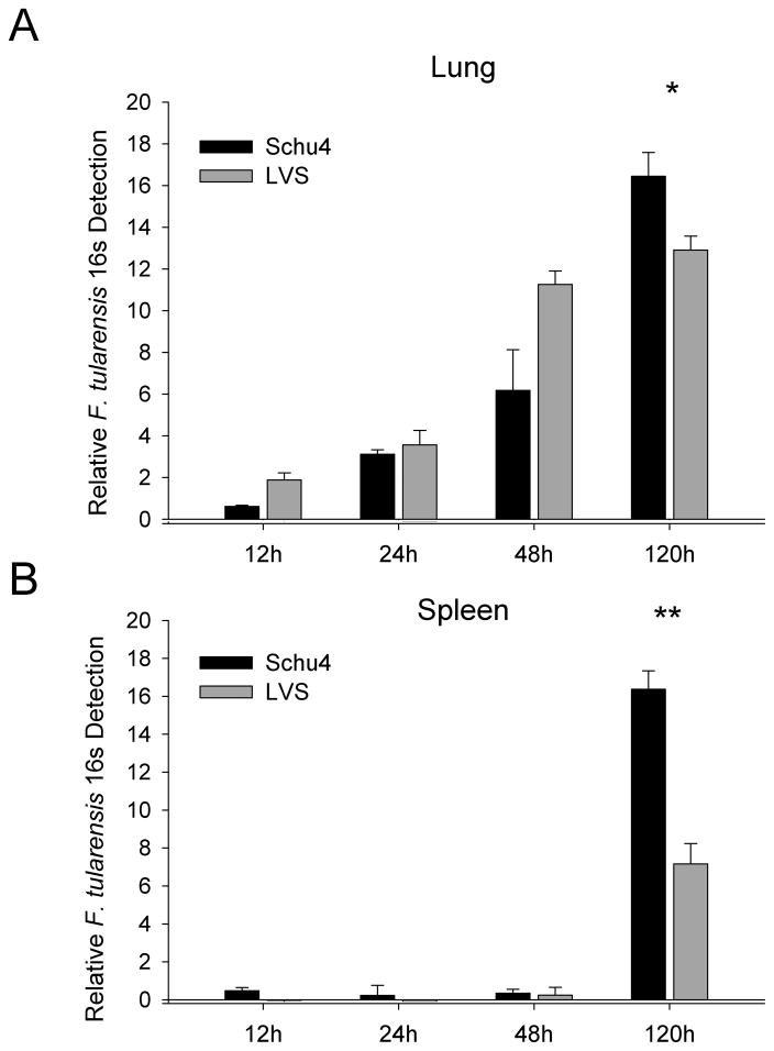
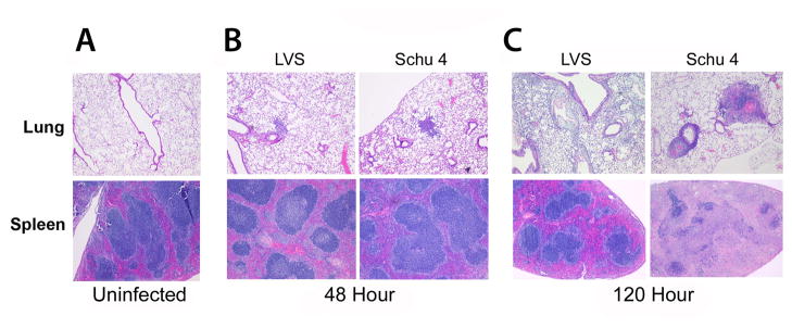
Francisella tularensis is a category A select agent based on its infectivity and virulence but disease mechanisms in infection remain poorly understood. Murine pulmonary models of infection were therefore employed to assess and compare dissemination and pathology and to elucidate the host immune response to infection with the highly virulent Type A F. tularensis strain Schu4 versus the less virulent Type B live vaccine strain (LVS). We found that dissemination and pathology in the spleen was significantly greater in mice infected with F. tularensis Schu4 compared to mice infected with F. tularensis LVS. Using gene expression profiling to compare the response to infection with the two F. tularensis strains, we found that there were significant differences in the expression of genes involved in the apoptosis pathway, antigen processing and presentation pathways, and inflammatory response pathways in mice infected with Schu4 when compared to LVS. These transcriptional differences coincided with marked differences in dissemination and severity of organ lesions in mice infected with the Schu4 and LVS strains. Therefore, these findings indicate that altered apoptosis, antigen presentation and production of inflammatory mediators explain the differences in pathogenicity of F. tularensis Schu4 and LVS.
Copyright © 2010 Institut Pasteur. Published by Elsevier SAS. All rights reserved.
Figures
Similar articles
-
Interleukin-17 protects against the Francisella tularensis live vaccine strain but not against a virulent F. tularensis type A strain.Infect Immun. 2013 Sep;81(9):3099-105. doi: 10.1128/IAI.00203-13. Epub 2013 Jun 17. Infect Immun. 2013. PMID: 23774604 Free PMC article.
-
Oral immunization of mice with the live vaccine strain (LVS) of Francisella tularensis protects mice against respiratory challenge with virulent type A F. tularensis.Vaccine. 2007 May 10;25(19):3781-91. doi: 10.1016/j.vaccine.2007.02.014. Epub 2007 Feb 26. Vaccine. 2007. PMID: 17346863 Free PMC article.
-
Tularemia in BALB/c and C57BL/6 mice vaccinated with Francisella tularensis LVS and challenged intradermally, or by aerosol with virulent isolates of the pathogen: protection varies depending on pathogen virulence, route of exposure, and host genetic background.Vaccine. 2003 Sep 8;21(25-26):3690-700. doi: 10.1016/s0264-410x(03)00386-4. Vaccine. 2003. PMID: 12922099
-
Live Attenuated Tularemia Vaccines for Protection Against Respiratory Challenge With Virulent F. tularensis subsp. tularensis.Front Cell Infect Microbiol. 2018 May 15;8:154. doi: 10.3389/fcimb.2018.00154. eCollection 2018. Front Cell Infect Microbiol. 2018. PMID: 29868510 Free PMC article. Review.
-
Programmed cell death and the pathogenesis of tissue injury induced by type A Francisella tularensis.FEMS Microbiol Lett. 2009 Nov;301(1):1-11. doi: 10.1111/j.1574-6968.2009.01791.x. Epub 2009 Sep 17. FEMS Microbiol Lett. 2009. PMID: 19811540 Free PMC article. Review.
Cited by
-
TPR1, a novel rifampicin derivative, demonstrates efficacy alone and in combination with doxycycline against the NIAID Category A priority pathogen Francisella tularensis.JAC Antimicrob Resist. 2021 May 4;3(2):dlab058. doi: 10.1093/jacamr/dlab058. eCollection 2021 Jun. JAC Antimicrob Resist. 2021. PMID: 34223120 Free PMC article.
-
Alginate microencapsulation of an attenuated O-antigen mutant of Francisella tularensis LVS as a model for a vaccine delivery vehicle.PLoS One. 2022 Mar 11;17(3):e0259807. doi: 10.1371/journal.pone.0259807. eCollection 2022. PLoS One. 2022. PMID: 35275912 Free PMC article.
-
Detrimental Influence of Alveolar Macrophages on Protective Humoral Immunity during Francisella tularensis SchuS4 Pulmonary Infection.Infect Immun. 2018 Mar 22;86(4):e00787-17. doi: 10.1128/IAI.00787-17. Print 2018 Apr. Infect Immun. 2018. PMID: 29311236 Free PMC article.
-
The ly49 gene family. A brief guide to the nomenclature, genetics, and role in intracellular infection.Front Immunol. 2013 Apr 16;4:90. doi: 10.3389/fimmu.2013.00090. eCollection 2013. Front Immunol. 2013. PMID: 23596445 Free PMC article.
-
Formulation studies of InhA inhibitors and combination therapy to improve efficacy against Mycobacterium tuberculosis.Tuberculosis (Edinb). 2016 Dec;101:8-14. doi: 10.1016/j.tube.2016.07.016. Epub 2016 Aug 3. Tuberculosis (Edinb). 2016. PMID: 27865404 Free PMC article.
References
-
- Dennis DT, Inglesby TV, Henderson DA, Bartlett JG, Ascher MS, Eitzen E, Fine AD, Friedlander AM, Hauer J, Layton M, Lillibridge SR, McDade JE, Osterholm MT, O’Toole T, Parker G, Perl TM, Russell PK, Tonat K. Tularemia as a biological weapon: medical and public health management. JAMA. 2001;285:2763–2773. - PubMed
-
- Petersen JM, Schriefer ME. Tularemia: emergence/re-emergence. Vet Res. 2005;36:455–467. - PubMed
-
- Edwin LO, Tigertt WD, Paul JK, Martha KW, Charkes ND, Robert MR, Theodore ES, Mallory S. An analysis of forty-two cases of laboratory-acquired tularemia: Treatment with broad spectrum antibiotics. The American journal of medicine. 1961;30:785–806. - PubMed
-
- Chocarro A, Gonzalez A, Garcia I. Treatment of Tularemia with Ciprofloxacin. Clinical Infectious Diseases. 2000;31:623–623. - PubMed
Publication types
MeSH terms
Grants and funding
LinkOut - more resources
Full Text Sources
Molecular Biology Databases
