. 2010 Nov 10;18(11):1420-30.
doi: 10.1016/j.str.2010.08.018.

Engagement of arginine finger to ATP triggers large conformational changes in NtrC1 AAA+ ATPase for remodeling bacterial RNA polymerase

Affiliations

Engagement of arginine finger to ATP triggers large conformational changes in NtrC1 AAA+ ATPase for remodeling bacterial RNA polymerase

Baoyu Chen et al. Structure. .

Abstract

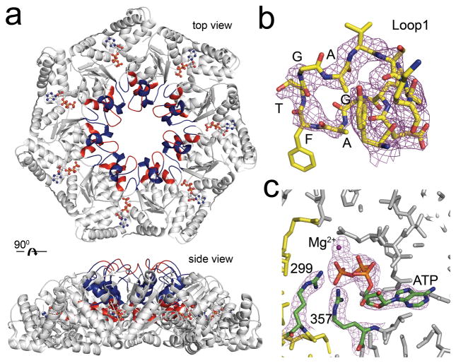
The NtrC-like AAA+ ATPases control virulence and other important bacterial activities through delivering mechanical work to σ54-RNA polymerase to activate transcription from σ54-dependent genes. We report the first crystal structure for such an ATPase, NtrC1 of Aquifex aeolicus, in which the catalytic arginine engages the γ-phosphate of ATP. Comparing the new structure with those previously known for apo and ADP-bound states supports a rigid-body displacement model that is consistent with large-scale conformational changes observed by low-resolution methods. First, the arginine finger induces rigid-body roll, extending surface loops above the plane of the ATPase ring to bind σ54. Second, ATP hydrolysis permits Pi release and retraction of the arginine with a reversed roll, remodeling σ54-RNAP. This model provides a fresh perspective on how ATPase subunits interact within the ring-ensemble to promote transcription, directing attention to structural changes on the arginine-finger side of an ATP-bound interface.


Conflict of interest statement

COMPETING INTERESTS STATEMENT

The authors declare that they have no competing financial interests.

Figures

Figure 1
Figure 1
Properties of amino acid variant NtrC1C,E239A. (a) Size-exclusion chromatography profiles illustrate the ability of NtrC1C,E239A to self-assemble and to bind to σ54 in the presence of Mg/ATP. Elution profiles are shown for ATPase alone (green); ATPase with nucleotide (red); and ATPase plus excess σ54 with nucleotide (black). SDS-PAGE analyses (not shown) confirmed the co-elution of both proteins in the complex peak (black arrow). (b) Control experiments with NtrC1C,WT using the ATP transition state analog ADPAlFx. (c) Solution X-ray scattering profiles (arbitrary intensity scale versus momentum transfer Q) of different nucleotide states of NtrC1C,E239A (upper curves: APO – pink; Mg/ADP – red; Mg/ADPBeFx – cyan; Mg/ADPAlFx – medium blue; Mg/ATP – dark blue) and NtrC1C,WT (lower curves: APO – light green; Mg/ADPAlFx – dark green) reveal similar conformational changes for wild-type and mutant protein. For clarity, data for NtrC1C,WT is offset by -0.8 a.u., and the enlargement shows the high precision of the data – each horizontal bar being ± std err. (d) Distance distribution function p(R) for scattering data from NtrC1C,E239A (same colors as in c). Rg and Dmax values from these distribution functions are: 45.7 ± 0.1 Å, 136 Å; 45.4 ± 0.1 Å, 136 Å; 43.3 ± 0.1 Å, 129 Å; 42.8 ± 0.1 Å, 127 Å; 45.1 ± 0.1 Å, 135 Å for NtrC1C,E239A and 47.5 ± 0.1 Å, 142 Å; 43.8 ± 0.1 Å, 135 Å for NtrC1C,WT. (e) Ab initio solution structures (transparent red envelopes) for the NtrC1C,E239A or NtrC1C,WT superimposed on the new crystal structure of ATP-bound NtrC1C,E239A (ribbons; final NSD values: E239A - ADP 1.625, ATP 1.224; WT – ADP 1.487, ADPAlFx 1.262). See also Table S1 and Figures S1 and S2.
Figure 2
Figure 2
Mg2+/ATP-bound, Rfinger-engaged structure of NtrC1C,E239A. (a) Top and side views of heptamer ring. Two rigid bodies (white – rigid-body 1, blue – rigid-body 2) and two flexible regions (red; see text and Figure 3) are symmetrically ordered with Mg2+/ATP bound at the cleft between protomers. (b) Resolve density map and final model of the GAFTGA-motif of chain A contoured at 2.4 sigma. (c) Simulated anneal density map calculated with model lacking Mg2+/ATP, contoured at 5 sigma. The ATP and Mg2+ ion were added after map calculation. Contact with the γ-phosphate of ATP is illustrated for the Rfinger (R299) and the arginine of Sensor II (R357), the latter of which also contacts the α-phosphate.
Figure 3
Figure 3
Relative displacement between ATP- and ADP-bound subunits. (a) To the left, a single protomer is shown identifying important structural elements (the L1-loop contains the GAFTGA-motif; the Rfinger residue R299 and residue R293 are from the otherwise not shown, adjacent protomer; secondary structure elements are numbered as in the structure for NtrC1RC, PDB code 1NY5, which contains both N-terminal receiver and central ATPase domains). To the right, ATP-bound (yellow) and ADP-bound (grey, chain A, PDB 1NY6) models aligned on P-loop residues 168–175. Arrow denotes displacement between ATP and ADP bound states (b) To the left, each column portrays the difference distance matrix assignments for the ATP-bound structure compared to one of the 14 ADP-bound subunits. Collectively they assign rigid-body 1 (off-white, defined as residues 138–194, 225–241, 253–260, 267–384); rigid-body 2 (blue, defined as residues 195–213, 222–224, 261–266); flexible region 1 (red, residues 214–221); and flexible region 2 (red, residues 242–247, 250–252). To the right, ATP-bound and ADP-bound models are aligned on rigid-body 1. Arrow denotes rigid-body roll between ATP and ADP bound states. See also Figure S2.
Figure 4
Figure 4
ATP driven reorganization of the subunit interface. (a) ADP- and ATP-bound interfaces between two subunits. Walker-subunit is colored grey and Rfinger-subunit yellow. (b) Close-up of the active site occupied with ATP showing contacts from the R-finger group. Potential hydrogen bonds are shown as dashed green lines. (c) Interactions of the R253 residue at the ATP-bound interface. Below are residues surrounding A239. (d) The L1-GAFTGA and L2 interface (rotated 180 degrees relative to a-c): two clusters of interactions reside above the active site. Those of group three are in front at the bottom – residues S198 and P200 on the Walker-subunit with K250 on the Rfinger-subunit. Those of group four are in back at the top - residues F226, L229 on the Walker-subunit with Y261 and R266 on the Rfinger-subunit. See also Figures S2 and S4.
Figure 5
Figure 5
Conformational change propagating from the engaged Rfinger. ADP-bound structures (left) are aligned to ATP-bound structures (right) on rigid-body 1. (a) Four groups of interactions (see text) are shown in green, magenta, cyan and yellow viewed from the Walker-subunit side of an interface near the pore of the ring and looking at the Rfinger-subunit. (b) Rfinger (R299) inserted into i±4 groove of helix H9 formed by E256 and L252 to engage γ-phosphate of ATP. (c) Top view from L1 to H9 in (a) showing that relocation of the side-chain of K250 accommodates rotation of F209 – the basal tip of rigid-body 2 maintained by hydrophobic and charge interactions that are conserved between ADP and ATP states, shown in (d). (e) Kink in helix H9 and relocation of the K250 side-chain re-defines the charge cluster between N-terminal ends of helixes H8 and H9, stabilizing unwinding and rolling of H8 (top), and also pushing helix H10 downward via a stable group of hydrophobic interactions (yellow residues). See the Supplemental video NtrC1C_engine.mov for a stereo rendition of the transitions from presumed APO to ATP-bound and ATP-bound to ADP-bound states for binding and then remodeling σ54, and Figures S2 and S4.
Figure 6
Figure 6
Communication of Rfinger-subunit’s engaged status to adjacent Walker-subunit. Rfinger (labeled 299) engages the γ-phosphate of ATP, promoting a tighter interface and structural rearrangements between two subunits (Walker-subunit, grey; Rfinger-subunit, yellow; the ADP state is represented by the same colors, but transparently). The Walker-subunit is superimposed on rigid-body 1 of an ADP-bound subunit. Also labeled are the secondary structure elements Linker 1, helixes H8, H9 and H11, L1- and L2-loops, and strands β7 and β8). Movement induced in the Walker-subunit (red arrows) in strand β7 and Linker 1 are associated with partial unwinding and translocation of helix H8, and with an up- and outward roll of the L1-loop and its σ54-binding GAFTGA-motif. The tightly connected Loop-L2 is accordingly moved right, with its supporting helix H9 and preceding linker slightly pressed downward.
Figure 7
Figure 7
Properties of substitution K250E. (a) Ring assembly and complex formation assayed by gel filtration for APO protein (top panels; K250E left, WT right) and protein incubated in the presence of Mg2+/ADPAlFx (lower panels). Assembled rings elute at ~12 ml (red arrow), and complexes elute at ~11 ml (black arrow). Samples are ATPase alone (red), σ54 alone (green), and a mixture (black; 7:2 ATPase:σ54). Similar data, not shown, were seen for Mg2+/ADPBeFx). (b) Filtered average of 50 ab initio GASBOR models from SAXS data measured for K250E (top) and WT (bottom) protein at 7 mg/ml concentration in APO, Mg2+/ADPBeFx and Mg2+/ADPAlFx states (red) superimposed on ATP-bound crystal structure of NtrC1C,E239A (blue). From top left to bottom right, Rg and Dmax values from the p(R) functions were 43.9 ± 0.1 Å, 129 Å; 41.8 ± 0.1 Å, 127 Å; 48.3 ± 0.1 Å, 151 Å; 46.6 ± 0.1 Å, 142 Å; 42.0 ± 0.1 Å, 134 Å; 42.5 ± 0.1 Å, 135 Å. (c) Light scattering and images of cuvettes containing translucent or turbid solutions show precipitation of K250E upon addition of Mg2+/ADP (1 mM) – circles, first 700 sec. Diluting 1:2 with 2X Mg2+/AlFx (triangles) but not buffer (circles, second 700 sec) brought the protein back into solution. Diluting with 2X Mg2+/BeFx had the same affect (not shown).
Figure 8
Figure 8
Schematic model of hypothetical motor mechanism. Conformational changes induced in the Rfinger-subunit upon engagement with the γ-phosphate of ATP, its hydrolysis, and Pi release. Upon binding ATP, the Rfinger engages γ-phosphate, kinking helix H9 and permitting rigid-body roll of L1/L2 that extends GAFTGA-motif for binding to σ54. Upon binding σ54 and the power stroke associated with hydrolysis, return of the Rfinger to its resting position, and Pi release, the sigma factor is remodeled (σ54*) so that promoter opening can occur.

Comment in

References

    1. Adams PD, Afonine PV, Bunkoczi G, Chen VB, Davis IW, Echols N, Headd JJ, Hung L-W, Kapral GJ, Grosse-Kunstleve RW, et al. PHENIX: a comprehensive Python-based system for macromolecular structure solution. Acta Crystallographica. 2010;D66:213–221. - PMC - PubMed
    1. Batchelor JD, Sterling HJ, Hong E, Williams ER, Wemmer DE. Receiver domains control the active-state stoichiometry of Aquifex aeolicus σ54 activator NtrC4, as revealed by electrospray ionization mass spectrometry. J Mol Biol. 2009;393:634–643. - PMC - PubMed
    1. Bose D, Pape T, Burrows PC, Rappas M, Wigneshweraraj SR, Buck M, Zhang X. Organization of an acitvator-bound RNA polymerase holoenzyme. Mol Cell. 2008;32:337–346. - PMC - PubMed
    1. Brunger AT, Adams PD, Clore GM, DeLano WL, Gros P, Grosse-Kunstleve RW, Jiang J-S, Kuszewski J, Nilges N, Pannu NS, et al. Crystallography and NMR system (CNS): A new software system for macromolecular structure determination. Acta Crystallographica. 1998;D54:905–921. - PubMed
    1. Chen B, Doucleff M, Wemmer DE, De Carlo S, Huang HC, Nogales E, Hoover TR, Kondrashkina E, Guo L, Nixon BT. ATP Ground- and Transition States of Bacterial Enhancer Binding AAA+ ATPases Support Complex Formation with Their Target Protein, σ54. Structure. 2007;15:429–440. - PMC - PubMed

Publication types

MeSH terms