. 2010 Nov 12;40(3):465-80.
doi: 10.1016/j.molcel.2010.10.021.

A stress-responsive system for mitochondrial protein degradation

Affiliations

A stress-responsive system for mitochondrial protein degradation

Jin-Mi Heo et al. Mol Cell. .

Abstract

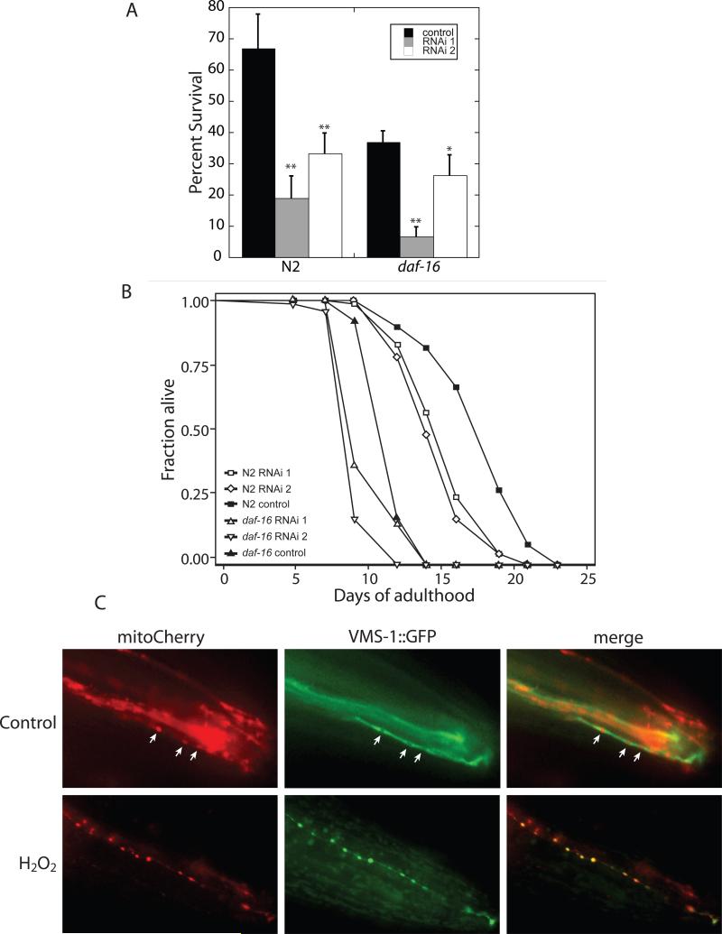
We show that Ydr049 (renamed VCP/Cdc48-associated mitochondrial stress-responsive--Vms1), a member of an unstudied pan-eukaryotic protein family, translocates from the cytosol to mitochondria upon mitochondrial stress. Cells lacking Vms1 show progressive mitochondrial failure, hypersensitivity to oxidative stress, and decreased chronological life span. Both yeast and mammalian Vms1 stably interact with Cdc48/VCP/p97, a component of the ubiquitin/proteasome system with a well-defined role in endoplasmic reticulum-associated protein degradation (ERAD), wherein misfolded ER proteins are degraded in the cytosol. We show that oxidative stress triggers mitochondrial localization of Cdc48 and this is dependent on Vms1. When this system is impaired by mutation of Vms1, ubiquitin-dependent mitochondrial protein degradation, mitochondrial respiratory function, and cell viability are compromised. We demonstrate that Vms1 is a required component of an evolutionarily conserved system for mitochondrial protein degradation, which is necessary to maintain mitochondrial, cellular, and organismal viability.


Figures

Figure 1
Figure 1. Vms1 exhibits stress-responsive mitochondrial translocation and loss of Vms1 causes hypersensitivity to hydrogen peroxide and rapamycin
(A) The vms1Δ strain containing both a plasmid expressing mito-RFP (a fusion of the N. crassa Su9 presequence to RFP) and a plasmid expressing Vms1-GFP under the native VMS1 promoter was grown in SD-Ura-Leu medium. Upon reaching mid-log phase, the culture was either treated with vehicle (top) or with compounds as indicated and subjected to fluorescence microscopy. The 2nd row shows representative images of Vms1-GFP localization in an vms1Δ rhoo strain (lacking the mitochondrial genome) in log phase. The field shown in the top image was selected to show the weak mitochondrial localization of Vms1-GFP in the absence of stressor in a small percentage of cells (indicated with an arrow). Representative images are shown. (B) WT, vms1Δ, sod2Δ, and vms1Δ sod2Δ strains were grown to saturation in SD-Ura. Serial 5-fold dilutions of each culture were spotted on both SD-Ura (top) and SD-Ura + 3mM hydrogen peroxide (bottom) plates and grown at 30°C for 2 days. (C) WT and vms1Δ strains were transformed with empty vector (ev), a plasmid containing the yeast VMS1 gene (pVMS1), or a plasmid containing the human VMS1 gene under the control of the yeast GPD promoter (pHsVMS1). Each strain was streaked on an SD-Ura plate without (top) or with 30ng/ ml rapamycin (bottom) and grown at 30°C for 2 days (top) or 5 days (bottom).
Figure 2
Figure 2. Deletion of VMS1 induces mitochondrial dysfunction
(A) The vms1Δ strain containing plasmids expressing mito-RFP and Vms1-GFP was grown in SD-Leu-Ura for 1.5 days and subjected to fluorescence imaging. (B) The vms1Δ strain containing a plasmid expressing C-terminally HA-tagged Vms1 was grown for 1.5 days and subjected to differential centrifugation. The mitochondria enriched fraction (mito) was then fractionated on a sucrose cushion and subjected to SDS-PAGE followed by western blot. Tom20 and Pgk1 were used as mitochondrial and cytop lasmic markers, respectively. (C and D) WT and vms1Δ strains grown in SD medium were harvested at either log phase or at day 1.5 of culture and were subjected to oxygen consumption assay (C) and aconitase activity assay (D). Mean ± s.d of three independent cultures is shown. (E) WT and vms1Δ strains in the W303 background, grown in synthetic complete glucose (SD) media for 1.5, 3.5, 5.5, or 8.5 days, were 5-fold serially diluted and equivalent cell numbers of each strain were spotted on both YPAD and YPAGlycerol plates and grown at 30°C. (F) WT and two independent vms1Δ strains in the BY4741 background were grown in synthetic complete glucose (SD) media for 1, 3, or 8 days. 500 cells from each culture were plated on YPAD and grown at 30°C for determination of colony formation. Colony forming units were determined for at least three independent cultures per strain and mean ± s.d. is shown. (G) WT and vms1Δ strains in the BY4741 background were grown in synthetic complete glucose (SD) media for 4 days and stained with dihydroethidium. Ethidium fluorescence was determined by FACS analysis for each strain. For each strain, three independent cultures were tested and mean ± s.d. is shown. (H) WT, vms1Δ, sod2Δ, and vms1Δ sod2Δ strains were transformed with empty vector (ev), a plasmid containing the SOD2 gene (pSOD2) or a plasmid containing the VMS1 gene (pVMS1). Each strain was grown to saturation in SD-Ura media. Serial 5-fold dilutions of each culture were then spotted on an SGlycerol-Ura plate, and grown at 30°C.
Figure 3
Figure 3. C. elegans vms-1 is required for wild-type lifespan and hydrogen peroxide resistance
(A) Wild-type (N2) or daf-16 mutant worms were grown to the first day of egglaying on bacteria expressing vector control or either of two independent vms-1 RNAi constructs, and their survival was scored after 5 h in 20mM H2O2 at room temperature. Results shown are the average ± s.d. of three replicate experiments comprised of 100 animals each. * p<0.05 and ** p<0.01. (B) Wild-type (N2) or daf-16 mutant worms grown as in (A) were assayed for lifespan. Mean lifespans were as follows: wild type on vector control was 18.0 ± 0.1d, wild type on RNAi 1 was 15.5 ± 0.2 d (p<0.0001), wild type on RNAi 2 was 15.0 ± 0.2 d (p<0.0001), daf-16 on vector control was 12.1 ± 0.1 d, daf-16 on RNAi 1 was 10.3 ± 0.1 d (p<0.0001), and daf-16 on RNAi 2 was 9.3 ± 0.1d (p<0.0001). p values reflect statistical significance of each RNAi treatment compared to vector control for each strain. (C) Subcellular localization of VMS-1::GFP in dendritic processes of amphid neurons was imaged in worms co-expressing Pvms-1::VMS-1::GFP and the mito-mCherry mitochondrial marker (Pegl-3::DIC-1::mCherry). Worms were treated for 1 h in either M9 as a control (top) or 200mM H2O2 dissolved in M9 (bottom). Representative images for each population are shown. Three sites of mito-mCherry localization and VMS-1::GFP exclusion in the control images are indicated with arrows.
Figure 4
Figure 4. Vms1 constitutively interacts with Cdc48 and Npl4
(A) Strains expressing either untaggedor TAP-tagged Vms1 under the native VMS1 promoter were grown to late log phase and subjected to TAP purification. The final eluates from each of two independent purifications for each strain were analyzed by SDS-PAGE and Coomassie staining. The major unique bands from the TAP-tagged Vms1 purification were identified by mass spectrometry as Cdc48 and Vms1-TAP as indicated. (B) WT, vms1Δ, cdc48Δ, and vmsΔ cdc48Δ strains bearing plasmids expressing either Vms1 or Vms1-HA and Cdc48, Cdc48-myc, or Cdc48(S565G)-myc were grown to late log phase and subjected to immunoprecipitation with anti-HA antibody. Immunoblots of crude lysates and immunoprecipitates were developed with the indicated antibodies. (C) The vms1Δ, vms1Δ npl4Δ, ufd1Δ, and vms1Δ ufd1Δ strains containing plasmids expressing either Vms1 or Vms1-HA and either Npl4-myc or Ufd1-myc were grown to late log phase and subjected to immunoprecipitation and immunoblot as in (B). (D) The ufd1Δ, ufd1Δ npl4Δ, vms1Δ, and ufd1Δ vms1Δ strains containing plasmids expressing either Ufd1 or Ufd1-HA and either Npl4-myc or Vms1-myc were analyzed as in (B). (E) The vms1Δ strain containing plasmids expressing either untagged full-length (-) or HA-tagged full-length (FL), C-terminus-only (ΔN) or N-terminus-only (ΔC) Vms1 deletion mutants and either untagged or myc-tagged Cdc48 were analyzed as in (B). (F) The vms1Δ strain containing plasmids expressing either untagged full-length (-) or HA-tagged full-length (FL), C-terminus-only (ΔN) or N-terminus-only (ΔC) Vms1 deletion mutants and either untagged or myc-tagged Npl4 were analyzed as in (B).
Figure 5
Figure 5. Vms1 interacts with Cdc48/Npl4 through a VIM and this interaction is required for Vms1 function
(A) Sequence alignment showing the VCP Interacting Motif (VIM) from known VCP interacting proteins and VMS1 orthologs. The amino acids mutated in human Vms1 for use in (F) are bracketed above the sequence. The amino acids deleted in yeast Vms1 for use in (B, C, D) are bracketed below the sequence. (B) The cdc48Δ vms1Δ strain containing plasmids expressing either Vms1, Vms1-HA, or Vms1 (VIMΔ)-HA and either Cdc48 or Cdc48-myc was grown to late log phase and subjected to immunoprecipitation using anti-HA antibody. Immunoblots of crude lysates and immunoprecipitates were developed with the indicated antibodies. (C) The vms1Δ npl4Δ strain containing plasmids expressing either Vms1, Vms1-HA or Vms1 (VIMΔ)-HA and either Npl4 or Npl4-myc was grown to late log phase and analyzed as in (B). (D) WT and vms1Δ strains transformed with empty vector or centromeric plasmids expressing Vms-HA or Vms1 (VIMΔ)-HA from the endogenous VMS1 promoter were grown to saturation in SD-Ura medium. 5-fold serial dilution of equivalent cell numbers were then spotted on both SD-Ura (left) and SD-Ura + 20ng/ ml rapamycin (right) plates and grown at 30°C. (E) C2C12 cells stably expressing either C-terminally Flag-HA tagged VMS1 (+) or empty vector (-) were harvested and subjected to two-step affinity purification: immunoprecipitation with anti-Flag antibody and eluted with triple-Flag peptide; and immunoprecipitation with anti-HA antibody and eluted with HA peptide. Equivalent amounts of eluates were separated by SDS-PAGE and subjected to silver staining. (F) Control C2C12 cells or cells stably expressing either mouse VMS1-Flag-HA or VMS1(VIMΔ)-Flag-HA wherein VIM residues 684-686 (ERR—as indicated in (A)) were mutated to AAA were harvested and the lysates were subjected to immunoprecipitation with anti-Flag antibody. Lysates (Lys), immunodepleted supernatants (Sup), and immunoprecipitates (IP) were analyzed by western blot for VMS1 and VCP. VMS1-Flag-HA and VMS1(VIMΔ)-Flag-HA were expressed at and immunoprecipitated in equal amounts (upper band; lower band is endogenous VMS1). The asterisk indicates a non-specific band. Tubulin was used as a loading control for lysates and immunodepleted supernatants.
Figure 6
Figure 6. Vms1 is required for the mitochondrial translocation of Cdc48
(A) WT and vms1Δ strains expressing a functional Cdc48-GFP fusion protein from the native CDC48 locus were transformed with mito-RFP plasmid and either empty vector or centromeric pVMS1-HA and cultured in SD-Leu-Ura at 30°C. Upon reaching mid-log phase, the culture was treated with either vehicle (left) or 3mM hydrogen peroxide for 3 hours (right) and imaged. Representative images are shown. (B) Ratio of mitochondria (mito-RFP) co-localized Cdc48-GFP to total cellular Cdc48-GFP signal (± s.e.m) is graphed for each strain and condition from (A). At least 50 blindly selected cells were analyzed for each strain and condition. (C) WT and vms1Δ strains transformed with either pRS426 (ev), pRS426-CDC48, pRS426-NPL4, or pRS426-UFD1 were grown to saturation in SD-Ura medium and serial 5-fold dilutions were spotted on both SD-Ura (left) and SD-Ura + 20ng/ ml rapamycin (right) plates and grown at 30°C.
Figure 7
Figure 7. Vms1 is required for normal ubiquitin-dependent mitochondrial protein degradation
(A) WT, vms1Δ, mdm30Δ, and vms1Δ mdm30Δ strains were grown in SD complete medium for 2.5 days and whole cell extracts were prepared and subjected to immunoblot using anti-Fzo1 antibody, with anti-Pgk1 being a loading control. (B) WT, vms1Δ, cdc48-S565G, and vms1Δ cdc48-S565G strains were treated as in (A). (C) WT and vms1Δ strains expressing an HA-tagged allele of Fzo1 were grown to log phase and treated with 0.1 mg/ ml cycloheximide. At the times indicated, samples were harvested and subjected to immunoblotting using anti-HA and anti-porin antibodies. Please note: cycloheximide causes rapid Vms1 translocation to mitochondria. NS indicates a non-specific band that is present in the absence of the plasmid encoding Fzo1-HA. (D) WT and vms1Δ strains expressing CPY*-HA were treated as in (C). (E) WT and ufd1-1 strains expressing CPY*-HA were treated as in (C). (F) WT and vms1Δ strains were grown in YPD medium for 5 days, harvested, and subjected to mitochondria isolation by differential centrifugation. The crude mitochondrial fraction (Crude Mito) was then loaded on a sucrose cushion to separate mitochondria (Pure Mito) from other membranes. Equivalent amounts of proteins were TCA precipitated and immunoblotted using anti-ubiquitin antibody. Anti-Pgk1, Cue1 and Tom20 were used as a marker and loading control for cytoplasm , ER and mitochondria, respectively. (G) WT and vms1Δ strains transformed with DHFR-GFP were grown in SD-Ura medium either to log phase or for 2.5 days. Equivalent numbers of cells were harvested, lysed, and subjected to SDS-PAGE followed by immunoblotting using anti-GFP and anti-Pgk1 (loading control) antibodies. The asterisk indicates a DHFR-GFP fragment that is targeted to mitochondria and is degraded upon mitophagy, similar to full-length DHFR-GFP. (H) Two independent cultures of WT and vms1Δ strains were grown in SD complete medium for 2.5 days and equivalent numbers of cells were harvested, lysed, and subjected to immunoblot using the indicated antibodies. (I) WT, vms1Δ, oma1Δ, and vms1Δ oma1Δ strains were grown in SD complete medium for 1.5 days. 5-fold serial dilution of equivalent numbers of cells were spotted on both YPAD and YPAGlycerol plates and grown at 30°C. (J) WT, vms1Δ, yme1Δ, and vms1Δ yme1Δ strains were analyzed as in (I).

Comment in

  • Stressed-out mitochondria get MAD.
    Chatenay-Lapointe M, Shadel GS. Chatenay-Lapointe M, et al. Cell Metab. 2010 Dec 1;12(6):559-60. doi: 10.1016/j.cmet.2010.11.018. Cell Metab. 2010. PMID: 21109188 Free PMC article.

Similar articles

Cited by

References

    1. Andreoli C, Prokisch H, Hortnagel K, Mueller JC, Munsterkotter M, Scharfe C, Meitinger T. MitoP2, an integrated database on mitochondrial proteins in yeast and man. Nucleic Acids Res. 2004;32:D459–462. - PMC - PubMed
    1. Azzu V, Brand MD. Degradation of an intramitochondrial protein by the cytosolic proteasome. J Cell Sci. 2010;123:578–585. - PMC - PubMed
    1. Azzu V, Mookerjee SA, Brand MD. Rapid turnover of mitochondrial uncoupling protein 3. Biochem J. 2010;426:13–17. - PMC - PubMed
    1. Bays NW, Hampton RY. Cdc48-Ufd1-Npl4: stuck in the middle with Ub. Curr Biol. 2002;12:R366–371. - PubMed
    1. Bestwick M, Khalimonchuk O, Pierrel F, Winge DR. The role of Coa2 in hemylation of yeast Cox1 revealed by its genetic interaction with Cox10. Mol Cell Biol. 2010;30:172–185. - PMC - PubMed

Publication types

MeSH terms