. 2011 Feb;32(4):1002-9.
doi: 10.1016/j.biomaterials.2010.10.020. Epub 2010 Nov 10.

Hydrogels with time-dependent material properties enhance cardiomyocyte differentiation in vitro

Affiliations

Hydrogels with time-dependent material properties enhance cardiomyocyte differentiation in vitro

Jennifer L Young et al. Biomaterials. 2011 Feb.

Abstract

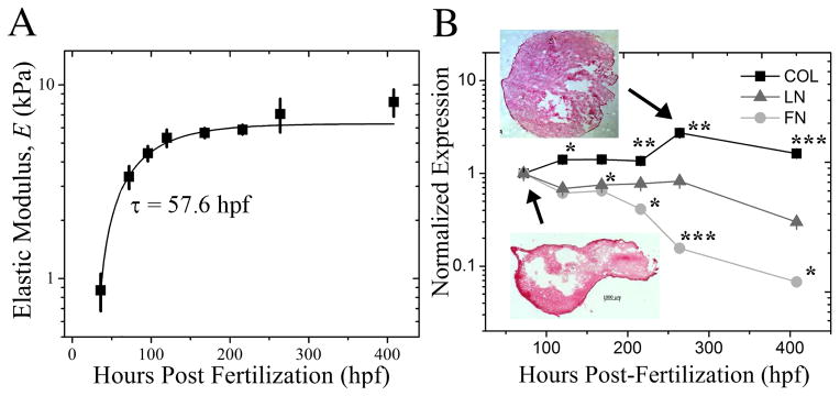
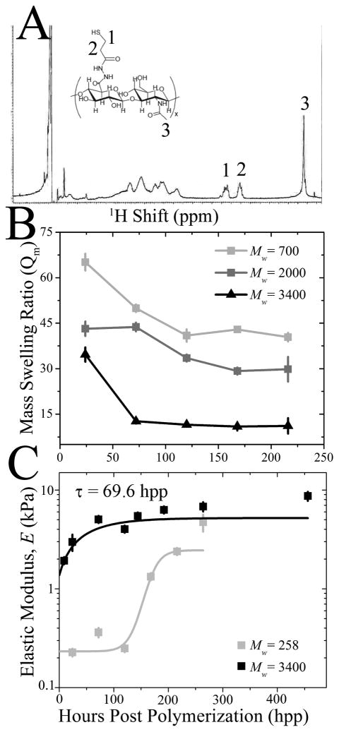
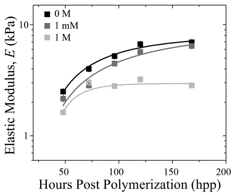
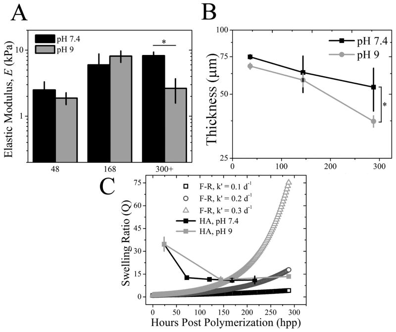
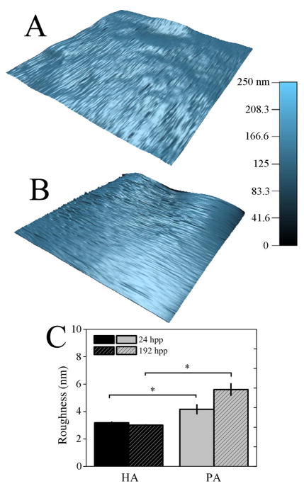
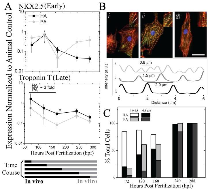
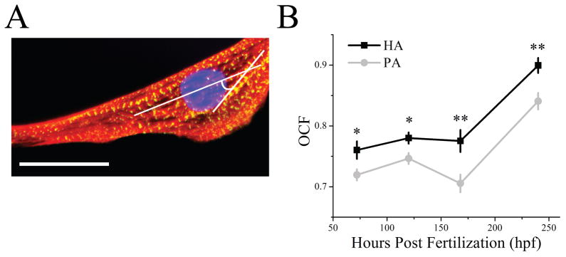
Tissue-specific elastic modulus (E), or 'stiffness,' arises from developmental changes in the extracellular matrix (ECM) and suggests that progenitor cell differentiation may be optimal when physical conditions mimic tissue progression. For cardiomyocytes, maturing from mesoderm to adult myocardium results in a 9-fold stiffening originating in part from a change in collagen expression and localization. To mimic this temporal stiffness change in vitro, thiolated-hyaluronic acid (HA) hydrogels were crosslinked with poly(ethylene glycol) diacrylate, and their dynamics were modulated by changing crosslinker molecular weight. With the hydrogel appropriately tuned to stiffen as heart muscle does during development, pre-cardiac cells grown on collagen-coated HA hydrogels exhibit a 3-fold increase in mature cardiac specific markers and form up to 60% more maturing muscle fibers than they do when grown on compliant but static polyacrylamide hydrogels over 2 weeks. Though ester hydrolysis does not substantially alter hydrogel stiffening over 2 weeks in vitro, model predictions indicate that ester hydrolysis will eventually degrade the material with additional time, implying that this hydrogel may be appropriate for in vivo applications where temporally changing material properties enhance cell maturation prior to its replacement with host tissue.


Figures

Fig. 1
Fig. 1. Characterizing Myocardial Development in the Chicken Embryo
(A) Myocardial elastic modulus was measured by atomic force microscopy from 36 to 408 hpf. Data was fit with an exponential curve, which exhibited a time constant, τ, of 57.6 hpf as indicated. (B) qPCR quantification of myocardial collagen (black squares), laminin (grey triangles) and fibronectin (light grey circles) expression was normalized to the initial time point at 72 hpf and plotted as a function of developmental time. Inset images show tissue samples isolated at the indicated time points and stained with Picrosirius Red to indicate collagen localization (red). *p < 0.05, **p < 0.01, *** p < 0.001 compared to the initial time point.
Fig. 2
Fig. 2. HA Hydrogel Stiffening can be Tuned by Molecular Weight
(A) 1H NMR spectroscopy of thiolated HA, indicating peaks from the thiolation (1, 2) and HA backbone (3) as shown on the inset schematic of thiolated HA. (B) Mass swelling ratio (Qm) was examined over 216 hpp among HA hydrogels prepared from PEGDA of Mw ~ 700 Da (light grey), 2000 Da (dark grey) and 3400 Da (black). (C) Elastic modulus of HA hydrogels made of Mw ~ 258 and 3400 Da PEGDA was determined by AFM as a function of time. 3400 Da hydrogels were fit with an exponential curve exhibiting a time constants, τ, of 65 hpp. 258 Da hydrogels were hyperbolically fit to indicate slower crosslinking.
Fig. 3
Fig. 3. Disulfide Bond Formation Does Not Substantially Contribute to Time-Dependent Stiffening
HA hydrogels made of Mw ~ 3400 Da PEGDA were polymerized, subsequently treated with 0 M (black), 1 mM (grey) and 1 M (light grey) DTT, and their elastic modulus was determined by AFM as a function of time. HA hydrogel data at 1 mM and 1 M were fitted with exponential curves, exhibiting time constants, τ, of 80 and 20 hpp, respectively.
Fig. 4
Fig. 4. HA Hydrogel Stiffening Initially Outcompetes Ester Hydrolysis
(A) HA hydrogel elastic modulus was measured in both pH conditions over time and was reduced only in pH 9 samples after 300+ hpp. *p < 0.05. (B) HA hydrogel thickness was examined over 288 hpp in pH 7.4 (black squares) and 9 (grey circles) by confocal microscopy. Modest hydrogel thinning was observed for pH 7.4 but was > 45% for pH 9. *p < 0.005. (C) Equilibrium mass swelling ratio of HA hydrogels at pH 7.4 (black) and 9 (light grey) was determined up to 288 hpp. Data is plotted as compared to a modified Flory-Rehner (F-R) model of hydrolysis for volumetric swelling where the F-R ester bond hydrolysis constant, k′, was varied. k′ = 0.1 day−1 (open black squares), k′ = 0.2 day−1 (open dark grey circles) and k′ = 0.3 day−1 (open light grey triangles).
Fig. 5
Fig. 5. HA Hydrogels Surface Topography Does Not Change Over Time
3-dimensional surface topography maps of HA (A) and PA (B) hydrogels at 196 hpp. (C) Surface roughness, as measured by root-mean-squared distance, was computed from topographical maps and plotted at both 24 and 192 hpp for HA and PA hydrogel systems. * p < 0.05.
Fig. 6
Fig. 6. Cardiomyocyte Maturation Is Improved on HA versus PA Hydrogels
(A) Cells from early through late myocardial development were plated onto HA (black squares) and PA (grey circles) hydrogels such that total cell age (in vivo and in vitro) was 312 hpf. Early and late cardiac markers NKX2.5 (top) and Troponin T (bottom) were measured by qPCR in cells plated on PA and HA hydrogels and normalized to the animal expression level at 312 hpf for the respective gene (see Supplemental Figure 5). Normalized expression below, at, or above 1 indicates lower, similar, or higher expression than the animal control, respectively. *p < 0.05 between HA and PA hydrogels. (B) Representative immunofluorescent images of cells stained for actin (red), alpha-actinin (green), and nuclei (blue) illustrate the stages of myofibril development in terms of striation length: premyofibrils at ~ 0.8 μm (i), maturing myofibrils at ~ 1.5 μm (ii), and mature myofibrils at ~2.0 μm (iii). Inset images indicate the myofibril that was examined in the corresponding intensity plot profile. Scale bar is 25 μm. (C) Cells were cultured in accordance to the time course in (A), and the percentage of cells containing nascent (1.0–1.8 μm; HA in white, PA in light grey) or mature myofibrils of (>1.8 μm; HA in black, PA in dark grey) was quantified. Premyofibrils (<1.0 μm), consist of the remaining percentage of cells, were not plotted. n > 25 cells were analyzed per time point per hydrogel.
Fig. 7
Fig. 7. Myofibrils are Most Oriented on HA Hydrogels
(A) Myofibril orientation was examined by calculating an orientation correlation function (OCF) where θ is the difference between the myofibril angle and the long axis of the cell as indicated in white in the image of a representative cell. Scale bar is 25 μm. (B) Quantification of OCF over time for cells cultured on HA (black squares) or PA hydrogels (grey circles). Note that OCF = 1 and 0.5 indicates parallel and diagonal alignment with respect to the long axis of the cell. * p < 0.05, ** p < 0.005 compared between HA and PA hydrogels.

References

    1. Discher DE, Mooney DJ, Zandstra PW. Growth factors, matrices, and forces combine and control stem cells. Science. 2009;324(5935):1673–1677. - PMC - PubMed
    1. Saha K, Keung AJ, Irwin EF, Li Y, Little L, Schaffer DV, et al. Substrate modulus directs neural stem cell behavior. Biophys J. 2008;95(9):4426–4438. - PMC - PubMed
    1. Engler AJ, Sen S, Sweeney HL, Discher DE. Matrix elasticity directs stem cell lineage specification. Cell. 2006;126(4):677–689. - PubMed
    1. Huebsch N, Arany PR, Mao AS, Shvartsman D, Ali OA, Bencherif SA, et al. Harnessing traction-mediated manipulation of the cell/matrix interface to control stem-cell fate. Nat Mater. 9(6):518–526. - PMC - PubMed
    1. Lutolf MP, Hubbell JA. Synthetic biomaterials as instructive extracellular microenvironments for morphogenesis in tissue engineering. Nat Biotechnol. 2005;23(1):47–55. - PubMed

Publication types