. 2011 Mar;39(6):2271-85.
doi: 10.1093/nar/gkq1124. Epub 2010 Nov 10.

The AddAB helicase-nuclease catalyses rapid and processive DNA unwinding using a single Superfamily 1A motor domain

Affiliations

The AddAB helicase-nuclease catalyses rapid and processive DNA unwinding using a single Superfamily 1A motor domain

Joseph T P Yeeles et al. Nucleic Acids Res. 2011 Mar.

Abstract

The oligomeric state of Superfamily I DNA helicases is the subject of considerable and ongoing debate. While models based on crystal structures imply that a single helicase core domain is sufficient for DNA unwinding activity, biochemical data from several related enzymes suggest that a higher order oligomeric species is required. In this work we characterize the helicase activity of the AddAB helicase-nuclease, which is involved in the repair of double-stranded DNA breaks in Bacillus subtilis. We show that the enzyme is functional as a heterodimer of the AddA and AddB subunits, that it is a rapid and processive DNA helicase, and that it catalyses DNA unwinding using one single-stranded DNA motor of 3' → 5' polarity located in the AddA subunit. The AddB subunit contains a second putative ATP-binding pocket, but this does not contribute to the observed helicase activity and may instead be involved in the recognition of recombination hotspot sequences.


Figures

Figure 1.
Figure 1.
DNA unwinding models for SF1 DNA helicases. Various models have been suggested for how SF1 helicases may catalyse DNA unwinding. They have been proposed to function as both monomers (A) and dimers (B). It has also been shown that multiple SF1 helicase monomers may cooperate to facilitate unwinding of the same DNA molecule (C). A bipolar helicase (e.g. RecBCD) utilizes two SF1 helicase motors of opposite polarity to catalyse processive DNA unwinding (D). The DNA unwinding activity of many SF1 enzymes is enhanced through interactions with specific partner proteins (E). SF1 helicase domains are represented by ovals with white arrows and accessory protein(s) are shown as squares. See the text for references and further discussion of these mechanisms.
Figure 2.
Figure 2.
RecBCD- and AddAB-type helicase–nucleases contain Superfamily 1 helicase domains. (A) Primary structure diagrams highlighting the positions of the SF1 helicase domains and Walker A motifs in RecBCD- and AddAB-type helicase–nuclease enzymes. (B) Sequence alignment of the seven SF1 helicase motifs (1) of E. coli UvrD, Rep, RecB and RecD and B. subtilis PcrA and AddA. B. subtilis AddB has a conserved Walker A motif (helicase motif 1) but apparently lacks the remaining helicase motifs. Conserved residues are shown in bold.
Figure 3.
Figure 3.
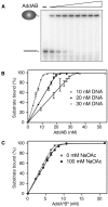
EMSA analysis of the stoichiometry of AddAB binding to dsDNA ends. (A) EMSA assays were conducted with 10 nM 32P-labelled Bind15 substrate in the standard dsDNA end-binding buffer with varying AddAB concentrations (0, 3.6, 7.2, 9, 10.8, 13.5. 18, 27, 36 nM). The positions of the free DNA and AddAB:DNA complex are indicated. (B) EMSA experiments were conducted with 10, 20 or 30 nM Bind15 as indicated. The proportions of the substrate bound by varying concentrations of AddAB were calculated for two independent repeats. Error bars represent the standard error of the mean (SEM). The data were fit to Equation (1). (C) Quantification of EMSA data characterizing the binding of AddANBN to Cy5Bind15. Samples contained 5 nM Cy5Bind15 in the standard dsDNA end-binding buffer in the presence or absence of 100 mM sodium acetate. The proportion of the bound substrate was calculated for two repeats. The error bars represent the SEM and the data were fit to Equation (1).
Figure 4.
Figure 4.
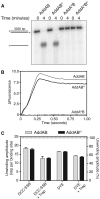
The AddAB heterodimer is the active form of the enzyme during dsDNA unwinding. (A) Schematic representation of the DNA substrate used in real time helicase assays. The position of a 5′-biotin is denoted by a B, while S represents a streptavidin molecule. (B) Fluorescent SSB assays were conducted in the standard buffer with a DCC-SSB concentration of 200 nM. AddANBN enzymes were pre-bound at the indicated concentrations to the substrate DNA (1 nM) that was blocked at the distal end with streptavidin. Substrate unwinding was initiated by rapid mixing of the DNA:AddANBN complex against an equal volume of 0.5 mM ATP and 100 nM AddAHB trap in reaction buffer. The data have been normalized by subtracting the initial fluorescence values for the individual traces. (C) The final amplitude of unwinding (measured as a change in fluorescence at 5 s) is plotted for each AddANBN concentration. The fluorescence was calibrated using heat-denatured substrate DNA and data were fit to Equation (2).
Figure 5.
Figure 5.
A single helicase motor catalyses processive DNA unwinding by AddAB. (A) dsDNA processing by wild-type AddAB and the Walker A mutant enzymes. The processing of the Chi-free DNA substrate pADGF0 was investigated under conditions of limited free magnesium that suppress AddAB nuclease activity. The positions of the dsDNA substrate and the unwound full-length ssDNA reaction products are indicated. (B) Comparison of AddAB and AddABH DNA unwinding using the DCC-SSB helicase assay. The DNA substrate used is shown in Figure 4A. Saturating (2.5 nM) AddAB and AddABH were pre-bound to 1 nM DNA substrate that was blocked at the distal end with streptavidin. Reactions were initiated with 0.5 mM ATP and 100 nM AddAHB. For the AddAHB trace, the experiment was conducted as described for AddAB and AddABH, however no helicase was pre-bound to the DNA substrate. The data have been normalized by subtracting the initial fluorescence values for the individual traces. (C) Analysis of the processivity of the wild-type AddAB and AddABH. The proportions of the ∼48.5-kb Phage λ DNA molecule unwound by AddAB and AddABH were measured using the DCC-SSB and dye-displacement assays. The data are the average of at least two repeats and the error bars represent the SEM.
Figure 6.
Figure 6.
The AddA subunit is a 3′→5′ ssDNA translocase and 3′ → 5′ DNA helicase. (A) Streptavidin displacement from 3′- and 5′-biotinylated oligonucleotides. Reactions were conducted at 37°C for 60 min. PcrA and Dda are included as controls for 3′→5′ and 5′→3′ translocation respectively. The positions of the free oligonucleotides and the streptavidin bound oligonucleotides are indicated, as is the translocation directionality that the substrates report on. (B) Streptavidin displacement time courses for the various different enzyme and substrate combinations indicated. Reactions were conducted at 37°C and aliquots were removed at the indicated time-points. (C) Strand-displacement helicase assays. Reactions were conducted at 20°C for 4 min. Dda and PcrA are included as controls for 5′→3′ and 3′→5′ helicase activity, respectively. The positions of the substrates and displaced oligonucleotides are indicated to the left of the gels, while the polarities on which the substrates report on are indicated to the right. (D) Reaction time courses of helicase assays. Reactions were incubated at 20°C and aliquots were withdrawn and quenched at the indicated times. The displaced oligonucleotide is shown as a proportion of the total DNA in each lane.
Figure 7.
Figure 7.
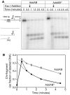
Mutation of the AddB Walker A motif reduces the stability of the AddAB:Chi complex. The stability of the complexes formed between Chi and the AddAB or AddABH enzymes were investigated using an exonuclease chase experiment. (A) Tailed 5′-32P-labelled pAGD6406 (Chi +) (1.6 nM) was incubated with 3.2 nM AddAB in standard high magnesium reaction buffer. Following a 30-s incubation, Exo1 was added and aliquots were removed at the indicated times. The positions of the substrate and reaction products are indicated to the left of the gel. The PAS site is a region of secondary structure that is resistant to Exo1 degradation. (B) The proportions of Chi-fragments remaining at each time point were quantified. The data represents the average of four repeats with the error bars showing the SEM. The dotted lines show fits of the data to a single exponential decrease (forced to decay to zero) to yield apparent unbinding rate constants for the AddAB-Chi fragment interaction. In performing this fit, we are interested in determining the rate of decay of the ExoI-protected (AddAB bound) Chi fragment. Therefore, the 0.5-min sample (which is taken before ExoI chase addition) is taken as an approximation of the 0-min timepoint. This is because the 0.5-min sample measures total Chi fragment (both ExoI-protected and ExoI-sensitive) which is equivalent to the starting yield of ExoI-protected fragment formed within the first few seconds of the reaction. Measurements of the intensity of the Chi band made after the addition of the ExoI chase only reflect ExoI-protected material.

References

    1. Gorbalenya AE, Koonin EV. Helicases - amino-acid-sequence comparisons and structure-function-relationships. Curr. Opin. Struct. Biol. 1993;3:419–429.
    1. Singleton MR, Dillingham MS, Wigley DB. Structure and mechanism of helicases and nucleic acid translocases. Annu. Rev. Biochem. 2007;76:23–50. - PubMed
    1. Subramanya HS, Bird LE, Brannigan JA, Wigley DB. Crystal structure of a DExx box DNA helicase. Nature. 1996;384:379–383. - PubMed
    1. Kim JL, Morgenstern KA, Griffith JP, Dwyer MD, Thomson JA, Murcko MA, Lin C, Caron PR. Hepatitis C virus NS3 RNA helicase domain with a bound oligonucleotide: the crystal structure provides insights into the mode of unwinding. Structure. 1998;6:89–100. - PubMed
    1. Singleton MR, Wigley DB. Modularity and specialization in superfamily 1 and 2 helicases. J. Bacteriol. 2002;184:1819–1826. - PMC - PubMed

Publication types