. 2011 Mar;39(4):e19.
doi: 10.1093/nar/gkq1074. Epub 2010 Nov 11.

Digital MDA for enumeration of total nucleic acid contamination

Affiliations

Digital MDA for enumeration of total nucleic acid contamination

Paul C Blainey et al. Nucleic Acids Res. 2011 Mar.

Abstract

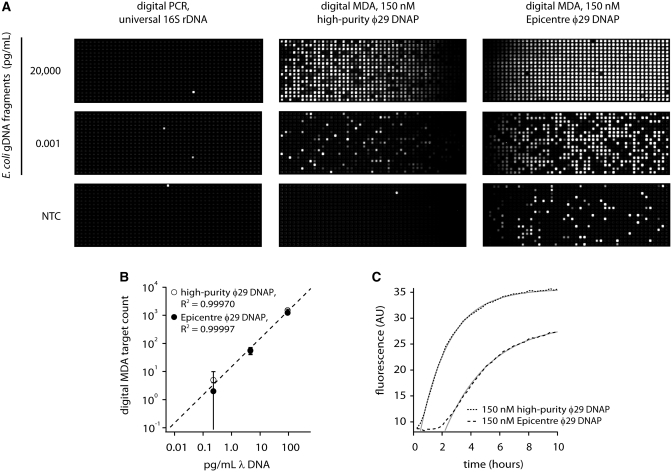
Multiple displacement amplification (MDA) is an isothermal, sequence-independent method for the amplification of high molecular weight DNA that is driven by φ29 DNA polymerase (DNAP). Here we report digital MDA (dMDA), an ultrasensitive method for quantifying nucleic acid fragments of unknown sequence. We use the new assay to show that our custom φ29 DNAP preparation is free of contamination at the limit of detection of the dMDA assay (1 contaminating molecule per assay microliter). Contamination in commercially available preparations is also investigated. The results of the dMDA assay provide strong evidence that the so-called 'template-independent' MDA background can be attributed to high-molecular weight contaminants and is not primer-derived in the commercial kits tested. dMDA is orders of magnitude more sensitive than PCR-based techniques for detection of microbial genomic DNA fragments and opens up new possibilities for the ultrasensitive quantification of DNA fragments in a wide variety of application areas using MDA chemistry and off-the-shelf hardware developed for digital PCR.


Figures

Figure 1.
Figure 1.
Expression and activity of the high-purity φ29 DNAP. (A) Denaturing acrylamide gel following expression and purification of affinity-tagged φ29 DNAP. Lanes show content of crude lysate, column flow-throughs (FT) and eluates. The arrow indicates the expression target. Bands in product below 63 kDa marker in the final product (second Ni affinity elution) are most likely C-terminal deletions of the designed product: no effort was made to remove these. (B) Agarose gel showing MDA products from μl-scale reactions using 10 000 GE E. coli genomic DNA as template. The variously sourced enzymes all produced high molecular weight DNA products.
Figure 2.
Figure 2.
Digital MDA versus dPCR quantification of 3–5 kb E. coli genomic DNA fragments. (A) Digital assay panels, 765 reactions per panel. Digital PCR does not effectively detect the E. coli genome fragments (positive control panel with amplicon template not shown). All three digital PCR panels and the positive control reaction were prepared from the same reaction mix and run in a single chip. Digital MDA easily detects E. coli genome fragments down to femptograms per microliter. The Epicentre enzyme shows higher background in the no template control than the high-purity enzyme prepared in-house. All six digital MDA panels shown were prepared using the same reaction mix (except for the enzyme) and run in a single chip. (B) Calibration curve showing quantification of denatured λ DNA. Panels were scored at 1.5 h for high-purity φ29 DNAP reactions and at 4 h for Epicentre φ29 DNAP reactions. Each datapoint corresponds to a single digital assay panel with error bars that represent 95% confidence intervals based on event counting statistics. The coefficient of determination, R2, exceeded 0.999 in both cases. (C) Time-dependent appearance of digital MDA signal in digital amplification of denatured λ DNA. Both the high-purity and Epicenter φ29 DNAP preparations show strong MDA activity. All the data presented in parts (B) and (C) were obtained from a single chip run.
Figure 3.
Figure 3.
Digital MDA on Fluidigm 12.765 digital array reveals varying levels of contamination in φ29 DNAP from three commercial providers. φ29 DNAP prepared in-house shows little contamination. A single reaction mix including exceptionally clean lots of the GE MDA ‘reaction’ and ‘sample’ buffers were used in all cases. (A) Example image of dMDA assay endpoint (all four panels are from a single chip run). Readout is digital, indicating contaminants, but not primer-derived products, underlie background amplification. (B) Quantification of dMDA assay results, indicating contaminant levels in the various enzyme preparations. The boxplots show data quartile ranges, median (black line) and outlier values (crosses). Results from several enzyme lots are pooled for each manufacturer, with 9–13 total dMDA assays per enzyme source.

References

    1. Dean FB, Nelson JR, Giesler TL, Lasken RS. Rapid amplification of plasmid and phage DNA using Phi 29 DNA polymerase and multiply-primed rolling circle amplification. Genome Res. 2001;11:1095–1099. - PMC - PubMed
    1. Dietmaier W, Hartmann A, Wallinger S, Heinmoller E, Kerner T, Endl E, Jauch K, Hofstadter F, Ruschoff J. Multiple mutation analyses in single tumor cells with improved whole genome amplification. Am. J. Pathol. 1999;154:83. - PMC - PubMed
    1. Telenius H, Carter N, Bebb C. Degenerate oligonucleotide-primed PCR: general amplification of target DNA by a single degenerate primer. Genomics. 1992;13:718–725. - PubMed
    1. Zhang L, Cui X, Schmitt K, Hubert R, Navidi W, Arnheim N. Whole genome amplification from a single cell: implications for genetic analysis. Proc. Natl Acad. Sci. 1992;89:5847. - PMC - PubMed
    1. Lao K, Xu N, Straus N. Whole genome amplification using single-primer PCR. Biotechnol. J. 2008;3:378–382. - PubMed

Publication types

MeSH terms

Substances