. 2011 Mar;39(6):2367-77.
doi: 10.1093/nar/gkq1118. Epub 2010 Nov 11.

A conserved structure within the HIV gag open reading frame that controls translation initiation directly recruits the 40S subunit and eIF3

Affiliations

A conserved structure within the HIV gag open reading frame that controls translation initiation directly recruits the 40S subunit and eIF3

Nicolas Locker et al. Nucleic Acids Res. 2011 Mar.

Abstract

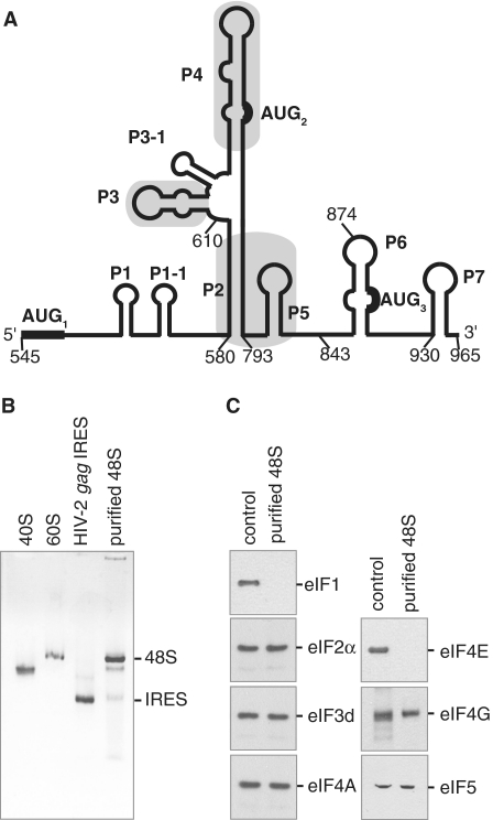
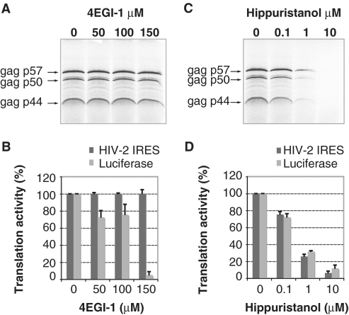
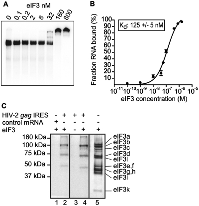
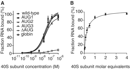
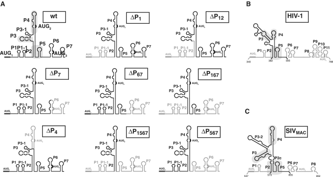
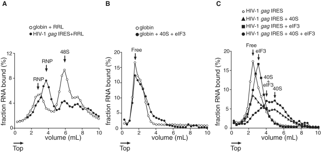
Translation initiation on HIV genomic RNA relies on both cap and Internal Ribosome Entry Site (IRES) dependant mechanisms that are regulated throughout the cell cycle. During a unique phenomenon, the virus recruits initiation complexes through RNA structures located within Gag coding sequence, downstream of the initiation codon. We analyzed initiation complexes paused on the HIV-2 gag IRES and revealed that they contain all the canonical initiation factors except eIF4E and eIF1. We report that eIF3 and the small ribosomal subunit bind HIV RNA within gag open reading frame. We thus propose a novel two step model whereby the initial event is the formation of a ternary eIF3/40S/IRES complex. In a second step, dependent on most of the canonical initiation factors, the complex is rearranged to transfer the ribosome on the initiation codons. The absolute requirement of this large structure for HIV translation defines a new function for a coding region. Moreover, the level of information compaction within this viral genome reveals an additional level of evolutionary constraint on the coding sequence. The conservation of this IRES and its properties in rapidly evolving viruses suggest an important role in the virus life cycle and highlight an attractive new therapeutic target.


Figures

Figure 1.
Figure 1.
HIV-2 gag IRES ribosomal 48S complexes contain most of the canonical initiation factors except for eIF4E and eIF1. (A) Schematic representation of the HIV-2 gag IRES secondary structure adapted from (15). Pairings are designated by Pn in order of appearance from 5′ to 3′. In frame AUG codons leading to the different Gag isoforms are indicated and numbered. Positions of the deletions for binding studies are indicated. (B) The mobility of 48S complexes assembled onto HIV-2 gag IRES purified on streptomycin derived sepharose beads is compared to the mobility of 40S, 60S subunits as well as HIV-2 gag IRES RNA as indicated. (C) The composition of 48S complexes assembled onto HIV-2 gag RNA was assessed by comparing western blots of purified complexes (purified 48S) to those of total Rabbit Reticulocyte Lysate (control). Initiation factor specificity of the primary antibody is indicated.
Figure 2.
Figure 2.
Translation of the three Gag isoforms in the presence of inhibitors of translation. (A, C) Autoradiogram showing products of in vitro translation of HIV-2 gag transcripts. Transcripts (10 ng/µl) were translated in RRL in the presence of increasing concentration of 4EGI-1 (0, 50, 100 and 150 µM) (A) or Hippuristanol (0, 0.1, 1 and 10 µM) (C) and their products resolved on a 20% SDS–PAGE gel. Gag isoforms from the three in frame AUG codons are indicated. (B, D) Relative intensity, expressed as a percentage of the initial product translated in the absence of 4EGI-1 (B) or in the absence of hippuristanol (D), of the three Gag isoforms (dark grey) compared to that of canonical luciferase (light grey) performed under the same experimental conditions. Data are representative of three independent experiments. Error bars are standard deviation of the mean.
Figure 3.
Figure 3.
The initiation factor eIF3 directly binds to the HIV-2 gag IRES. (A) Autoradiograph of 4% acrylamide native gel with 0.5 nM of 32P labeled HIV-2 gag IRES in the absence or in the presence of increasing quantities (from 0.1 nM to 0.8 µM) of purified eIF3. (B) Binding curve of 32P-labeled HIV-2 gag IRES RNA to purified eIF3. Binary complexes were formed under the conditions used for native gels and analyzed by filter binding assay. (C) The determination of the eIF3 subunits involved in HIV-2 gag IREs binding was performed by crosslinking experiments. Binary complexes between eIF3 and 32P-labeled globin mRNA (lane 1) or 32P-labeled HIV-2 gag IRES RNA (lane 2 and 4) or HIV-2 gag IRES RNA alone (lane 3) were resolved by gel electrophoresis after UV cross-linking, and RNase digestion. Binary complexes are formed under the conditions used for native gels. The positions of molecular weight markers are indicated to the left and eIF3 subunits were visualized by silver staining as a reference (lane 5) [(note that eIF3a, a 170-kDa protein, is classically proteolysed as a 100- to 130-kDa protein in RRL (23,48)].
Figure 4.
Figure 4.
HIV-2 gag IRES directly binds to the 40S subunit. (A) Binding curves of 32P-labeled wild-type, AUG mutants and globin control to purified 40S subunits. (B) Stoichiometric binding assays of 32P-labeled HIV-2 gag IRES to 40S subunit. Results are from three independent experiments.
Figure 5.
Figure 5.
eIF3 and the 40S ribosomal subunit bind to the conserved core of the HIV-2 IRES. (A) Secondary structure schematic representation of the mutants used in eIF3 and 40S subunit binding studies. Deleted domains are in light grey. Positions of the deletion are noted on the structures. Secondary structure schematic representation of the HIV-1 gag IRES (B) and SIVMac (C), conserved structural elements between primates lentivirus are boxed in grey. The stabilities of each structures as calculates by Mfold are HIV-1 = −58.38 KICal, HIV-2 = −72.37 and SIVMAC = −60.18 Kcal. Note that this portion of HIV-1 is only 60% homologous to HIV-2, while SIVMAC is 80% homologous to HIV-2.
Figure 6.
Figure 6.
Mobility of ribosomal complexes during centrifugation through 10–30% linear sucrose density gradients. (A) Fractionation by sucrose density gradient of initiation complexes formed after incubation of capped and polyadenylated globin mRNA (open circles) or HIV-1 (closed circles) RNA with RRL pre-treated with GMP-PNP. (B) Fractionation by sucrose density gradient centrifugation of capped and globin mRNA in the absence (open circles) or presence (closed circles) of excess purified eIF3 and 40S subunits. (C) Fractionation by sucrose density gradient centrifugation of HIV-1 RNA (open circles) in the presence of excess 40S subunits (triangles), eIF3 (squares) or eIF3 and 40S subunits (rounds). Arrows indicate whether the RNA is free or associated with 40S subunits and/or eIF3. The percent of RNA bound represents the mean of at least two independent experiments.

Similar articles

Cited by

References

    1. Balvay L, Lastra ML, Sargueil B, Darlix JL, Ohlmann T. Translational control of retroviruses. Nat. Rev. Microbiol. 2007;5:128–140. - PMC - PubMed
    1. Butsch M, Boris-Lawrie K. Destiny of unspliced retroviral RNA: ribosome and/or virion? J. Virol. 2002;76:3089–3094. - PMC - PubMed
    1. Berkhout B. Structure and function of the human immunodeficiency virus leader RNA. Prog. Nucleic Acid Res. Mol. Biol. 1996;54:1–34. - PubMed
    1. Dirac AM, Huthoff H, Kjems J, Berkhout B. Regulated HIV-2 RNA dimerization by means of alternative RNA conformations. Nucleic Acids Res. 2002;30:2647–2655. - PMC - PubMed
    1. James L, Sargueil B. RNA secondary structure of the feline immunodeficiency virus 5'UTR and Gag coding region. Nucleic Acids Res. 2008;36:4653–4666. - PMC - PubMed

Publication types

MeSH terms

Substances