. 2011 Jan 28;286(4):2607-16.
doi: 10.1074/jbc.M110.177014. Epub 2010 Nov 11.

Autophosphorylation activates Dictyostelium myosin II heavy chain kinase A by providing a ligand for an allosteric binding site in the alpha-kinase domain

Affiliations

Autophosphorylation activates Dictyostelium myosin II heavy chain kinase A by providing a ligand for an allosteric binding site in the alpha-kinase domain

Scott W Crawley et al. J Biol Chem. .

Abstract

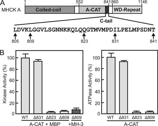
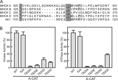
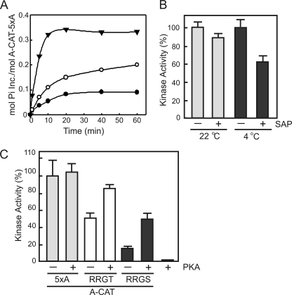
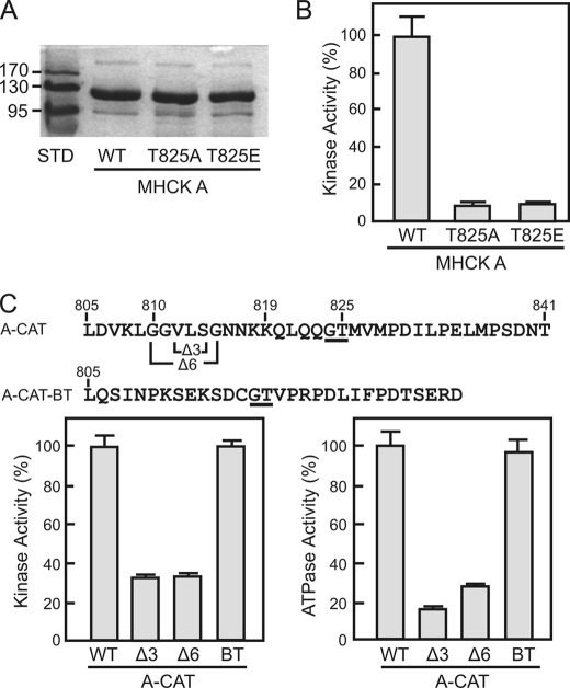
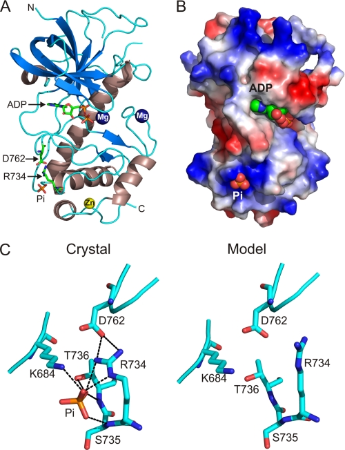
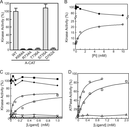
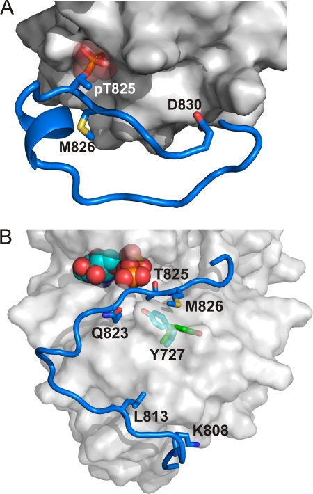
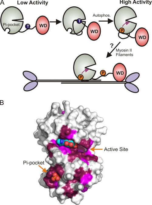
Dictyostelium discoideum myosin II heavy chain kinase A (MHCK A), a member of the atypical α-kinase family, phosphorylates sites in the myosin II tail that block filament assembly. Here we show that the catalytic activity of A-CAT, the α-kinase domain of MHCK A (residues 552-841), is severely inhibited by the removal of a disordered C-terminal tail sequence (C-tail; residues 806-841). The key residue in the C-tail was identified as Thr(825), which was found to be constitutively autophosphorylated. Dephosphorylation of Thr(825) using shrimp alkaline phosphatase decreased A-CAT activity. The activity of a truncated A-CAT lacking Thr(825) could be rescued by P(i), phosphothreonine, and a phosphorylated peptide, but not by threonine, glutamic acid, aspartic acid, or an unphosphorylated peptide. These results focused attention on a P(i)-binding pocket located in the C-terminal lobe of A-CAT. Mutational analysis demonstrated that the P(i)-pocket was essential for A-CAT activity. Based on these results, it is proposed that autophosphorylation of Thr(825) activates ACAT by providing a covalently tethered ligand for the P(i)-pocket. Ab initio modeling studies using the Rosetta FloppyTail and FlexPepDock protocols showed that it is feasible for the phosphorylated Thr(825) to dock intramolecularly into the P(i)-pocket. Allosteric activation is predicted to involve a conformational change in Arg(734), which bridges the bound P(i) to Asp(762) in a key active site loop. Sequence alignments indicate that a comparable regulatory mechanism is likely to be conserved in Dictyostelium MHCK B-D and metazoan eukaryotic elongation factor-2 kinases.


Figures

FIGURE 1.
FIGURE 1.
Truncation of the C-tail inhibits the kinase and ATPase activities of A-CAT. A, schematic diagram showing the domain organization of MHCK A. A-CAT encompasses the entire α-kinase domain and part of the unstructured sequence (C-tail) linking the WD-repeat domain. The amino acid sequence of the C-tail is shown with the experimental sites of truncation indicated. B, the kinase (left panel) and ATPase (right panel) activities of wild-type A-CAT (WT) and A-CAT truncated at residues 831 (Δ831), 823 (Δ823), or 809 (Δ809) were determined. Removal of residues 823 to 831 from the C-tail resulted in a large decrease in kinase and ATPase activities. Activities were determined from time courses performed as described under “Experimental Procedures” using either MBP or the MH-3 peptide as substrate as indicated. Activities are reported as a percentage of the wild-type activity. Error bars represent the standard deviation.
FIGURE 2.
FIGURE 2.
The conserved Thr825 residue in the C-tail is required for A-CAT activity. A, a multiple sequence alignment of the C-tail regions of D. discoideum MHCK A, MHCK B, MHCK C, MHCK D, and AK1. Conserved residues are shaded in gray. A Gly-Thr motif (residues 824 and 825 in MHCK A) is present in all of the C-tail sequences. The NCBI accession numbers are: MHCK A, XP_635119; MHCK B, XP_636368; MHCK C, XP_635600; MHCK D, XP_640080; and AK1, XP_629868. B, the kinase (left panel) and ATPase (right panel) activities of wild-type A-CAT (WT), A-CAT-5xA, and the indicated Thr825 mutants were determined. Mutation of Thr825 severely inhibited both the kinase and ATPase activities of A-CAT. Activities were determined from time courses performed as described under “Experimental Procedures” and the activities are reported as a percentage of A-CAT activity. Error bars represent the standard deviation.
FIGURE 3.
FIGURE 3.
Phosphorylation of the C-tail activates A-CAT. A, time course of the autophosphorylation of A-CAT-5xA prior to treatment (●) and following treatment for 1 h with calf intestinal alkaline phosphatase (○) or for 5 h with SAP (▾). The low level of 32P incorporation indicates that Thr825 is highly phosphorylated and is resistant to dephosphorylation by calf intestinal alkaline phosphatase and SAP. Autophosphorylation assays were performed by incubating A-CAT-5xA with [γ-32P]ATP as described under “Experimental Procedures.” B, the kinase activity of A-CAT-5xA incubated for 5 h in the presence or absence of SAP was assayed at 22 and 4 °C. The assays at 4 °C, which limits the ability of A-CAT-5xA to autophosphorylate Thr825, showed that dephosphorylation of Thr825 by SAP decreased the kinase activity of A-CAT-5xA. Activities were determined from time courses performed as described under “Experimental Procedures” and are reported as a percentage of the untreated A-CAT-5xA activity. Error bars represent the standard deviation. C, the kinase activities of A-CAT-5xA (5xA) or A-CAT-5xA with the QQGT sequence in the C-tail mutated to RRGT or RRGS were determined before and after incubation with PKA as indicated. Activities were determined from time courses performed as described under “Experimental Procedures” with the MH-3 peptide as substrate. The MH-3 peptide was not a substrate for PKA. Incubation with PKA activated the RRGT and RRGS mutants, but not A-CAT-5xA, showing that phosphorylation of the Thr825 site enhances kinase activity. Activities are reported as a percentage of A-CAT-5xA activity. Error bars represent the standard deviation.
FIGURE 4.
FIGURE 4.
Regulation of A-CAT and MHCK A by Thr825. A, the Coomassie Blue-stained SDS gel shows wild-type MHCK A (WT) and the MHCK A T825A and T825E mutants purified from D. discoideum as described under “Experimental Procedures.” B, the kinase activities of MHCK A (WT) and the MHCK A T825A and T825E mutants were assayed as described under “Experimental Procedures.” Mutation of Thr825 severely inhibited the kinase activity of MHCK A. Activities are reported as a percentage of MHCK A activity. Error bars represent the standard deviation. C, the top line shows the sequence of the A-CAT C-tail and indicates the residues deleted to generate the Δ3 and Δ6 constructs. The second line shows the sequence of the MHCK B C-tail (residues 326–354) that was fused to Leu805 of A-CAT to generate the A-CAT-BT chimera. The conserved Gly-Thr sequence is underlined. The kinase (left panel) and ATPase (right panel) activities of wild-type A-CAT (WT), the Δ3 and Δ6 constructs, and the A-CAT-BT chimera (BT) were determined from time courses performed as described under “Experimental Procedures.” The C-tail of MHCK B rescues the activity of the truncated A-CAT. Activities are reported as a percentage of wild-type A-CAT activity. Error bars represent the standard deviation.
FIGURE 5.
FIGURE 5.
X-ray crystal structure of A-CAT-Δ809. A, the structure of A-CAT-Δ809 is shown with β-strands in blue and α-helices in strawberry. Mg2+ ions are rendered as dark blue spheres and the zinc atom as a yellow sphere. Arg734, Asp762, ADP, and Pi are shown as sticks with the C, O, N, and P atoms colored green, red, blue, and orange, respectively. N, N terminus; C, C terminus. B, the distribution of electrostatic potential is shown on the molecular surface of A-CAT-Δ809, with blue indicating areas of net positive charge and red areas of net negative charge. ADP and Pi are rendered as spheres. The view is rotated 40° to the right from the view in panel A. C, detailed view of the Pi-pocket. The crystal structure of A-CAT (left panel) shows that the Pi molecule interacts with the side chains of Arg734, Lys684, and Thr736 and the main chain carbonyl of Ser735 (dotted lines). The bent Arg734 side chain bridges the Pi molecule to Asp762 in an active site loop. Ab initio modeling of the Pi-pocket in the absence of Pi (right panel) shows that the side chain of Arg734 can switch from the bent to a straight configuration that does not interact with Asp762. Although the straight conformation of Arg734 can occur when modeling in the presence of a ligand, it is favored by an empty Pi-pocket. The conformational change in Arg734 is postulated to act as an allosteric switch that controls the activity of A-CAT in response to Pi binding.
FIGURE 6.
FIGURE 6.
Ligand binding to the Pi-pocket activates A-CAT. A, the kinase activity of wild-type A-CAT (WT) and the indicated Pi-pocket mutants were determined as described under “Experimental Procedures.” Error bars represent the standard deviation. Mutation of residues that bind Pi resulted in a severe loss in kinase activity. B, the kinase activity of A-CAT (closed symbols) and A-CAT-Δ809 (open symbols) were assayed in the presence of NaH2PO4. The addition of Pi enhanced the kinase activity of A-CAT-Δ809. A hyperbolic curve fit to the A-CAT-Δ809 data yielded a Ka = 440 ± 150 μm and a Vmax = 22 ± 3%. Activities are reported as a percentage of the A-CAT activity in the absence of Pi. C, the kinase activity of A-CAT (closed symbols) and A-CAT-Δ809 (open symbols) were assayed in the presence of phosphothreonine (squares), the QQG(p)TMVMPD peptide (circles), threonine (triangles), and the unphosphorylated QQGTMVMPD peptide (inverted triangles). The A-CAT-Δ809-R734A mutant was assayed in the presence of phosphothreonine (diamonds). Hyperbolic curves fit to the A-CAT-Δ809 data yielded a Ka = 260 ± 80 μm and a Vmax = 65 ± 6% for phosphothreonine and a Ka = 35 ± 12 μm and a Vmax = 64 ± 3% for the QQG(p)TMVMPD peptide. Activities are reported as a percentage of the A-CAT activity in the absence of any additions. D, the ATPase activity of A-CAT-Δ809 was assayed in the presence of phosphothreonine (squares), QQG(p)TMVMPD peptide (circles), or threonine (triangles). Hyperbolic curves fit to the data yielded a Ka = 360 ± 40 μm and a Vmax = 93 ± 2% for phosphothreonine and a Ka = 120 ± 40 μm and a Vmax = 97 ± 8% for the QQG(p)TMVMPD peptide. The kinase or ATPase activity shown for each ligand concentration in panels B–D was determined from a time course performed as described under “Experimental Procedures.”
FIGURE 7.
FIGURE 7.
Models of the C-tail bound to the Pi-pocket and active site. The Rosetta FloppyTail and FlexPepDock protocols were used to model the C-tail (residues 806–830) using the procedure described under “Experimental Procedures.” A, model of the C-tail with the phosphorylated Thr825 bound into the Pi-pocket. The C-tail is colored blue, key residues in the C-tail that interact with the α-kinase domain are shown in stick representation, and the original Pi in the crystal structure is represented as transparent spheres. The surface of A-CAT (PBD code 3LLA, chain B) is shown in gray. The model shows that the Thr825 phosphoryl group can act as an intramolecular ligand for the Pi-pocket. B, model of the C-tail with the unphosphorylated Thr825 at the active site. The C-tail is colored blue, residues in the C-tail that interact with the α-kinase domain are shown in stick representation, and ATP (from PBD code 3LMI) is shown as spheres. The surface of A-CAT is shown in gray and is transparent to illustrate the position of Tyr727 predicted by the model (cyan sticks) and in the crystal structure (green sticks). The model illustrates that it is feasible for Thr825 to be autophosphorylated via an intramolecular mechanism.
FIGURE 8.
FIGURE 8.
A conserved activation mechanism for the MHCK/eEF2K family. A, model for the activation of MHCK A. Only the α-kinase (green) and WD-repeat (red) domains of MHCK A are shown. MHCK A is in a low-activity state when the Pi-pocket in the α-kinase domain is empty. The intramolecular autophosphorylation of Thr825 in the disordered sequence connecting the α-kinase and WD-repeat domains provides a ligand for the Pi-pocket and induces a conformational change (purple arrow) that converts MHCK A to a high activity state. MHCK A is targeted to its physiological substrate, myosin II filaments, by the WD-repeat domain. It is speculated that the Pi-pocket could bind Thr(P) residues in the myosin II tail and promote the phosphorylation of adjacent threonine residues. B, the ConSurf server was used to map the conservation of amino acids, derived from a multiple sequence alignment of 20 MHCK/eEF2K homologues, onto the surface structure of A-CAT (PDB code 3LKM). Invariant residues (conservation score of 9) are colored purple and highly conserved residues (conservation score of 8) are colored violet. The Pi-pocket, the active site, and the N/D-loop represent the most highly conserved parts of the surface.

References

    1. Côte G. P., Bukiejko U. (1987) J. Biol. Chem. 262, 1065–1072 - PubMed
    1. De La Roche M. A., Smith J. L., Betapudi V., Egelhoff T. T., Côté G. P. (2002) J. Muscle Res. Cell Motil. 23, 703–718 - PubMed
    1. Vaillancourt J. P., Lyons C., Côté G. P. (1988) J. Biol. Chem. 263, 10082–10087 - PubMed
    1. Lück-Vielmetter D., Schleicher M., Grabatin B., Wippler J., Gerisch G. (1990) FEBS Lett. 269, 239–243 - PubMed
    1. Kolman M. F., Futey L. M., Egelhoff T. T. (1996) J. Cell Biol. 132, 101–109 - PMC - PubMed

Publication types

MeSH terms

Substances

Associated data