. 2011 Feb;300(2):G303-15.
doi: 10.1152/ajpgi.00368.2010. Epub 2010 Nov 11.

Paracrine modulation of cholangiocyte serotonin synthesis orchestrates biliary remodeling in adults

Affiliations

Paracrine modulation of cholangiocyte serotonin synthesis orchestrates biliary remodeling in adults

Alessia Omenetti et al. Am J Physiol Gastrointest Liver Physiol. 2011 Feb.

Abstract

Paracrine signaling between cholangiocytes and stromal cells regulates biliary remodeling. Cholangiocytes have neuroepithelial characteristics and serotonin receptor agonists inhibit their growth, but whether they are capable of serotonin biosynthesis is unknown. We hypothesized that cholangiocytes synthesize serotonin and that cross talk between liver myofibroblasts (MF) and cholangiocytes regulates this process to influence biliary remodeling. Transwell cultures of cholangiocytes ± MF, and tryptophan hydroxylase-2 knockin (TPH2KI) mice with an inactivating mutation of the neuronal tryptophan hydroxylase (TPH) isoform, TPH2, were evaluated. Results in the cell culture models confirm that cholangiocytes have serotonin receptors and demonstrate for the first time that these cells express TPH2 and produce serotonin, which autoinhibits their growth but stimulates MF production of TGF-β(1). Increased TGF-β(1), in turn, counteracts autocrine inhibition of cholangiocyte growth by repressing cholangiocyte TPH2 expression. Studies of TPH2KI mice confirm that TPH2-mediated production of serotonin plays an important role in remodeling damaged bile ducts because mice with decreased TPH2 function have reduced biliary serotonin levels and exhibit excessive cholangiocyte proliferation, accumulation of aberrant ductules and liver progenitors, and increased liver fibrosis after bile duct ligation. This new evidence that cholangiocytes express the so-called neuronal isoform of TPH, synthesize serotonin de novo, and deploy serotonin as an autocrine/paracrine signal to regulate regeneration of the biliary tree complements earlier work that revealed that passive release of serotonin from platelets stimulates hepatocyte proliferation. Given the prevalent use of serotonin-modulating drugs, these findings have potentially important implications for recovery from various types of liver damage.


Figures

Fig. 1.
Fig. 1.
Cholangiocytes express HTR1A and respond to 5-HT treatment by reducing proliferative activity. A: type 1A 5-HT receptor (HTR1A) immunostaining in representative sections from patient with primary biliary cirrhosis (PBC) or healthy control subject (NL; inset). Arrows indicate HRT1A-positive bile ducts/ductules and point at HTR1A-positive stromal cell. Original magnification ×40. B and C: immunocytochemistry of murine 603B line (B) and rat normal rat cholangiocyte (NRC) line (C) demonstrating that cholangiocytes uniformly express HT1RA. Inset in B shows 603B cholangiocytes exposed to nonimmune sera as negative (NEG) control. Original magnification ×63. D: treatment of murine cholangiocytes with exogenous 5-HT caused inhibition of cell proliferation, as demonstrated by the a dose-dependent decrease of 5-bromo-2′-deoxyuridine (BrdU) labeling index (top, and bottom left) and ELISA BrdU incorporation assay (bottom right). Representative pictures of chamber slides of cholangiocytes exposed to exogenous 5-HT (0–6-60–180 ng/ml) for 48 h are shown at ×10 original magnification. Quantitative data are expressed as means ± SE and differences between groups are evaluated by 2-tailed Student's t-test. *P < 0.05, **P < 0.001.
Fig. 2.
Fig. 2.
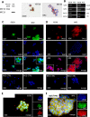
Cholangiocytes express enzymes for 5-HT biosynthesis, catabolism, and transport. A: Western blot (left) and immunocytochemical (middle and right) analyses show that both murine 603B and rat NRC cholangiocytes homogeneously express the ductular marker keratin-19 (KRT19). Original magnification ×63. B: 2% agarose gel of quantitative real-time reverse transcription-polymerase chain reaction (QRT-PCR) amplicons demonstrates expression of tryptophan hydroxylase (tph)-1 and -2, htr1A and B, monoamine oxidase (mao) A and B and serotonin (5-HT) transporter (sert) in 603B cholangiocytes. C and D: immunofluorescence cell staining for TPH1 in green (C) and TPH2 in red (D) showing that both murine (603B, C and D, left) and rat (NRC, D and E, right) express the rate-limiting enzymes for 5-HT biosynthesis. 4′,6-Diamidino-2-phenylindole (DAPI) demonstrates nuclei. 603B and NRC incubated with nonimmune sera plus secondary antibodies are shown as negative controls. Original magnification ×40 (C and D). Coimmunofluorescence of TPH2 in red with the ductular marker γ-glutamyl transpeptidase (GGT) in green is displayed in 603B (E) and NRC (F) cholangiocytes. Original magnification ×63.
Fig. 3.
Fig. 3.
Myofibroblast (MF)-derived TGF-β inhibits Angpt1/Tie2/TPH2 axis and 5-HT production in neighboring cholangiocytes. A: HPLC was employed to detect 5-HT content in the culture medium from monocultures of 603B cholangiocytes (solid bars) or MF (open bars), and from cholangiocyte/MF cocultures (shaded bars). Data are expressed as means ± SE of the absolute concentration (ng/ml). B and C: QRT-PCR of monocultured and cocultured cholangiocytes (solid and shaded bars, respectively) shows differential expression of tph-1 and tph-2 (B) and angiopoietin 1 (angpt1) and tie-2 (C). Data are normalized to monocultured cholangiocytes and displayed as means ± SE. D: ELISA assay demonstrated TGF-β1 content in the culture medium from monocultured 603B cholangiocytes (solid bars) or MF (open bars), and in conditioned medium of cocultures (shaded bars). Results are normalized to values in cholangiocytes monocultures and expressed as means ± SE. E: 603B-cholangiocyte monocultures were exposed to recombinant TGF-β1 (2 ng/ml) or vehicle for 24 h in serum-deprived medium, and QRT-PCR analysis was performed to evaluate the expression of angpt1, tie2, and tph2. Data are normalized to vehicle-treated controls and displayed as means ± SE. F: MF monocultures were treated with exogenous 5-HT for 48 h and TGF-β1 cell induction was assessed by QRT-PCR. Results are normalized to vehicle-treated MF and expressed as means ± SE. For all data sets 2-tailed Student's t-test was employed to evaluate differences between groups. *P < 0.05, **P < 0.001.
Fig. 4.
Fig. 4.
Working model of paracrine mechanisms that modulate 5-HT-TGF-β1 production in liver MF and cholangiocytes. Cholangiocyte-derived 5-HT (red spheres) acts in an autocrine fashion to limit cholangiocyte growth (1) and functions in a paracrine fashion to stimulate production of TGF-β1 (blue spheres) by neighboring MF (2). The MF-derived TGF-β1, in turn, provides an autocrine stimulus for MF growth (3) but acts in a paracrine fashion to downregulate cholangiocyte 5-HT production (4). Reduction of 5-HT derepress cholangiocyte growth, permitting expansion of the ductular cell population (5). Proliferating cholangiocytes, in turn, produce soluble factors (e.g., Hedgehog ligands, PDGF-BB, TGF-β1) that sustain MF growth despite reductions in local 5-HT (6).
Fig. 5.
Fig. 5.
Mice with functional inactivation of TPH2 (TPH2KI) displayed exaggerated proliferation of ductular and hepatocytic cells in response to bile duct ligation (BDL). A: HPLC was performed to assess 5-HT content in bile from TPH2KI (n = 10) and wild-type (WT; n = 8) mice 2 wk after BDL surgery. Dots demonstrate the absolute biliary concentration of 5-HT (ng/ml) in each animal. Black bars show means ± SE of values for each group. B: hematoxylin and eosin (H&E) staining of liver sections from representative mice that were euthanized 2 wk after either Sham (left) or BDL (right) surgery. Original magnification ×20. C: representative pictures of keratin (KRT)-19 staining in WT (top) or TPH2KI (bottom) mice 2 wk after BDL surgery. Original magnification ×40. The graph on the right shows results of the computer-assisted morphometric evaluation of KRT19+ area in WT and TPH2KI mice 2 wk after Sham or BDL surgery. Data are normalized to WT Sham controls and displayed as means ± SE. D: 2 wk after surgery, mice were injected with BrdU and euthanized 2 h later; cholangiocyte BrdU labeling index was quantified in portal tracts (PT) from WT (top) and TPH2KI (bottom). Data are normalized to WT BDL and expressed as means ± SE. Original magnification ×40. Inset shows magnified image of small duct with BrdU positive nucleus. E: representative pictures of section from BDL TPH2KI mice display KRT19/BrdU costaining with BrdU localizing to KRT19+ ducts (top) and to KRT19 hepatocytic cells in periportal areas (top and bottom). F: hepatocytic cell BrdU labeling index was quantified in periportal areas from WT (top) and TPH2KI (bottom). Data are normalized to WT BDL and expressed as means ± SE. Original magnification ×40. All quantitative data are expressed as means ± SE and differences between groups are evaluated by 2-tailed Student's t-test. *P < 0.05, **P < 0.001.
Fig. 6.
Fig. 6.
Reduced TPH2 activity and biliary 5-HT content promote the outgrowth of liver progenitors. Immunohistochemical analysis of AE1/AE3 (which marks progenitor cell-associated cytokeratins) (A and B) and α-fetoprotein (AFP) (C and D) in representative sections of WT (left) and TPH2KI mice (right) 2 wk after sham (top) or BDL (bottom) surgery. Representative pictures are displayed at ×40 original magnification. B: AE1/AE3+ area was quantified by computer-assisted morphometric evaluation. Data are normalized to WT Sham controls and displayed as means ± SE. D: AFP+ hepatocytic cells near portal tracts were counted in immunostained liver sections (n = 5/group) under ×40 magnification. Mean ± SE results are shown. *P < 0.05, **P < 0.001.
Fig. 7.
Fig. 7.
Increased hepatic fibrogenesis in TPH2KI mice after BDL. A: whole liver tissues from TPH2KI (shaded bars) and WT (solid bars) mice 2 wk after BDL were analyzed by QRT-PCR to assess changes in fibrogenic markers [platelet-derived growth factor BB (PDGFBB), TGF-β1, collagen 1α(I), α-smooth muscle actin (αSMA)]. Data are normalized to WT-BDL values and displayed as means ± SE. Differences between groups are evaluated by 2-tailed Student's t-test. *P < 0.05. B: Sirius red staining was done to demonstrate differences in fibrous matrix. Representative photomicrographs from each group are shown. Original magnification ×20. C: computer-assisted quantification of picrosirius red+ area in liver sections from WT (solid bars) and TPH2KI (shaded bars) mice (N = 5 per group), 2 wk BDL surgery. Results are normalized to WT-BDL values and displayed as means ± SE. Differences between groups are evaluated by 2-tailed Student's t-test. *P < 0.05.

Similar articles

Cited by

References

    1. Beaulieu JM, Zhang X, Rodriguiz RM, Sotnikova TD, Cools MJ, Wetsel WC, Gainetdinov RR, Caron MG. Role of GSK3 beta in behavioral abnormalities induced by serotonin deficiency. Proc Natl Acad Sci USA 105: 1333–1338, 2008 - PMC - PubMed
    1. Bochkis IM, Rubins NE, White P, Furth EE, Friedman JR, Kaestner KH. Hepatocyte-specific ablation of Foxa2 alters bile acid homeostasis and results in endoplasmic reticulum stress. Nat Med 14: 828–836, 2008 - PMC - PubMed
    1. Clotman F, Jacquemin P, Plumb-Rudewiez N, Pierreux CE, Van der Smissen P, Dietz HC, Courtoy PJ, Rousseau GG, Lemaigre FP. Control of liver cell fate decision by a gradient of TGF beta signaling modulated by Onecut transcription factors. Genes Dev 19: 1849–1854, 2005 - PMC - PubMed
    1. Dewachter L, Adnot S, Fadel E, Humbert M, Maitre B, Barlier-Mur AM, Simonneau G, Hamon M, Naeije R, Eddahibi S. Angiopoietin/Tie2 pathway influences smooth muscle hyperplasia in idiopathic pulmonary hypertension. Am J Respir Crit Care Med 174: 1025–1033, 2006 - PubMed
    1. Eddahibi S, Guignabert C, Barlier-Mur AM, Dewachter L, Fadel E, Dartevelle P, Humbert M, Simonneau G, Hanoun N, Saurini F, Hamon M, Adnot S. Cross talk between endothelial and smooth muscle cells in pulmonary hypertension: critical role for serotonin-induced smooth muscle hyperplasia. Circulation 113: 1857–1864, 2006 - PubMed

Publication types

MeSH terms

LinkOut - more resources