. 2011 Jan;300(1):G170-80.
doi: 10.1152/ajpgi.00406.2010. Epub 2010 Nov 11.

Characterization of silent afferents in the pelvic and splanchnic innervations of the mouse colorectum

Affiliations

Characterization of silent afferents in the pelvic and splanchnic innervations of the mouse colorectum

Bin Feng et al. Am J Physiol Gastrointest Liver Physiol. 2011 Jan.

Abstract

Hypersensitivity in inflammatory/irritable bowel syndrome is contributed to in part by changes in the receptive properties of colorectal afferent endings, likely including mechanically insensitive afferents (MIAs; silent afferents) that have the ability to acquire mechanosensitivity. The proportion and attributes of colorectal MIAs, however, have not previously been characterized. The distal ∼3 cm of colorectum with either pelvic (PN) or lumbar splanchnic (LSN) nerve attached was removed, opened longitudinally, pinned flat in a recording chamber, and perfused with oxygenated Krebs solution. Colorectal receptive endings were located by electrical stimulation and characterized as mechanosensitive or not by blunt probing, mucosal stroking, and circumferential stretch. MIA endings were tested for response to and acquisition of mechanosensitivity by localized exposure to an inflammatory soup (IS). Colorectal afferents were also tested with twin-pulse and repetitive electrical stimulation paradigms. PN MIAs represented 23% of 211 afferents studied, 71% (30/42) of which acquired mechanosensitivity after application of IS to their receptive ending. LSN MIAs represented 33% of 156 afferents studied, only 23% (11/48) of which acquired mechanosensitivity after IS exposure. Mechanosensitive PN endings uniformly exhibited significant twin-pulse slowing whereas LSN endings showed no significant twin-pulse difference. PN MIAs displayed significantly greater activity-dependent slowing than LSN MIAs. In conclusion, significant proportions of MIAs are present in the colorectal innervation; significantly more in the PN than LSN acquire mechanosensitivity in an inflammatory environment. This knowledge contributes to our understanding of the possible roles of MIAs in colon-related disorders like inflammatory/irritable bowel syndrome.


Figures

Fig. 1.
Fig. 1.
Distributions of electrical stimulus thresholds (A and B) and conduction velocities (C and D) of pelvic nerve (PN) and lumbar splanchnic nerve (LSN) endings in the mouse colorectum. CV, conduction velocity.
Fig. 2.
Fig. 2.
Mouse colorectal pelvic nerve (A) and lumbar splanchnic nerve (B) endings identified by electrical stimulation (e-stim; leftmost column; arrow indicates stimulus artifact) and classified based on responses to mechanical stimuli. All mechanosensitive endings responded to probing (0.4–1.4 g); muscular endings were also activated by circumferential stretch (0–170 mN), mucosal endings also by stroking (10 mg), and muscular/mucosal endings also by both stretch and stroking. Serosal endings were activated only by probing. Mechanically insensitive afferents (MIA) did not respond to mechanical stimuli. Muscular/mucosal endings were unique to the pelvic pathway; mesenteric endings were unique to the splanchnic pathway (0.4 g probing).
Fig. 3.
Fig. 3.
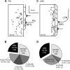
Distributions of MIA receptive endings (A and C) and proportions of afferent classes (B and D) of PN and LSN endings innervating the mouse colorectum. Squares represent the location of MIA endings. The proportions of MIAs (exploded black pie segments) did not differ between the pelvic and lumbar splanchnic nerve innervations. MPG, major pelvic ganglion; IMG, inferior mesenteric ganglion.
Fig. 4.
Fig. 4.
Electrical stimulus thresholds (A) and conduction velocities (B and C) of colorectal endings in the PN and LSN pathways. *Significantly different (P < 0.05, 1-way ANOVA with Dunn's post hoc multiple comparison) from respective MIAs. B and C: scatterplot of conduction velocities; horizontal line represents mean conduction velocity for each class of ending.
Fig. 5.
Fig. 5.
Representative examples of responses of MIAs to chemical stimuli and acquisition of mechanosensitivity. A: most MIAs that acquired mechanosensitivity (23/31) did not respond to direct application of inflammatory soup (IS) or capsaicin (Cap) but responded to 1.4 g probing. B: some MIAs (8/31) that acquired mechanosensitivity also responded directly to the application of IS and/or Cap. C: a small proportion of MIAs (4/64 studied and exclusive to the splanchnic pathway) responded to chemical stimuli (IS or Cap) but did not acquire mechanosensitivity. Subgroups of MIAs are activated (IS+) or not (IS) by IS and acquired (M+) or not (M) mechanosensitivity to probing.
Fig. 6.
Fig. 6.
Summary of activation and/or acquisition of mechanosensitivity of MIAs by IS in pelvic (A) and lumbar splanchnic (B) pathways. Responses of MIAs to IS and/or Cap are summarized in C.
Fig. 7.
Fig. 7.
Afferent responses to twin-pulse and repetitive stimulation paradigms. A: typical recordings showing slowing (top) and speeding (bottom) following an electrical twin-pulse stimulation (50-ms interpulse interval) at the colorectal receptive field. The second conduction latency is 1.6 ms longer than the first in the top trace and 0.6 ms shorter in the bottom trace, corresponding to twin-pulse slowing of 5.8% and speeding of 1.7%, respectively. Arrows indicate stimulus artifacts. B: typical change of latency in an afferent with repetitive stimulation of 2 Hz for 3 min. The final slowing is quantified as percentage increase of the last conduction latency relative to the first one. C and D: twin-pulse differences plotted against final slowing for the 44 PN (C) and 27 LSN (D) afferents tested. diff, Difference.

Similar articles

Cited by

References

    1. Anand U, Otto WR, Facer P, Zebda N, Selmer I, Gunthorpe MJ, Chessell IP, Sinisi M, Birch R, Anand P. TRPA1 receptor localisation in the human peripheral nervous system and functional studies in cultured human and rat sensory neurons. Neurosci Lett 438: 221–227, 2008 - PubMed
    1. Berthoud HR, Blackshaw LA, Brookes SJ, Grundy D. Neuroanatomy of extrinsic afferents supplying the gastrointestinal tract. Neurogastroenterol Motil 16, Suppl 1: 28–33, 2004 - PubMed
    1. Bostock H, Campero M, Serra J, Ochoa J. Velocity recovery cycles of C fibres innervating human skin. J Physiol 553: 649–663, 2003 - PMC - PubMed
    1. Brierley SM, Carter R, Jones W, III, Xu L, Robinson DR, Hicks GA, Gebhart GF, Blackshaw LA. Differential chemosensory function and receptor expression of splanchnic and pelvic colonic afferents in mice. J Physiol 567: 267–281, 2005 - PMC - PubMed
    1. Brierley SM, Hughes PA, Page AJ, Kwan KY, Martin CM, O'Donnell TA, Cooper NJ, Harrington AM, Adam B, Liebregts T, Holtmann G, Corey DP, Rychkov GY, Blackshaw LA. The ion channel TRPA1 is required for normal mechanosensation and is modulated by algesic stimuli. Gastroenterology 137: 2084–2095, 2009 - PMC - PubMed

Publication types

LinkOut - more resources