. 2010 Nov 12;330(6006):985-9.
doi: 10.1126/science.1196554.

Essential regulation of CNS angiogenesis by the orphan G protein-coupled receptor GPR124

Affiliations

Essential regulation of CNS angiogenesis by the orphan G protein-coupled receptor GPR124

Frank Kuhnert et al. Science. .

Abstract

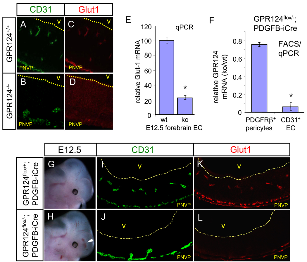
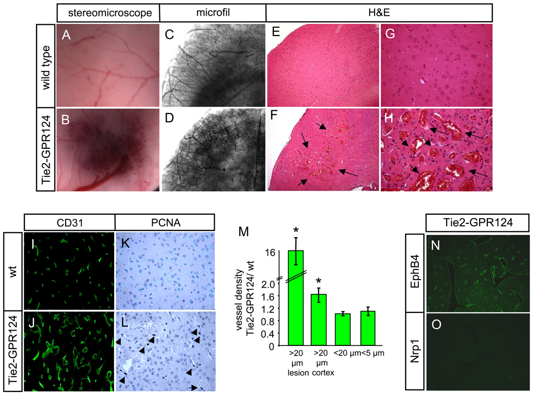
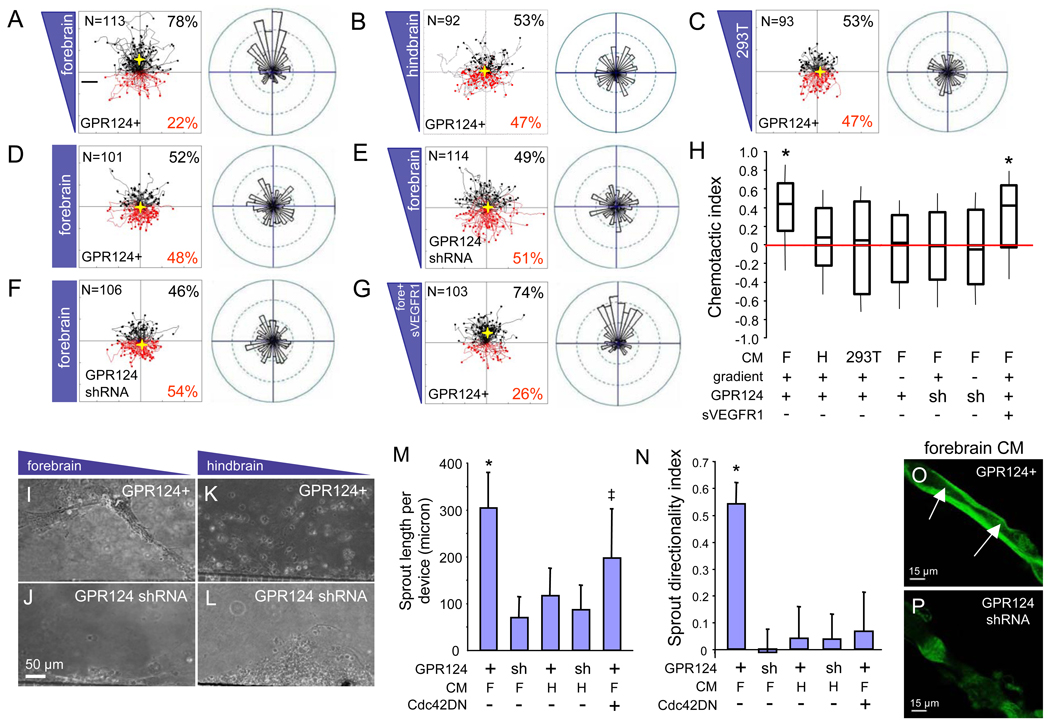
The orphan G protein-coupled receptor (GPCR) GPR124/tumor endothelial marker 5 is highly expressed in central nervous system (CNS) endothelium. Here, we show that complete null or endothelial-specific GPR124 deletion resulted in embryonic lethality from CNS-specific angiogenesis arrest in forebrain and neural tube. Conversely, GPR124 overexpression throughout all adult vascular beds produced CNS-specific hyperproliferative vascular malformations. In vivo, GPR124 functioned cell-autonomously in endothelium to regulate sprouting, migration, and developmental expression of the blood-brain barrier marker Glut1, whereas in vitro, GPR124 mediated Cdc42-dependent directional migration to forebrain-derived, vascular endothelial growth factor-independent cues. Our results demonstrate CNS-specific angiogenesis regulation by an endothelial receptor and illuminate functions of the poorly understood adhesion GPCR subfamily. Further, the functional tropism of GPR124 marks this receptor as a therapeutic target for CNS-related vascular pathologies.


Figures

Fig. 1
Fig. 1
(A to X) CNS vascular patterning defects in GPR124−/− embryos. GPR124−/− embryos manifested progressive hemorrhage confined to forebrain telencephalon (arrowheads) and neural tube (*) [(A) to (D)]. Wild type E11.5 embryos exhibited efficient sprouting angiogenesis (arrows) from the perineural vascular plexus (PNVP) into the telencephalon [(E) and (G)], which was completely ablated in GPR124−/− embryos that exhibited PNVP thickening only [(F) to (H)], Green- CD31 IF. Large, glomeruloid vascular malformations (arrowheads) and absence of a vascular network were observed in E11.5 GPR124−/− forebrain ganglionic eminences and ventral neural tube [(I) to (L), CD31 IF]. By E14.5, GPR124−/− telencephalon was essentially devoid of a mature capillary network with glomeruloid malformations abutting the pia/PNVP, while the diencephalon was unaffected [(M) to (N), transverse; (O) to (P), sagittal; vascular malformations (arrows), (Q) and (R), CD31 IF]. Glomeruloid malformations were irregular, multi-layer endothelial aggregates versus single layer capillaries in controls [(S) and (T), EM 4300x], with normal peripheral PDGFRβ+ pericyte investment [(U) and (V)] but lacked ventricularly-directed endothelial filopodia (arrows) [(W) and (X), CD31 IF]; VD, ventricle diencephalons; VT, ventricle telencephalon; blue, DAPI.
Fig. 2
Fig. 2
(A to L) GPR124 functions cell-autonomously in endothelium. Expression of Glut1 is lost in GPR124−/− CNS endothelium, with compensatory Glut1 upregulation in neuroepithelium, [(A) to (D)], E12.5. Glut1 is strongly down-regulated in FACS-isolated, GPR124−/− telencephalic endothelium. qPCR, n=9, E12.5 [(E)]. Selective deletion of GPR124 in FACS-isolated forebrain CD31+ endothelium but not PDGFRβ+ pericytes. qPCR, n=6, E 12.5 [(F)]. E12.5 GPR124flox/−; PDGFB-iCre embryos exhibit forebrain hemorrhaging, formation of PNVP--associated glomeruloid vascular malformations and endothelial Glut1 downregulation [(G) to (L)], recapitulating the global GPR124 deletion phenotype. PNVP, perineural vascular plexus. Error bars are +/− 1 S. D.
Fig. 3
Fig. 3
(A to O) CNS vascular malformations in adult (12 months old) Tie2-GPR124 transgenic animals. Focally increased red blood cell accumulation in Tie2-GPR124 cerebral cortex [(A) and (B)] corresponded to increased vascular density and tortuous, enlarged vessels upon microfil vascular casting [(C) and (D)]. H&E staining of a hypervascular area (arrows) in Tie2-GPR124 cortex, 100x [(E) and (F)] revealed dramatically enlarged, engorged thin-walled vessels with intervening neural tissue reminiscent of venous angiomas, 400x [(G) and (H)]. Tie2-GPR124 transgenics exhibited increased CD31+ microvessel density and markedly enlarged [(I) to (J), (M)] and hyperproliferative vascular malformations versus wild type controls [(K) and (L), arrowheads denote PCNA+ cells]. Tie2-GPR124 CNS vascular malformations expressed the venous marker EphB4, but not the arterial receptor Nrp1 [(N) and (O)]. Error bars are +/− 1 S. D.
Fig. 4
Fig. 4
(A to O) GPR124 regulates CNS endothelial migration and sprout formation in vitro. Motility of the GPR124-expressing brain endothelial line bEND3 was monitored by real-time video microscopy in a microfluidic migration chamber allowing extremely stable, shear-minimized linear chemoattractant gradients. GPR124-overexpressing bEND3 cells (GPR124+) exhibited directed migration towards gradients of conditioned medium (CM) from cultured E12.5 forebrain cortical cells, but migrated randomly to equivalent hindbrain or HEK 293T CM [(A) to (C), tracks of individual cells (left) and angular histograms (right) are shown]. No directed migration was observed with even distribution of forebrain CM across the microfluidic chamber (no gradient), or GPR124 shRNA [(D) to (F)]; migration was further insensitive to a recombinant soluble VEGFR1 ectodomain (400 ng/ml), indicating VEGF-independence [(G)]. Box plot of chemotactic indices of bEND3 migration [(H), center line: median, boxes: upper and lower quartiles, *= p<0.001]. In a sprout formation assay GPR124+ bEND3 displayed collective cell migration and robust formation of stable sprouts towards forebrain CM gradients, but not to hindbrain CM nor upon GPR124 shRNA knockdown [(I) to (L), (M)]. Dominant-negative Cdc42N17 inhibited sprout directionality but not elongation [(M) to (N), *= p<0.05, ‡ = N.S.]. GPR124+ sprouts towards forebrain CM contained stable, contiguous lumens, while rare GPR124 shRNA sprouts were unstable and failed to lumenize. Confocal microscopy [(O) to (P)]. F, forebrain CM; H, hindbrain CM. Error bars are +/− 1 S. E. *= p<0.05. N= 3–5 chambers per condition.

References

    1. Carmeliet P. Nature. 2005 Dec 15;438:932. - PubMed
    1. Aird WC. Circ Res. 2007 Feb 2;100:174. - PubMed
    1. Ballabh P, Braun A, Nedergaard M. Neurobiol Dis. 2004 Jun;16:1. - PubMed
    1. Pardridge WM. Methods Mol Med. 2003;89:385. - PubMed
    1. Shen Q, et al. Science. 2004 May 28;304:1338. - PubMed

Publication types

MeSH terms

Substances