. 2010 Nov 30;107(48):20726-31.
doi: 10.1073/pnas.1007295107. Epub 2010 Nov 11.

Cell-free fusion of bacteria-containing phagosomes with endocytic compartments

Affiliations

Cell-free fusion of bacteria-containing phagosomes with endocytic compartments

Ulrike Becken et al. Proc Natl Acad Sci U S A. .

Abstract

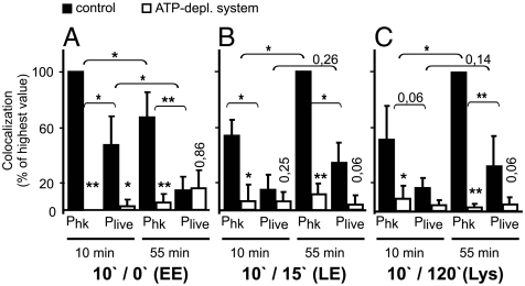
Uptake of microorganisms by professional phagocytic cells leads to formation of a new subcellular compartment, the phagosome, which matures by sequential fusion with early and late endocytic compartments, resulting in oxidative and nonoxidative killing of the enclosed microbe. Few tools are available to study membrane fusion between phagocytic and late endocytic compartments in general and with pathogen-containing phagosomes in particular. We have developed and applied a fluorescence microscopy assay to study fusion of microbe-containing phagosomes with different-aged endocytic compartments in vitro. This revealed that fusion of phagosomes containing nonpathogenic Escherichia coli with lysosomes requires Rab7 and SNARE proteins but not organelle acidification. In vitro fusion experiments with phagosomes containing pathogenic Salmonella enterica serovar Typhimurium indicated that reduced fusion of these phagosomes with early and late endocytic compartments was independent of endosome and cytosol sources and, hence, a consequence of altered phagosome quality.


Conflict of interest statement

The authors declare no conflict of interest.

Figures

Fig. 1.
Fig. 1.
Preparation of endocytic vesicles. Macrophages were incubated with ferrofluid and magnetic compartments were isolated from a PNS. (A) Western blot analysis of PNS and purified endocytic compartments. Fifty or 10 μg protein of PNS, 10 μg protein for each purified fraction were added. Pulse/chase times for ferrofluid are indicated. Arrowheads indicate the positions of cathepsin D (Cat D) precursor (p), intermediate (i), and mature (m) forms. (B) Quantification of specific lysosomal acid β-galactosidase activities (U/mg) and of fluorescence (FU) of lysosomal BSA rhodamine. Shown are means and standard deviations of three independent experiments.
Fig. 2.
Fig. 2.
Assay for cell-free phagosome–endosome fusion. (A) Schematic representation of the cell-free phagosome–endosome fusion assay. See text for details. (B) Micrograph showing a representative image section of an in vitro ECP-lysosome fusion reaction prepared for fluorescence microscopy. In the overlay, phagosomes containing E. coli are shown in green, lysosomes in red. Enlargements show bacteria not colocalizing with lysosomal fluor (Upper) or colocalizing with lysosomal fluor (Lower). (Right) False color-coded (open arrowhead) images of FRET intensity for each pixel (black: low intensity; white: high intensity). Bar: 2 μM.
Fig. 3.
Fig. 3.
In vitro fusion of ECPs or LBPs with lysosomes. Fusion rates under standard conditions (control) were set as 100%. This translates to 3–12% of the bacteria or latex beads being positive for the lysosomal fluor. Means and standard deviations of ≥3 independent experiment are shown. p < 0.05, ∗∗p < 0.01 compared to control. (A) Time course of phagosome–lysosome fusion. Fusion by 75 min (ECPs) or by 60 min (LBPs) was set as 100%. Fusion reactions contained ECPs (open circles, error bars downward) or LBPs (closed squares, error bars upward). (B and C) Compounds added at the beginning of the fusion reaction: 4 mM NEM, 10 mM BAPTA, 0.5 mg/mL RabGDI, 40 nM bafilomycin A1, 1 μM nigericin, 20 μM nocodazole. Reactions contained ECPs (B) or LBPs (C). (DF) Reactions contained ECPs. (D) Colocalization of bacteria with lysosomal BSA rhodamine and percentage of bacteria with mean FRET intensity > 0 were calculated and expressed in percent of the respective control. One hundred percent (SD ± 73, n = 7) of colocalizing bacteria showed mean FRET intensity > 0. (E) Affinity purified anti-Rab7 or (F) GST-Rab7 WT or GST-Rab7T22N were added at the indicated final concentrations.
Fig. 4.
Fig. 4.
SNARE proteins in cell-free ECP-lysosome fusion. (A) Cell-free fusion of ECPs with lysosomes was carried out in the presence of single solSNAREs or combinations of solSNARES as indicated. Preincubations of solSNAREs for 90 min on ice were done (pre) where indicated or solSNAREs were added directly at the start of the fusion reaction (no pre). Data are means and standard deviations of ≥3 independent experiments. p < 0.05, ∗∗p < 0.01 compared to control. (B) Combinations of purified recombinant solSNAREs were mixed as indicated in equimolar ratios (12 μM each) and incubated for 90 min on ice. Samples were incubated with SDS-sample buffer without boiling and analyzed by Western blotting using an antibody to Stx8. The white arrowhead indicates the running position of solStx8; the black arrowhead indicates SDS resistant four-helix bundles. (C) PNS and purified lysosomes were analyzed for enrichment or depletion of various compartmental markers by Western blotting.
Fig. 5.
Fig. 5.
In vitro fusion of Salmonella-containing phagosomes with different-aged endocytic compartments. In vitro fusion experiments were carried out with early (10 min) or late (55 min) phagosomes containing live (Plive) or heat-killed (Phk) S. enterica. Fusion rates of phagosomes with endosomes of different age cannot be directly compared, because fluor contents and volume of endocytic organelles likely change with maturation. Therefore, only fusion reactions with same-stage endocytic compartments were compared. Only phagosomes containing an intact membrane (bacteria were not antibody accessible) were considered. (A) Fusion reactions contained 10/0 endosomes (early endosomes, EE), (B) 10/15 endosomes (late endosomes, LE), or (C) 10/120 endosomes (lysosomes, Lys). Data are the means and standard deviations of ≥3 independent experiments. Asterisks or values above open bars without brackets mark p values comparing adjacent open and filled bars. ∗∗p < 0.01; *: p < 0, 05.

References

    1. Haas A. The phagosome: Compartment with a license to kill. Traffic. 2007;8:311–330. - PubMed
    1. Desjardins M, Huber LA, Parton RG, Griffiths G. Biogenesis of phagolysosomes proceeds through a sequential series of interactions with the endocytic apparatus. J Cell Biol. 1994;124:677–688. - PMC - PubMed
    1. Jahraus A, et al. In vitro fusion of phagosomes with different endocytic organelles from J774 macrophages. J Biol Chem. 1998;273:30379–30390. - PubMed
    1. Zerial M, McBride H. Rab proteins as membrane organizers. Nat Rev Mol Cell Biol. 2001;2:107–117. - PubMed
    1. Alvarez-Dominguez C, et al. Phagocytosed live Listeria monocytogenes influences Rab5-regulated in vitro phagosome-endosome fusion. J Biol Chem. 1996;271:13834–13843. - PubMed

Publication types

MeSH terms

LinkOut - more resources