. 2011 Jan;62(2):735-48.
doi: 10.1093/jxb/erq309. Epub 2010 Nov 11.

Endosperm development in Brachypodium distachyon

Affiliations

Endosperm development in Brachypodium distachyon

Magdalena Opanowicz et al. J Exp Bot. 2011 Jan.

Abstract

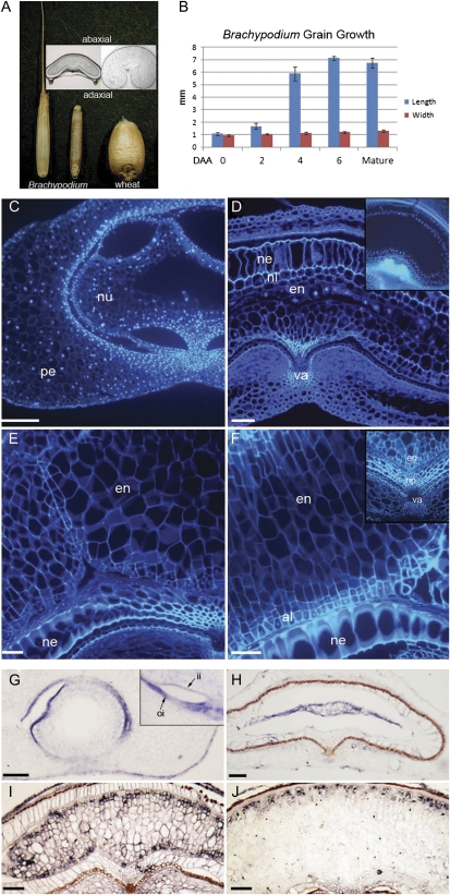
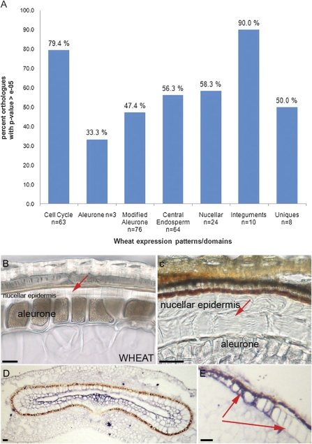
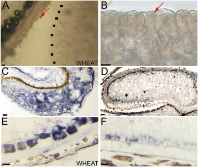
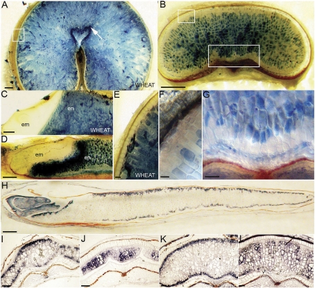
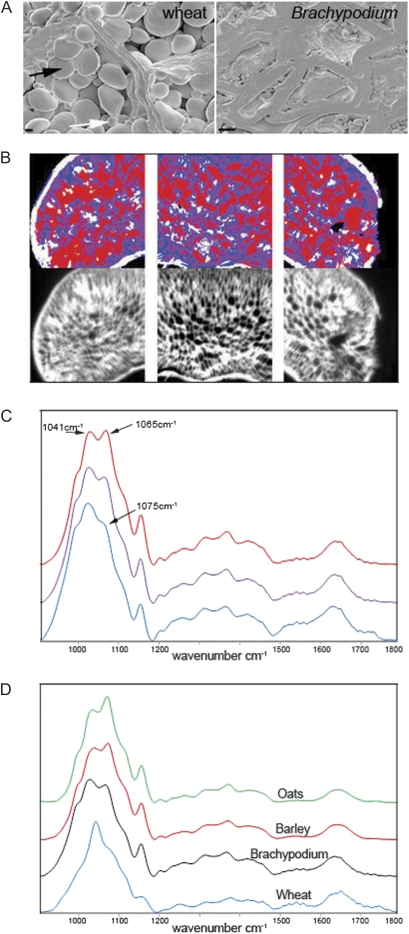
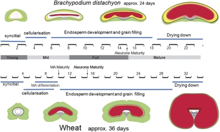
Grain development and its evolution in grasses remains poorly understood, despite cereals being our most important source of food. The grain, for which many grass species have been domesticated, is a single-seeded fruit with prominent and persistent endosperm. Brachypodium distachyon, a small wild grass, is being posited as a new model system for the temperate small grain cereals, but little is known about its endosperm development and how this compares with that of the domesticated cereals. A cellular and molecular map of domains within the developing Brachypodium endosperm is constructed. This provides the first detailed description of grain development in Brachypodium for the reference strain, Bd21, that will be useful for future genetic and comparative studies. Development of Brachypodium grains is compared with that of wheat. Notably, the aleurone is not regionally differentiated as in wheat, suggesting that the modified aleurone region may be a feature of only a subset of cereals. Also, the central endosperm and the nucellar epidermis contain unusually prominent cell walls that may act as a storage material. The composition of these cell walls is more closely related to those of barley and oats than to those of wheat. Therefore, although endosperm development is broadly similar to that of temperate small grain cereals, there are significant differences that may reflect its phylogenetic position between the Triticeae and rice.


Figures

Fig. 1.
Fig. 1.
Development of the Brachypodium grain. (A) Brachypodium grain with and without husk compared with wheat grain. (B) Measurements of length and width of Brachypodium grain through development showing rapid elongation early in development; n = 6 and error bars represent ±1 SD. (C) DAPI-stained sections of Brachypodium grains at 3 DAA (days after anthesis); (D) 5 DAA; (E) 8 DAA; (F) 15 DAA; pe, pericarp; nu, nucellar tissue; ne, nucellar epidermis; nl, nucellar lysate; np, nucellar projection; en, endosperm; va, vascular tissue; al, aleurone. (G) Histone H4 mRNA ISH in grain sections at 3 DAA; (H) 5 DAA; (I) 8 DAA; (J) 15 DAA; ii, inner integument; oi, outer integument; all scale bars, 50 μm
Fig. 2.
Fig. 2.
Analysis of nucellar tissue in Brachypodium grains. (A) Summary of BLASTN results for wheat sequences (showing tissue-specific gene expression patterns; Drea et al., 2005b) compared with the Brachypodium genome. (B) Brightfield micrograph of peripheral grain cell layers in mature wheat. (C) Brachypodium grains. Nucellar epidermis layers are indicated with red arrows and both nucellar epidermis and aleurone are labelled. (D) ISH of BdC13 showing expression in nucellar lysate of a 4 DAA (days after anthesis) cross-section; (E) BdC13 in the abaxial nucellar epidermis in a 15 DAA grain cross-section. Scale bars, 20 μm.
Fig. 3.
Fig. 3.
Analysis of aleurone tissue in Brachypodium and wheat grains. (A) Adherence of aleurone layer to the maternal tissues in wheat. (B) Adherence of aleurone layer to the endosperm in Brachypodium grains when grains are soaked in water and thin sections made with a sharp blade. The dotted line indicates the edge of the central endosperm. (C) ISH of BdPPDK showing expression in the pericarp of 4 DAA (days after anthesis) grains; (D) BdPPDK in the peripheral endosperm of 15 DAA grains. (E) Specific comparison of wheat PPDK with (F) BdPPDK in the aleurone layers. Scale bars, 20 μm.
Fig. 4.
Fig. 4.
Cellular and molecular domains within the Brachypodium endosperm. (A) Mature wheat and (B) Brachypodium grain cross-sections stained with Evans Blue. Boxed regions are shown in more detail in E, F, and G. Scale bars, 200 μm. (C) Wheat and (D) Brachypodium embryos are clear of staining. em, embryo; en; endosperm. Scale bars, 500 μm. (E) Wheat and (F) Brachypodium peripheral aleurone layers are clear of staining. Scale bars, 10 μm and 50 μm. (G) Brachypodium endosperm cells above the vascular/crease region are unstained. Scale bar, 50 μm. (H) BdGLO1expression in a mature grain long section. Scale bar, 500 μm. (I) BdGLO1expression at 7 DAA (days after anthesis) and (J) 20 DAA in grain cross-sections. (K) BdGLO2 expression at 7 DAA and (L) 20 DAA in grain cross-sections. Scale bars I–L, 50 μm.
Fig. 5.
Fig. 5.
Composition of the mature Brachypodium grain. (A) Scanning electron micrographs of grain cross-sections showing packed starch grains in wheat in contrast to the small and few starch grains in Brachypodium and the very thick cell walls of its central endosperm. Large and small starch granules in wheat are indicated by black and white arrows, respectively. Scale bars, 5 μm. (B) FT-IR spectroscopic images above the visible images for a thin cell-wall-only cross-section of a Brachypodium grain. Blue pixels indicate spectra with a shoulder at 1075 cm−1, purple pixels a peak at 1065 cm−1, and red a peak at 1065 cm−1 greater in absorbance than the peak at 1041 cm−1. (C) The average spectra for each colour-coded region. (D) Average spectra for the endosperm cell walls of wheat, Brachypodium, barley, and oats.
Fig. 6.
Fig. 6.
Schematic summarizing grain development. Brachypodium grain development compared with wheat focusing on the key stages in endosperm development—illustrations are not to scale and grain tissues are indicated by colour: pericarp, green; integuments; black; nucellar epidermis, yellow; nucellar lysate, blue; nucellar projection, grey; aleurone, pink; central endosperm, red; MA, modified aleurone. Numbers indicate DAA (days after anthesis). Stages indicated as young, mid, and full mature correspond to the material used in RT-PCR in Supplementary Fig. S2 at JXB online.

References

    1. Akiyama T, Kaku H, Shibuya N. A cell wall-bound beta-glucosidase from germinated rice: purification and properties. Phytochemistry. 1998;48:49–54. - PubMed
    1. Becraft PW. Cell fate specification in the cereal endosperm. Seminars in Cell and Developmental Biology. 2001;12:387–394. - PubMed
    1. Bewley JD, Black M. Seeds. Physiology of development and germination. New York: Plenum Press; 1994.
    1. Brown R, Lemmon B, Olsen O-A. Development of the endosperm in rice (Oryza sativa L.): cellularization. Journal of Plant Research. 1996;109:301–313.
    1. Chastain C, Heck J, Colquhoun T, Voge D, Gu X-Y. Posttranslational regulation of pyruvate, orthophosphate dikinase in developing rice (Oryza sativa) seeds. Planta. 2006;224:924–934. - PubMed

Publication types

MeSH terms