. 2010 Dec;30(12):2400-7.
doi: 10.1161/ATVBAHA.110.216531. Epub 2010 Nov 11.

Properties of procoagulant platelets: defining and characterizing the subpopulation binding a functional prothrombinase

Affiliations

Properties of procoagulant platelets: defining and characterizing the subpopulation binding a functional prothrombinase

Ammon M Fager et al. Arterioscler Thromb Vasc Biol. 2010 Dec.

Abstract

Objective: The goal of this study was to define and characterize the subpopulation of platelets capable of regulating the functional interactions of factors Va (FVa) and Xa (FXa) on the thrombin-activated platelet surface.

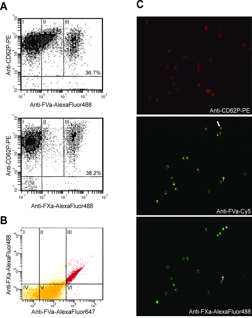
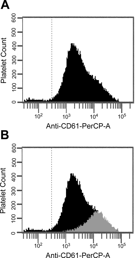
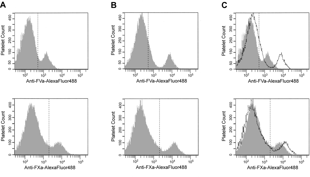
Methods and results: Flow cytometric analyses were used to define and characterize platelet subpopulations. At a concentration of thrombin known to elicit maximal platelet activation, platelet-derived FVa release, and prothrombinase assembly/function, only a subpopulation of platelets was positive for FVa and FXa binding. An additional subpopulation bound lower levels of FVa but little, if any, FXa. Fluorescence microscopy analyses confirmed these data. Phenotypically, platelets capable of binding FXa were more highly reticulated and demonstrated significantly increased expression of several key adhesion molecules, including P-selectin, glycoprotein Ibα, and integrins α(IIb) and β(3). This platelet subpopulation was also defined by the expression of a nondissociable, membrane-bound pool of functional platelet-derived FVa, which made up ≈35% to 50% of the total membrane-bound cofactor.

Conclusions: The ability of activated platelets to support thrombin generation is defined by a subpopulation of platelets expressing a nondissociable pool of platelet-derived FVa and increased adhesive receptor density. This subpopulation is hypothesized to play a significant role in regulating both normal hemostasis and pathological thrombus formation because the adherent properties of platelets and their ability to mount and sustain a procoagulant response are crucial steps in both of these processes.


Figures

Figure 1
Figure 1. FXa binding defines a unique procoagulant platelet subpopulation dependent upon, but not described by, FVa binding
Washed platelets were activated with thrombin (50 nM) and subsequently incubated with saturating concentrations of plasma-derived FVa and FXa (5 nM) as detailed in Methods. (A) Following fixation, platelets were stained with PE-conjugated anti-CD62P to identify activated platelets (y-axes) and specific AlexaFluor488-labeled antibodies to detect FVa (upper panel) and FXa (lower panel) binding (x-axes). The subpopulations of activated platelets that I) do not bind FVa or FXa, II) bind intermediate levels of FVa, or III) bind substantial amounts of FVa or FXa (“high binders”), are indicated by their relative levels of immunostaining. The percent of platelets binding “high” levels of FVa or FXa is indicated in the corresponding panel. (B) Similarly prepared platelets were simultaneously labeled with anti-CD62-PE, anti-FVa-AlexaFluor647, and anti-FXa-AlexaFluor488 to quantify FVa and FXa binding to the CD62+ platelet population. Panels I-III = FXa binding; panels II and V = “intermediate” FVa binding; panels III and VI = “high” FVa binding. For confocal microscopic analyses (C), platelets were activated as above, and cytocentrifuge specimens were prepared and visualized as described in Methods. These images represent a qualitative assessment of the ability of activated platelets to bind FVa and FXa. The laser intensity of the confocal microscope was set to a low level such that only those platelets capable of binding “high” levels of FVa were visualized. Shown here is a single field simultaneously immunostained with three different antibodies to define platelet activation (top panel), “high” FVa binding (middle panel), and FXa binding (bottom panel). The asterisk (*) indicates a platelet which stains for FVa and FXa, without evidence of P-selectin staining, reflecting the variance in P-selectin expression in the procoagulant population subpopulation (Supplemental Figure V). Also indicated are a platelet which expressed “high” levels of FVa but did not bind FXa (arrow), and an apparent anti-FXa-AlexaFluor488 antibody aggregate (†).
Figure 2
Figure 2. Integrin β3 expression is increased on the surface of procoagulant platelets
(A) Activated platelets capable of binding FXa were identified as described in Figure 1. Adhesive receptor density in the entire activated platelet population was defined using anti-CD61-PerCP. (B) To determine the portion of total integrin β3 expression contributed by the subpopulation of platelets capable of binding FXa, FXa-positive platelets were defined using anti-FXa-AlexaFluor488 and analyzed using anti-CD61-PerCP. The integrin β3 expression of the FXa-positive platelets (grey histogram) is shown as an overlay over the integrin β3 expression of the entire activated platelet population.
Figure 3
Figure 3. Identification of a functional, non-dissociable, platelet-derived FVa pool
Activated platelets were washed repeatedly (0 – 5 times) in the absence of Ca2+ as described in Methods. The remaining platelet-bound FVa heavy chain (HC) and light chain (LC) were evaluated by immunoblotting under reducing conditions (A) and quantified by densitometric analyses (B). Subsequent to washing, the remaining platelet-bound FVa activity was determined using a prothrombin time-based clotting assay specific for factor V (C). Data are expressed as the mean ± SEM (n =7) of the % FVa activity remaining after each wash as compared to the membrane-bound activity assessed immediately following platelet activation. Inset: Activated platelets were washed in the presence (♦) or absence (■) of Ca2+, and FVa activity was determined as above (n=1).
Figure 4
Figure 4. Procoagulant platelet subpopulation formation is defined by a single population capable of binding/expressing both dissociable and non-dissociable pools of platelet-derived FVa
FVa (top row) and FXa (bottom row) binding to thrombin-activated human platelets in the absence of exogenous FVa (A), or in the presence of saturating concentrations of plasma-derived FVa (B) were determined by flow cytometric analyses as in Figure 1. Panel (C) depicts a merged image of A & B with the transparent histogram indicating the additional FVa or FXa binding obtained in the presence of the added plasma-derived cofactor.

References

    1. Kalafatis M, Egan JO, van't Veer C, Cawthern KM, Mann KG. The regulation of clotting factors. Crit Rev Eukaryot Gene Expr. 1997;7:241–280. - PubMed
    1. Strukova SM. Thrombin as a regulator of inflammation and reparative processes in tissues. Biochemistry (Mosc) 2001;66:8–18. - PubMed
    1. Martorell L, Martinez-Gonzalez J, Rodriguez C, Gentile M, Calvayrac O, Badimon L. Thrombin and protease-activated receptors (PARs) in atherothrombosis. Thromb Haemost. 2008;99:305–315. - PubMed
    1. Osterud B, Rapaport SI, Lavine KK. Factor V activity of platelets: evidence for an activated factor V molecule and for a platelet activator. Blood. 1977;49:819–834. - PubMed
    1. Bouchard BA, Williams JL, Meisler NT, Long MW, Tracy PB. Endocytosis of plasma-derived factor V by megakaryocytes occurs via a clathrin-dependent, specific membrane binding event. J Thromb Haemost. 2005;3:541–551. - PubMed

Publication types

MeSH terms