. 2011 Feb;31(2):399-407.
doi: 10.1161/ATVBAHA.110.215939. Epub 2010 Nov 11.

Compartmentalized connexin 43 s-nitrosylation/denitrosylation regulates heterocellular communication in the vessel wall

Affiliations

Compartmentalized connexin 43 s-nitrosylation/denitrosylation regulates heterocellular communication in the vessel wall

Adam C Straub et al. Arterioscler Thromb Vasc Biol. 2011 Feb.

Abstract

Objective: To determine whether S-nitrosylation of connexins (Cxs) modulates gap junction communication between endothelium and smooth muscle.

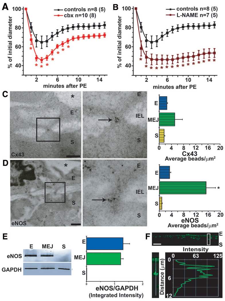
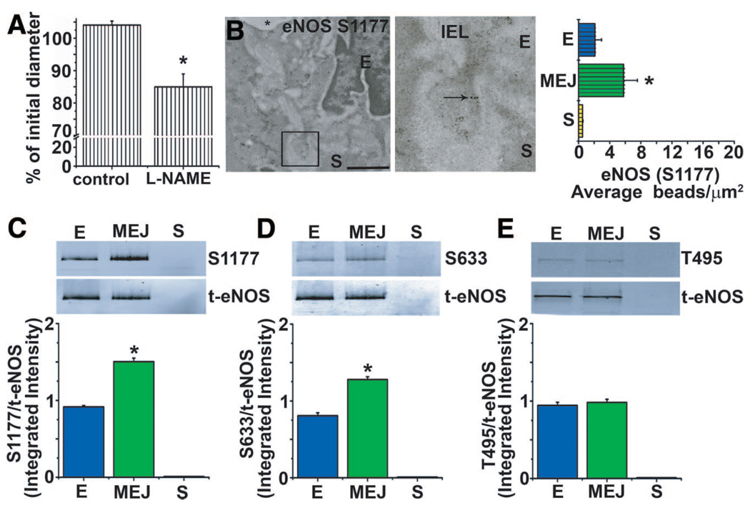
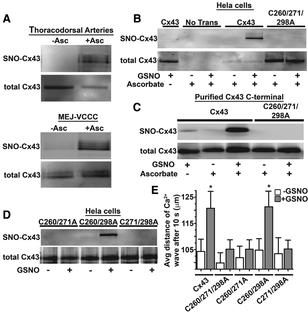
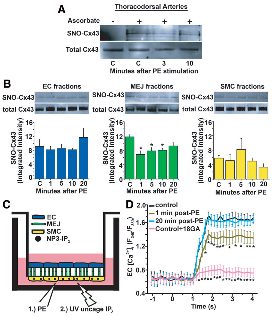
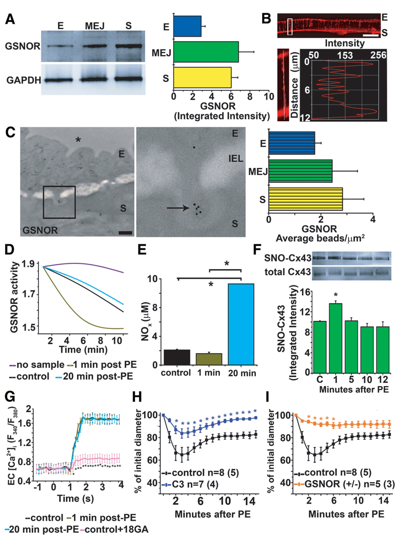
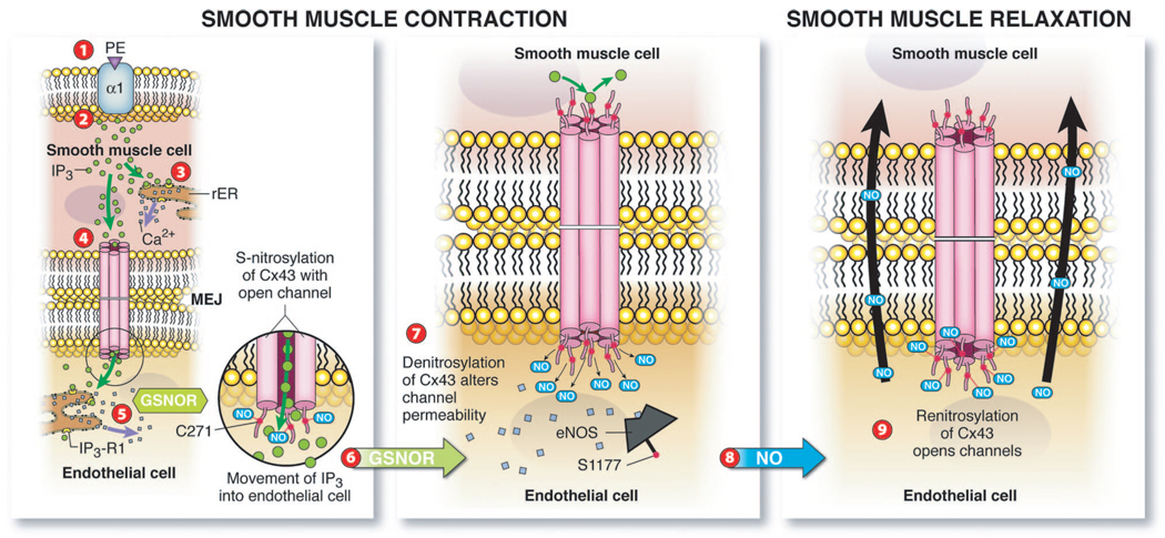
Methods and results: Heterocellular communication is essential for endothelium control of smooth muscle constriction; however, the exact mechanism governing this action remains unknown. Cxs and NO have been implicated in regulating heterocellular communication in the vessel wall. The myoendothelial junction serves as a conduit to facilitate gap junction communication between endothelial cells and vascular smooth muscle cells within the resistance vasculature. By using isolated vessels and a vascular cell coculture, we found that Cx43 is constitutively S-nitrosylated on cysteine 271 because of active endothelial NO synthase compartmentalized at the myoendothelial junction. Conversely, we found that stimulation of smooth muscle cells with the constrictor phenylephrine caused Cx43 to become denitrosylated because of compartmentalized S-nitrosoglutathione reductase, which attenuated channel permeability. We measured S-nitrosoglutathione breakdown and NO(x) concentrations at the myoendothelial junction and found S-nitrosoglutathione reductase activity to precede NO release.

Conclusions: This study provides evidence for compartmentalized S-nitrosylation/denitrosylation in the regulation of smooth muscle cell to endothelial cell communication.


Figures

Figure 1
Figure 1
Vasoreactivity is altered by inhibition of GJ communication and NO correlating with Cx43 and eNOS expression at the MEJ. Mouse TD arteries were cannulated, pressurized, and stimulated with 50-µmol/L PE. A and B, Application of carbenoxolone (50 µmol/L, A) and L-NAME (100 µmol/L, B) significantly enhanced PE-induced vasoconstriction in the TD arteries. C and D, Immuno-TEM analysis of Cx43 (C) and eNOS (D) localization labeled with 10-nm gold beads (arrows) at MEJs from the TD arteries quantified the number of beads per micrometer squared. E, Isolated EC, MEJ, and SMC protein fractions from the VCCC blotted for eNOS and normalized to GAPDH. F, Immunocytochemistry of transverse sections from a VCCC were labeled for eNOS (green). The white box illustrates an enlarged MEJ with a line scan measuring fluorescence down the pore. Data are represented as the mean±SE. (C, n=8; D, n=6; E, n=4). Significant differences (*P<0.05) were analyzed using a 2-way ANOVA (A and B) or a 1-way ANOVA (C–E). In A and B, n is the number of vessels and the value in parentheses is the number of mice. E indicates endothelial cell; IEL, internal elastic lamina; S, smooth muscle cell; *, lumen. The scale bar in C and D is 0.5 µm; and F, 10 µm. In C through E, the open bars indicate in vitro measurements; and bars with horizontal lines, in vivo measurements.
Figure 2
Figure 2
eNOS is differentially phosphorylated at the MEJ. Mouse TD arteries were cannulated and pressurized. A, The basal vascular tone was attenuated in the presence of L-NAME (100 µmol/L). B, Phosphorylated eNOS S1177 localization using 10-nm gold beads (arrow) in TD arteries using immuno-TEM and quantified as the number of beads per micrometer squared. C through E, Isolated EC, MEJ, and SMC fractions from the VCCC blotted for phosphorylated eNOS at sites S1177, S633, and T495 in EC, MEJ, and SMC fractions. Data are represented as the mean±SE. (A, n=5; B–E, n=4). Significant differences (*P<0.05) were analyzed using a 1-way ANOVA (A–D). E, endothelial cell; IEL, internal elastic lamina; S, smooth muscle cell; *, lumen. The scale bar is 0.5 µm (B). In A through E, open bars indicate in vitro measurements; and bars with horizontal lines, in vivo measurements.
Figure 3
Figure 3
Cx43 is S-nitrosylated on cysteine 271. A, Unstimulated TD arteries or MEJ fractions analyzed by the biotin switch assay for Cx43, in which ascorbate-dependent labeling demonstrates the presence of an S-nitrosylated cysteine residue(s). B, Nontransfected and transfected HeLa cells with Cx43 or Cx43C260/271298A were treated with or without 100-µmol/L GSNO for 1 hour. C, Biotin switch assay of purified Cx43 C-terminal or Cx43C260/271298A C-terminal peptides treated with or without 100-µmol/L GSNO for 1 hour. D, HeLa cells transfected with Cx43C260/271A, Cx43C260/298A, or Cx43C271/298A and treated with 100-µmol/L GSNO for 1 hour were lysed and subjected to the biotin switch assay. E, Uncaging of NPE-IP3 and analysis of calcium wave propagation and transfected HeLa cells with Cx43, Cx43C260/271/298A, Cx43C260/271A, Cx43C260/298A, and Cx43C271/298A. Data are represented as the mean±SE (n=6 to 8). Significant differences (*P<0.05) were analyzed using a 1-way ANOVA.
Figure 4
Figure 4
PE promotes denitrosylation of Cx43 and alters channel permeability. A and B, Immunoblots of isolated TD arteries or EC, MEJ, and SMC fractions identifying S-nitrosylated Cx43 using the biotin switch assay from VCCCs stimulated with PE. C, Schematic illustration of the experimental protocol used for measuring EC [Ca2+]i using an initial stimulus of PE, followed by uncaging of NPE-IP3 in SMCs using UV flash at specific points after PE stimulation. D, The maximum values of EC [Ca2+]i were measured at control, 0, 1, and 20 minutes after PE stimulation or with the addition of 18GA. Data are represented as the mean±SE (n=3). Significant differences (*P<0.05) were analyzed using a 1-way ANOVA. Open bars in B indicate in vitro experiments.
Figure 5
Figure 5
GSNOR regulates heterocellular communication. A, Quantitative Western blot analysis of GSNOR expression in isolated EC, MEJ, and SMC protein fractions from the VCCC normalized to GAPDH. Immunocytochemistry of transverse sections of a VCCC labeled for GSNOR (red). B, The white box illustrates an enlarged MEJ with a line scan measuring fluorescence down the pore. C, Immuno-TEM analysis of GSNOR expression labeled with 10-nm gold beads (arrows) at MEJs from the TD arteries and quantified as the number of beads per micrometer squared. D, Measurement of GSNOR activity by breakdown of GSNO in MEJ fractions at 1 and 20 minutes after PE stimulation. E, Identification of total NOx in MEJ fractions at 1 and 20 minutes after PE stimulation. F, Immunoblot of S-nitrosylated Cx43 from in vitro MEJ fractions pretreated with C3 inhibitor and then stimulated with PE for 0, 1, 5, 10, and 20 minutes. G, Measurement of maximum values of EC [Ca2+]i after UV uncaging is plotted at 0, 1, and 20 minutes after PE stimulation from the VCCCs pretreated with C3. H and I, Vasoconstriction response measuring percentage change of initial diameter to PE in TD arteries pretreated with C3 in wild-type mice (H) and GSNOR−/+ mice (I). Data are represented as the mean±SE. (A, n=5; C, n=5; E, n=2; and F, n=3). In H and I, n is the number of vessels and the value in parentheses is the number of mice. Significant differences (*P<0.05) were analyzed using a 1-way (E–G) or a 2-way (H–I) ANOVA. The scale bar in B is 10 µm; and in C, 0.5 µm. E indicates endothelial cell; IEL, internal elastic lamina; S, smooth muscle cell; *, lumen. Open bars indicate in vitro measurements (A, E, and F); and bars with horizontal lines, in vivo measurements (C).
Figure 6
Figure 6
Schematic summary of Cx43 S-nitrosylation/denitrosylation regulating heterocellular communication in the vessel wall. Application of PE stimulates the α1 receptor (1), followed by the induction of IP3 release in SMCs (2). The release of IP3 activates intracellular calcium stores in endoplasmic reticulum promoting SMC contraction (3). In addition, IP3 traverses S-nitrosylated Cx43 GJ channels at the MEJ to stimulate IP3 receptors in ECs, inducing calcium release (5). GSNOR activity increases (6), which promotes denitrosylation of Cx43, altering channel permeability (7). Calcium and phosphorylation activate eNOS, resulting in released NO (8) to promote SMC relaxation and renitrosylation of Cx43 to open GJ channels (9).

References

    1. Rhodin JA. The ultrastructure of mammalian arterioles and precapillary sphincters. J Ultrastruct Res. 1967;18:181–223. - PubMed
    1. Sandow SL, Hill CE. Incidence of myoendothelial gap junctions in the proximal and distal mesenteric arteries of the rat is suggestive of a role in endothelium-derived hyperpolarizing factor-mediated responses. CircRes. 2000;86:341–346. - PubMed
    1. Taugner R, Kirchheim H, Forssmann WG. Myoendothelial contacts in glomerular arterioles and in renal interlobular arteries of rat, mouse and Tupaia belangeri. Cell Tissue Res. 1984;235:319–325. - PubMed
    1. Isakson BE, Duling BR. Heterocellular contact at the myoendothelial junction influences gap junction organization. Circ Res. 2005;97:44–51. - PubMed
    1. Yashiro Y, Duling BR. Participation of intracellular Ca2+ stores in arteriolar conducted responses. Am J Physiol Heart Circ Physiol. 2003;285:H65–H73. - PubMed

Publication types

MeSH terms