. 2010 Nov 3;5(11):e13826.
doi: 10.1371/journal.pone.0013826.

Contribution of plasminogen activation towards the pathogenic potential of oral streptococci

Affiliations

Contribution of plasminogen activation towards the pathogenic potential of oral streptococci

Andreas Itzek et al. PLoS One. .

Abstract

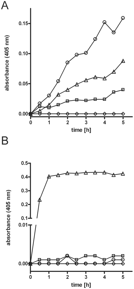
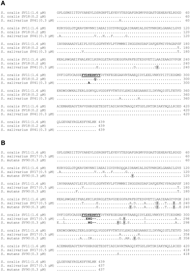
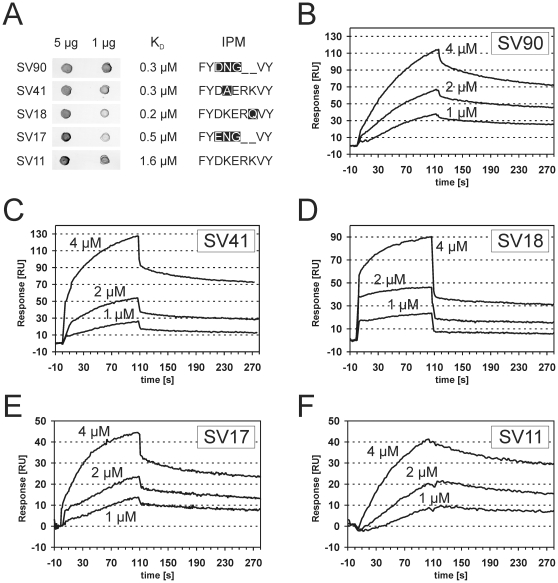
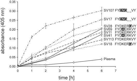
Oral streptococci are a heterogeneous group of human commensals, with a potential to cause serious infections. Activation of plasminogen has been shown to increase the virulence of typical human pathogenic streptococci such as S. pneumoniae. One important factor for plasminogen activation is the streptococcal α-enolase. Here we report that plasminogen activation is also common in oral streptococci species involved in clinical infection and that it depends on the action of human plasminogen activators. The ability to activate plasminogen did not require full conservation of the internal plasminogen binding sequence motif FYDKERKVY of α-enolase that was previously described as crucial for increased plasminogen binding, activation and virulence. Instead, experiments with recombinant α-enolase variants indicate that the naturally occurring variations do not impair plasminogen binding. In spite of these variations in the internal plasminogen binding motif oral streptococci showed similar activation of plasminogen. We conclude that the pathomechanism of plasminogen activation is conserved in oral streptococci that cause infections in human. This may contribute to their opportunistic pathogenic character that is unfurled in certain niches.


Conflict of interest statement

Competing Interests: The authors have declared that no competing interests exist.

Figures

Figure 1
Figure 1. Binding of human plasma proteins to Streptococcus oralis.
After incubation of Streptococcus oralis SV11 with human blood plasma (1), PBS containing purified human plasminogen (2), or PBS alone (3) proteins that were bound to the bacterial surface were eluted with glycine buffer (pH 2) and separated by 12% SDS-PAGE under reducing conditions. The proteins were stained with Coomassie Brilliant Blue. Mobility of marker proteins is indicated at the left and their molecular mass is given in kDa. Protein bands that were identified as plasminogen (Plg), human serum albumin (HSA), and immunoglobulin G (IgG) are marked. The control experiment (3) indicates proteins of bacterial origin.
Figure 2
Figure 2. Bacterial α-enolase and GAPDH in bacterial surface eluates of Streptococcus oralis.
Surface bound proteins of S. oralis SV11 were eluted with sodium carbonate buffer (pH 10), separated by 12% SDS-PAGE under reducing conditions and analysed by immunoblot with rabbit antisera specific for α-enolase (1) or GAPDH (2), respectively. Mobility of marker proteins is indicated at the left and their molecular mass is given in kDa.
Figure 3
Figure 3. Plasminogen activation by Streptococcus oralis.
(A) Bacterial cells from mid-logarithmic culture of S. oralis SV11 were incubated in human plasma () or PBS (). Plasma without bacteria was used as a control for auto-activation (). Incubation of plasma with 100 ng urokinase served as a positive control () (B) Bacterial cells from mid-logarithmic culture of Streptococcus oralis SV11 were incubated in PBS () or PBS containing purified human plasminogen (). Plasminogen without bacterial cells () served as a control for auto-activation. Incubation of human plasminogen with 100 ng urokinase served as a positive control (), demonstrating that the plasminogen could be activated. Plasminogen activation was measured using the colour reaction of plasmin-specific substrate (S-2251). The diagrams depict the absorbance of the liquid phase measured at a wavelength of 405 nm vs. time.
Figure 4
Figure 4. Alignment of recombinant α-enolase variants.
Proteins sequences of recombinant α-enolases originating from oral streptococcal strains (A) SV11, SV18, SV41 and from (B) SV11, SV17, SV90 were aligned using CLUSTALW 2.0.12. Streptococcal species and strain designation are indicated at the left, followed by the apparent dissociation constant for plasminogen interaction in brackets (see also: Fig. 5). Dots indicate amino acids that are identical in the upper sequence. Substitutions by lysine are highlighted in gray. The IPM and its variants are depicted in bold.
Figure 5
Figure 5. Interaction between human plasminogen and recombinant α-enolase variants.
Binding was analyzed in a ligand blot experiment (A) with immobilized recombinant α-enolase variants (5 µg and 1 µg) and human plasminogen as soluble analyte. Designation of the original oral streptococcal strain and the IPM are given together with the apparent dissociation constants, which where determined by surface plasmon resonance measurements (B–F) at different analyte concentrations (4 µM, 2 µM and 1 µM α-enolase). Although curve shapes suggested interactions of higher complexity, apparent dissociation constants were determined based on the Langmuir model for 1∶1 interaction.
Figure 6
Figure 6. Comparison of plasminogen activation in human blood plasma by oral streptococci.
Bacterial cells from mid-logarithmic culture of eight different oral streptococci strains with 4 different IPMs (Table 1) were incubated in human blood plasma ( and ). Plasma without bacterial cells () served as a control. Plasminogen activation was measured using the colour reaction of plasmin-specific substrate (S-2251). Absorbance of the liquid phase was measured over time at a wavelength of 405 nm. Bars represent the standard deviation of the experimental triplicates.

Similar articles

Cited by

References

    1. Claridge JE, 3rd, Attorri S, Musher DM, Hebert J, Dunbar S. Streptococcus intermedius, Streptococcus constellatus, and Streptococcus anginosus (“Streptococcus milleri group”) are of different clinical importance and are not equally associated with abscess. Clin Infect Dis. 2001;32:1511–1515. - PubMed
    1. Jacobs JA, Pietersen HG, Stobberingh EE, Soeters PB. Bacteremia involving the “Streptococcus milleri” group: analysis of 19 cases. Clin Infect Dis. 1994;19:704–713. - PubMed
    1. Whiley RA, Beighton D, Winstanley TG, Fraser HY, Hardie JM. Streptococcus intermedius, Streptococcus constellatus, and Streptococcus anginosus (the Streptococcus milleri group): association with different body sites and clinical infections. J Clin Microbiol. 1992;30:243–244. - PMC - PubMed
    1. Ruoff KL. Streptococcus anginosus (“Streptococcus milleri”): the unrecognized pathogen. Clin Microbiol Rev. 1988;1:102–108. - PMC - PubMed
    1. Colombo AP, Haffajee AD, Dewhirst FE, Paster BJ, Smith CM, et al. Clinical and microbiological features of refractory periodontitis subjects. J Clin Periodontol. 1998;25:169–180. - PubMed

Publication types

MeSH terms