

Midgut barrier imparts selective resistance to filarial worm infection in Culex pipiens pipiens
- PMID: 21072236
- PMCID: PMC2970536
- DOI: 10.1371/journal.pntd.0000875
Midgut barrier imparts selective resistance to filarial worm infection in Culex pipiens pipiens
Abstract
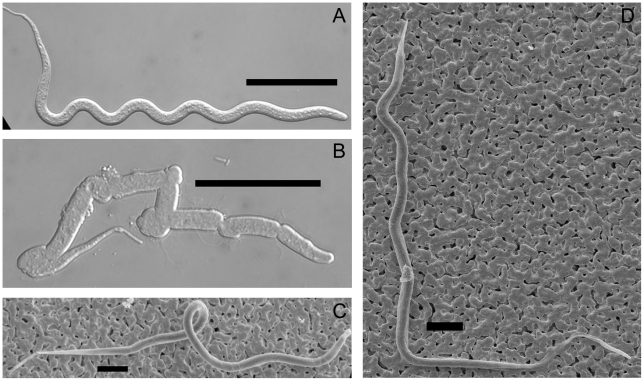
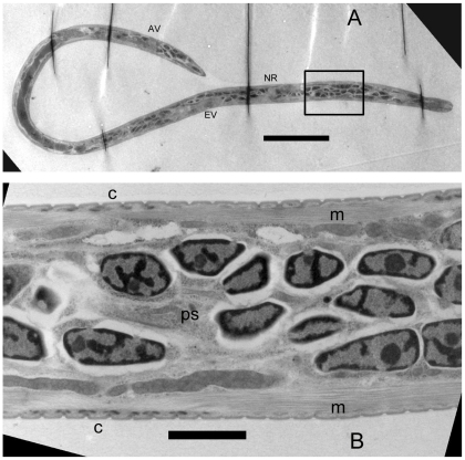
Mosquitoes in the Culex pipiens complex thrive in temperate and tropical regions worldwide, and serve as efficient vectors of Bancroftian lymphatic filariasis (LF) caused by Wuchereria bancrofti in Asia, Africa, the West Indies, South America, and Micronesia. However, members of this mosquito complex do not act as natural vectors for Brugian LF caused by Brugia malayi, or for the cat parasite B. pahangi, despite their presence in South Asia where these parasites are endemic. Previous work with the Iowa strain of Culex pipiens pipiens demonstrates that it is equally susceptible to W. bancrofti as is the natural Cx. p. pipiens vector in the Nile Delta, however it is refractory to infection with Brugia spp. Here we report that the infectivity barrier for Brugia spp. in Cx. p. pipiens is the mosquito midgut, which inflicts internal and lethal damage to ingested microfilariae. Following per os Brugia exposures, the prevalence of infection is significantly lower in Cx. p. pipiens compared to susceptible mosquito controls, and differs between parasite species with <50% and <5% of Cx. p. pipiens becoming infected with B. pahangi and B. malayi, respectively. When Brugia spp. mf were inoculated intrathoracically to bypass the midgut, larvae developed equally well as in controls, indicating that, beyond the midgut, Cx. p. pipiens is physiologically compatible with Brugia spp. Mf isolated from Cx. p. pipiens midguts exhibited compromised motility, and unlike mf derived from blood or isolated from the midguts of Ae. aegypti, failed to develop when inoculated intrathoracically into susceptible mosquitoes. Together these data strongly support the role of the midgut as the primary infection barrier for Brugia spp. in Cx. p. pipiens. Examination of parasites recovered from the Cx. p. pipiens midgut by vital staining, and those exsheathed with papain, suggest that the damage inflicted by the midgut is subcuticular and disrupts internal tissues. Microscopic studies of these worms reveal compromised motility and sharp bends in the body; and ultrastructurally the presence of many fluid or carbohydrate-filled vacuoles in the hypodermis, body wall, and nuclear column. Incubation of Brugia mf with Cx. p. pipiens midgut extracts produces similar internal damage phenotypes; indicating that the Cx. p. pipiens midgut factor(s) that damage mf in vivo are soluble and stable in physiological buffer, and inflict damage on mf in vitro.
Conflict of interest statement
The authors have declared that no competing interests exist.
Figures

References
-
- Michael E, Bundy DA, Grenfell BT. Reassessing the global prevalence and distribution of lymphatic filariasis. Parasitology. 1996;112:409–428. - PubMed
-
- Dreyer G, Noroes J, Figueredo-Silva J, Piessens W. Pathogenesis of lymphatic disease in bancroftian filariasis: a clinical perspective. Parasitol Today. 2000;16:544–548. - PubMed
-
- Krishna Kumari A, Harichandrakumar K, Das LK, Krishnamoorthy K. Physical and psychosocial burden due to lymphatic filariasis as perceived by patients and medical experts. Trop Med Int Health. 2005;10:567–573. - PubMed
-
- Bockarie M, Pedersen E, White G, Michael E. Role of vector control in the global program to eliminate lymphatic filariasis. Annu Rev Entomol. 2009;54:469–487. - PubMed
Publication types
MeSH terms
Grants and funding
LinkOut - more resources
Full Text Sources
Miscellaneous
