. 2010 Sep 21;3(4):270-82.

Targeting diphtheria toxin and TNF alpha expression in ovarian tumors using the H19 regulatory sequences

Targeting diphtheria toxin and TNF alpha expression in ovarian tumors using the H19 regulatory sequences

Aya Mizrahi et al. Int J Clin Exp Med. .

Abstract

Background: There are currently no effective therapies for the treatment of ovarian cancer ascites fluid (OCAF). H19 is an RNA oncofetal gene that is present at high levels in human cancer tissues (ovarian cancer and OCAF among them), while existing at a nearly undetectable level in the surrounding normal tissue. There is evidence for a synergistic effect in cell cytotoxicity mediated by TNFα and diphtheria toxin in sensitive and resistant human ovarian tumor cell line. Thus, we tested the cytotoxic effect of TNF-α cytokine, together with the diphtheria toxin, in the therapy of ovarian cancer.

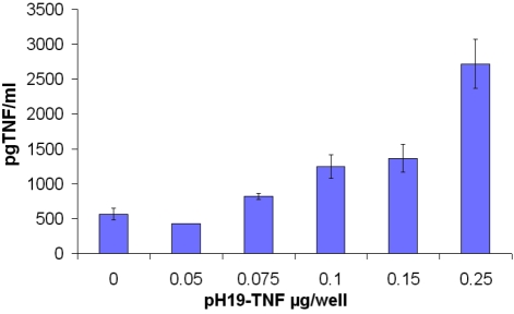
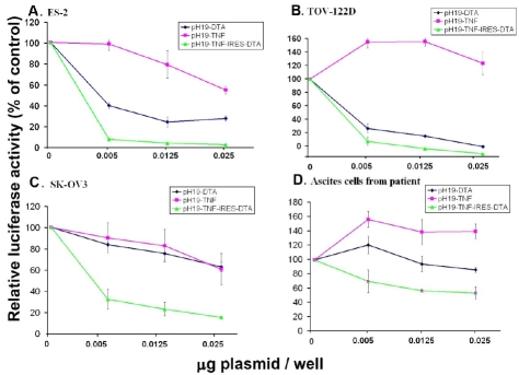
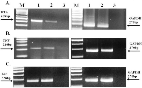
Methods: The therapeutic potential of toxin vectors carrying the DT-A gene alone (pH19-DTA), or in combination with the TNF-α gene (pH19-TNF-DTA), driven by H19 regulatory sequences were tested in ovarian carcinoma cell lines and in a heterotopic ovarian cancer model.

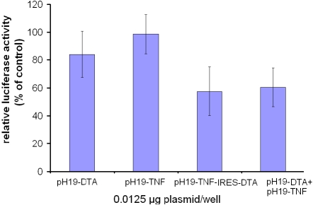
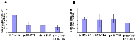
Results: The toxin vectors showed a high killing capacity when transfected into different ovarian cancer cell lines. In addition, intratumoral injection of the toxin vector into ectopically developed tumors caused 40% inhibition of tumor growth. The killing effect after injection of pH19-TNF-DTA plasmid into ectopically developed tumors was significantly higher than that showed by the pH19-DTA plasmid alone, particularly in diphtheria toxin and TNF resistant tumors.

Conclusions: These observations may be the first step towards a major breakthrough in the treatment of human ovarian cancer. It should enable us to identify likely non-responders in advance, and to treat patients who are resistant to all known therapies, thereby avoiding treatment failure coupled with unnecessary suffering and cost.

Keywords: DT-A; H19; IRES; TNF; ascites; ovarian cancer.


Figures

Figure 1
Figure 1
The amount of TNF-α protein secreted after transfection of TOV-122D cells with pH19 -TNF plas-mid determined by ELISA. Medium from TOV-122D transfected cell was collected after 48h. TNF-α protein level was analyzed in the supernatant using a TNF-specific ELISA. The amount of TNF-α protein was normalized to a standard curve with increasing concentrations of TNF-α.
Figure 2
Figure 2
The reduction of luciferase activity in ovarian cancer cell lines: ES-2, TOV-122D, SK-OV3 and in ascites cells of a patient due to co-transfection with the pH19-DTA, pH19-TNF or pH19-TNF-IRES-DTA vectors. The killing potential of each of the following vectors: pH19-DTA (blue), pH19-TNF (pink) and pH19-TNF-IRES-DTA (green) in (A) ES-2, (B) TOV-122D, (C) SK-OV3, (D) ascites cells isolated from a patient was measured as a reduction of luciferase activity induced by Luc-SV40. Cells were co-transfected with 2 ug/well of Luc-SV40 and the indicated concentrations of pH19 -DTA, pH19-TNFα, or pH19-TNF-IRES-DTA.
Figure 3
Figure 3
The reduction of luciferase activity in SK-OV3 cell line due to co-transfection with the pH19-DTA and pH19-TNF vectors, compared with transfection of the pH19-TNF-IRES-DTA vector. The killing potential of the pH19-DTA and pH19-TNF vectors or pH19-TNF-IRES-DTA was measured as a reduction of Luc-SV40 activity. Cells were transfected with Luc-SV40 (2 μg/well) and 0.0125 μg/well of each of the following vectors:pH19-DTA, pH19-TNF, orpH19-TNF -IRES-DTA, co-transfected with pH19-DTA and pH19-TNF 0.0125 μg/well of each plasmid). Cells were also transfected with LucSV40 alone. Transfection experiments were stopped after 48 hours and reporter gene activity was assessed.
Figure 4
Figure 4
The reduction of luciferase activity in SK-OV3 and ES-2 cell lines transfected with LucSV40 due to transfection with pH19-DTA, pH19-TNF-IRES-DTA or pH19-TNFrev-IRES-DTA vectors. The killing potential of the pH19-DTA and pH19-TNF-IRES-DTA or pH19-TNFrev-IRES-DTA was measured as a reduction of luciferase activity in SK-OV3 cell line (A) and in ES-2 cell line (B). Cells were transfected with 2 μg/well of Luc-SV40 and the indicated concentration of pH19-DTA, pH19-TNF-IRES-DTA, pH19-TNFrevDTA respectively. Cell lines were also transfected with Luc-SV40 alone. Transfection experiments were stopped after 48 hours and reporter gene activity was assessed. The luciferase activity in the co-transfected cells was compared to the luciferase activity of the cells transfected with the Luc-SV40 plas-mid alone.
Figure 5
Figure 5
The effect of intratumoral injection of the pH19-TNF-IRES-DTA, pH19 -TNFα, pH19-DTA or pH19-Luc plasmid on the growth of subcutaneous ovarian tumors in nude mice. After tumors developed, mice were divided into 4 groups of 6 mice. Mice received 4 injections of 25 μg of pH19-TNF-IRES-DTA, pH19 -TNFα, pH19-DTA or pH19-Luc plasmids complexed with PEI within two day intervals. One day after the last treatment animals were sacrificed. The tumor dimensions were measured in vivo prior to the first treatment and immediately before sacrifice. The mean fold increase of the final volume compared to the initial volume in the treated tumors was calculated. A and B represents tumors developed by ES-2 or by SK-OV3 cells respectively.
Figure 6
Figure 6
The relative RNA level of DTA, TNF and Luciferase after treatment of SK-OV3 subcutaneous tumors with pH19-DTA, pH19 -TNF-α, pH19-TNF-IRES-DTA or pH19-Luc plasmids determined by RT-PCR. 48h after the animals were treated with the plasmid; samples from the ovarian cancer subcutaneous tumors were excised and frozen immediately. 400 ng RNA from the tumors were used for the determination of DTA, TNF and Luc by RT-PCR reactions. A. Lane 1: DTA transcripts 48h after pH19-DTA plasmid injection, Lane 2: DTA transcripts 48h after pH19-TNF- IRES -DTA plasmid injection. B. Lane 1: TNF transcripts 48h after pH19-TNF plasmid injection, Lane 2: TNF transcripts 48h after pH19-TNF- IRES-DTA plasmid injection. C. Lane1-2: Luc transcripts 48h after pH19-Luc plasmid injection. The sizes of the PCR products are 468, 220, 328 and 270 bp for DTA, TNF, Luc and GAPDH internal control respectively. M-100 bp molecular weight marker.

Similar articles

Cited by

References

    1. Cannistra SA. Cancer of the ovary. N Engl J Med. 2004;351:2519–252. - PubMed
    1. Louis MH, Dutoit S, Denoux Y, Erbacher P, De-slandes E, Behr JP, P Gauduchon1, L Poulain. Intraperitoneal linear polyethylenimine (L-PEI)-mediated gene delivery to ovarian carcinoma nodes in mice. Cancer Gene Ther. 2006;13:367–74. - PubMed
    1. Jandu N, Richardson M, Singh G, Hirte H, Hatton MW. Human ovarian cancer ascites fluid contains a mixture of incompletely degraded soluble products of fibrin that collectively possess an antiangiogenic property. Int J Gynecol Cancer. 2006;16:1536–44. - PubMed
    1. Ruan H, Su H, Hu L, Lamborn KR, Kan YW, Deen DF. A hypoxia-regulated adeno-associated virus vector for cancer-specific gene therapy. Neoplasia. 2001;3:255–263. - PMC - PubMed
    1. Gu J, Fang B. Telomerase promoter-driven cancer gene therapy. Cancer Biol Ther. 2003;2:S64–S70. - PubMed

LinkOut - more resources