

Increased expression of TNF receptor-associated factor 6 after rat traumatic brain injury
- PMID: 21072581
- PMCID: PMC11498546
- DOI: 10.1007/s10571-010-9617-6
Increased expression of TNF receptor-associated factor 6 after rat traumatic brain injury
Abstract
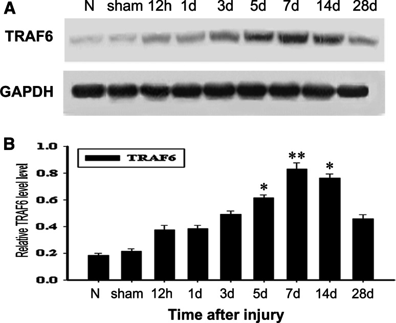
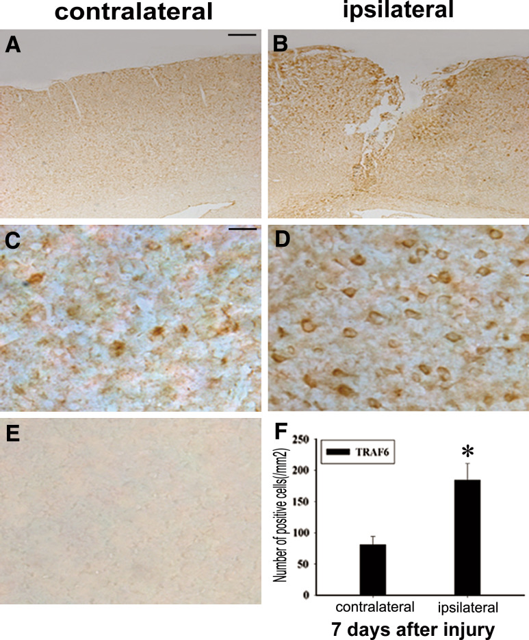
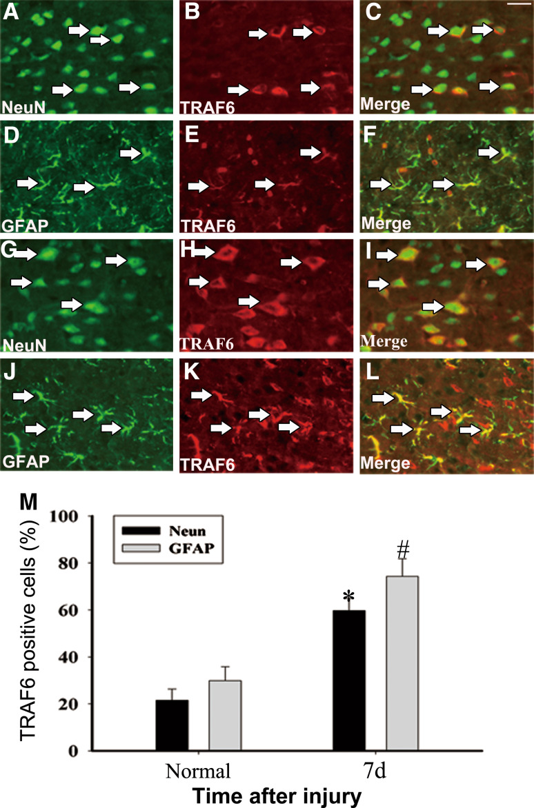
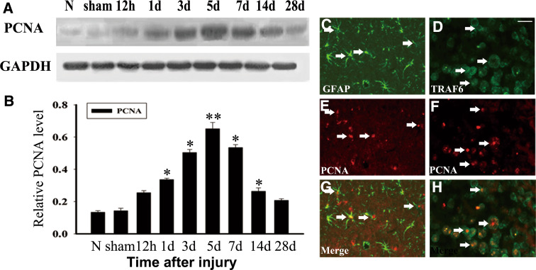
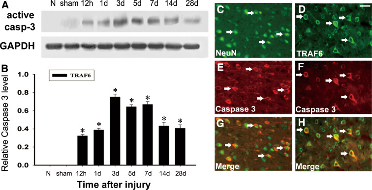
TRAF6 (TNF receptor-associated factor 6), a member of tumor necrosis factor receptor-associated factors family was identified as a molecule that binds to the cytoplasmic domain of CD40. TRAF6 functions as an adaptor, positively regulating the NF-κB, JNK pathway. Additionally, some studies have reported that TRAF6 is required for apoptosis within the developing CNS and regulates cell fate decisions by inducing caspase 8-dependent apoptosis. However, its distribution and function in the central nervous system (CNS) lesion are not well understood. In this study, we performed an acute traumatic brain injury model in adult rats. And we mainly examined protein expression and cellular localization of TRAF6 during rat traumatic brain injury (TBI). Western blot analysis showed TRAF6 level significantly improved at 7 days after injury, and then declined during the following days. The protein expression of TRAF6 was further analysed by immunohistochemistry. In comparison to contralateral cerebral cortex, we observed a highly significant accumulation of TRAF6 at the ipsilateral brain. Immunofluorescence double-labeling showed that TRAF6 was co-expressed with NeuN and GFAP. Besides, co-localization of TRAF6/active caspase 3 and TRAF6/proliferating cell nuclear antigen (PCNA) were detected in NeuN and GFAP, respectively. We also examined the expression profiles of proliferating cell nuclear antigen (PCNA) and active caspase 3 whose changes were correlated with the expression profiles of TRAF6. In conclusion, this is the first description of TRAF6 expression in traumatic brains. Our data suggested that TRAF6 might play important roles in CNS pathophysiology after TBI.
Figures
Similar articles
-
The expression pattern of ADP-ribosyltransferase 3 in rat traumatic brain injury.J Mol Histol. 2012 Feb;43(1):37-47. doi: 10.1007/s10735-011-9366-y. Epub 2011 Oct 26. J Mol Histol. 2012. PMID: 22037978
-
The cell-specific upregulation of bone morphogenetic protein-10 (BMP-10) in a model of rat cortical brain injury.J Mol Histol. 2012 Oct;43(5):543-52. doi: 10.1007/s10735-012-9431-1. Epub 2012 Jul 14. J Mol Histol. 2012. PMID: 22797972
-
Different functions of HIPK2 and CtBP2 in traumatic brain injury.J Mol Neurosci. 2013 Feb;49(2):395-408. doi: 10.1007/s12031-012-9906-2. Epub 2012 Oct 18. J Mol Neurosci. 2013. PMID: 23076816
-
Upregulation of p21-activated Kinase 6 in rat brain cortex after traumatic brain injury.J Mol Histol. 2011 Jun;42(3):195-203. doi: 10.1007/s10735-011-9324-8. Epub 2011 May 4. J Mol Histol. 2011. PMID: 21541790
-
Expression of PHB2 in rat brain cortex following traumatic brain injury.Int J Mol Sci. 2014 Feb 21;15(2):3299-318. doi: 10.3390/ijms15023299. Int J Mol Sci. 2014. PMID: 24566151 Free PMC article.
Cited by
-
Involvement of upregulated p53-induced death domain protein (PIDD) in neuronal apoptosis after rat traumatic brain injury.J Mol Neurosci. 2013 Nov;51(3):695-702. doi: 10.1007/s12031-013-0050-4. J Mol Neurosci. 2013. PMID: 23797734
-
Neurotrophin Signaling and Stem Cells-Implications for Neurodegenerative Diseases and Stem Cell Therapy.Mol Neurobiol. 2017 Nov;54(9):7401-7459. doi: 10.1007/s12035-016-0214-7. Epub 2016 Nov 5. Mol Neurobiol. 2017. PMID: 27815842 Review.
-
The blood-brain barrier and the neurovascular unit in subarachnoid hemorrhage: molecular events and potential treatments.Fluids Barriers CNS. 2022 Apr 11;19(1):29. doi: 10.1186/s12987-022-00312-4. Fluids Barriers CNS. 2022. PMID: 35410231 Free PMC article. Review.
-
Withania somnifera Extract Protects Model Neurons from In Vitro Traumatic Injury.Cell Transplant. 2017 Jul;26(7):1193-1201. doi: 10.1177/0963689717714320. Cell Transplant. 2017. PMID: 28933215 Free PMC article.
-
Different approaches for interpretation and reporting of immunohistochemistry analysis results in the bone tissue - a review.Diagn Pathol. 2014 Nov 29;9:221. doi: 10.1186/s13000-014-0221-9. Diagn Pathol. 2014. PMID: 25432701 Free PMC article. Review.
References
-
- Arch RH, Gedrich RW, Thompson CB (1998) Tumor necrosis factor receptor-associated factors (TRAFs) a family of adapter proteins that regulates life and death. Genes Dev 12:2821–2830 - PubMed
-
- Barnes PJ, Karin M (1997) Nuclear factor-kappaB: a pivotal transcription factor in chronic inflammatory diseases. N Engl J Med 336:1066–1071 - PubMed
-
- Cao Z, Xiong J, Takeuchi M, Kurama T, Goeddel DV (1996) TRAF6 is a signal transducer for interleukin-1. Nature 383:443–446 - PubMed
-
- Chung JY, Park YC, Ye H, Wu H (2002) All TRAFs are not created equal: common and distinct molecular mechanisms of TRAF-mediated signal transduction. J Cell Sci 115:679–688 - PubMed
-
- Deng L, Wang C, Spencer E, Yang L, Braun A, You J (2000) Activation of the IkappaB kinase complex by TRAF6 requires a dimeric ubiquitin-conjugating enzyme complex and a unique polyubiquitin chain. Cell 103:351–361 - PubMed
Publication types
MeSH terms
Substances
LinkOut - more resources
Full Text Sources
Research Materials
Miscellaneous
