

Total salvianolic acid improves ischemia-reperfusion-induced microcirculatory disturbance in rat mesentery
- PMID: 21072893
- PMCID: PMC2980679
- DOI: 10.3748/wjg.v16.i42.5306
Total salvianolic acid improves ischemia-reperfusion-induced microcirculatory disturbance in rat mesentery
Abstract
Aim: To investigate the effect of total salvianolic acid (TSA) on ischemia-reperfusion (I/R)-induced rat mesenteric microcirculatory dysfunctions.
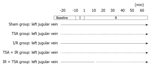
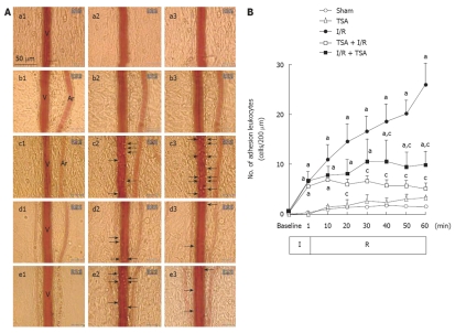
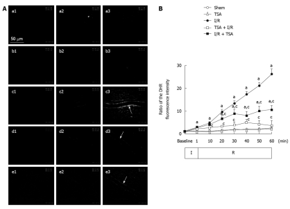
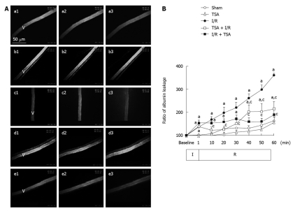
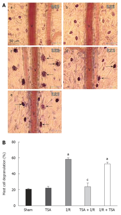
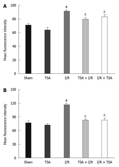
Methods: Male Wistar rats were randomly distributed into 5 groups (n = 6 each): Sham group and I/R group (infused with saline), TSA group, TSA + I/R group and I/R + TSA group (infused with TSA, 5 mg/kg per hour). Mesenteric I/R were conducted by a ligation of the mesenteric artery and vein (10 min) and subsequent release of the occlusion. TSA was continuously infused either starting from 10 min before the ischemia or 10 min after reperfusion. Changes in mesenteric microcirculatory variables, including diameter of venule, velocity of red blood cells in venule, leukocyte adhesion, free radicals released from venule, albumin leakage and mast cell degranulation, were observed through an inverted intravital microscope. Meanwhile, the expression of adhesion molecules CD11b/CD18 on neutrophils was evaluated by flow cytometry. Ultrastructural evidence of mesenteric venules damage was assessed after microcirculation observation.
Results: I/R led to multiple responses in mesenteric post-capillary venules, including a significant increase in the adhesion of leukocytes, production of oxygen radicals in the venular wall, albumin efflux and enhanced mast cell degranulation in vivo. All the I/R-induced manifestations were significantly reduced by pre- or post-treatment with TSA, with the exception that the I/R-induced increase in mast cell degranulation was inhibited only by pre-treatment with TSA. Moreover, pre- or post-treatment with TSA significantly attenuated the expression of CD11b/CD18 on neutrophils, reducing the increase in the number of caveolae in the endothelial cells of mesentery post-capillary venules induced by I/R.
Conclusion: The results demonstrated that TSA protects from and ameliorates the microcirculation disturbance induced by I/R, which was associated with TSA inhibiting the production of oxygen-free radicals in the venular wall and the expression of CD11b/CD18 on neutrophils.
Figures

Similar articles
-
Potential of 3,4-dihydroxy-phenyl lactic acid for ameliorating ischemia-reperfusion-induced microvascular disturbance in rat mesentery.Am J Physiol Gastrointest Liver Physiol. 2009 Jan;296(1):G36-44. doi: 10.1152/ajpgi.90284.2008. Epub 2008 Nov 13. Am J Physiol Gastrointest Liver Physiol. 2009. PMID: 19008340
-
Protective effects of dihydroxylphenyl lactic acid and salvianolic acid B on LPS-induced mesenteric microcirculatory disturbance in rats.Shock. 2008 Feb;29(2):205-11. doi: 10.1097/SHK.0b013e318070c61a. Shock. 2008. PMID: 17667359
-
Compound Danshen injection improves endotoxin-induced microcirculatory disturbance in rat mesentery.World J Gastroenterol. 2007 Jul 14;13(26):3581-91. doi: 10.3748/wjg.v13.i26.3581. World J Gastroenterol. 2007. PMID: 17659708 Free PMC article.
-
Improving effect of pretreatment with yiqifumai on LPS-induced microcirculatory disturbance in rat mesentery.Shock. 2009 Sep;32(3):310-6. doi: 10.1097/SHK.0b013e31819c36e8. Shock. 2009. PMID: 19174743
-
Ameliorating effects of compounds derived from Salvia miltiorrhiza root extract on microcirculatory disturbance and target organ injury by ischemia and reperfusion.Pharmacol Ther. 2008 Feb;117(2):280-95. doi: 10.1016/j.pharmthera.2007.09.008. Epub 2007 Oct 16. Pharmacol Ther. 2008. PMID: 18048101 Review.
Cited by
-
Phagocytosis, Degranulation and Extracellular Traps Release by Neutrophils-The Current Knowledge, Pharmacological Modulation and Future Prospects.Front Pharmacol. 2021 May 4;12:666732. doi: 10.3389/fphar.2021.666732. eCollection 2021. Front Pharmacol. 2021. PMID: 34017259 Free PMC article. Review.
-
Salvianolic acid A targeting the transgelin-actin complex to enhance vasoconstriction.EBioMedicine. 2018 Nov;37:246-258. doi: 10.1016/j.ebiom.2018.10.041. Epub 2018 Oct 23. EBioMedicine. 2018. PMID: 30361065 Free PMC article.
-
Total Salvianolic Acid Balances Brain Functional Network Topology in Rat Hippocampi Overexpressing miR-30e.Front Neurosci. 2018 Jul 5;12:448. doi: 10.3389/fnins.2018.00448. eCollection 2018. Front Neurosci. 2018. PMID: 30026682 Free PMC article.
-
Effect of salvianolic acid B on TNF-α induced cerebral microcirculatory changes in a micro-invasive mouse model.Chin J Traumatol. 2016 Apr 1;19(2):85-93. doi: 10.1016/j.cjtee.2015.07.011. Chin J Traumatol. 2016. PMID: 27140215 Free PMC article.
-
Total Salvianolic Acid Injection Prevents Ischemia/Reperfusion-Induced Myocardial Injury Via Antioxidant Mechanism Involving Mitochondrial Respiratory Chain Through the Upregulation of Sirtuin1 and Sirtuin3.Shock. 2019 Jun;51(6):745-756. doi: 10.1097/SHK.0000000000001185. Shock. 2019. PMID: 29863652 Free PMC article.
References
-
- Han JY, Fan JY, Horie Y, Miura S, Cui DH, Ishii H, Hibi T, Tsuneki H, Kimura I. Ameliorating effects of compounds derived from Salvia miltiorrhiza root extract on microcirculatory disturbance and target organ injury by ischemia and reperfusion. Pharmacol Ther. 2008;117:280–295. - PubMed
-
- Han JY, Miura S, Akiba Y, Higuchi H, Kato S, Suzuki H, Yokoyama H, Ishii H. Chronic ethanol consumption exacerbates microcirculatory damage in rat mesentery after reperfusion. Am J Physiol Gastrointest Liver Physiol. 2001;280:G939–G948. - PubMed
-
- Jones SP, Trocha SD, Strange MB, Granger DN, Kevil CG, Bullard DC, Lefer DJ. Leukocyte and endothelial cell adhesion molecules in a chronic murine model of myocardial reperfusion injury. Am J Physiol Heart Circ Physiol. 2000;279:H2196–H2201. - PubMed
-
- Lan W, Harmon D, Wang JH, Ghori K, Shorten G, Redmond P. The effect of lidocaine on in vitro neutrophil and endothelial adhesion molecule expression induced by plasma obtained during tourniquet-induced ischaemia and reperfusion. Eur J Anaesthesiol. 2004;21:892–897. - PubMed
-
- Kurose I, Wolf R, Cerwinka W, Granger DN. Microvascular responses to ischemia/reperfusion in normotensive and hypertensive rats. Hypertension. 1999;34:212–216. - PubMed
Publication types
MeSH terms
Substances
LinkOut - more resources
Full Text Sources
Research Materials
