Review
. 2011 Feb 15;44(2):134-46.
doi: 10.1021/ar100113q. Epub 2010 Nov 12.

New approaches toward recognition of nucleic acid triple helices

Affiliations
Review

New approaches toward recognition of nucleic acid triple helices

Dev P Arya. Acc Chem Res. .

Abstract

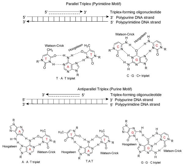
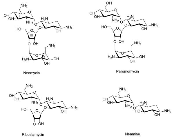
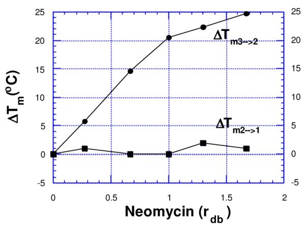
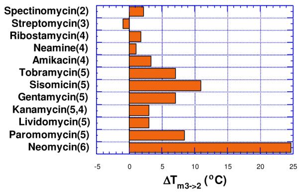
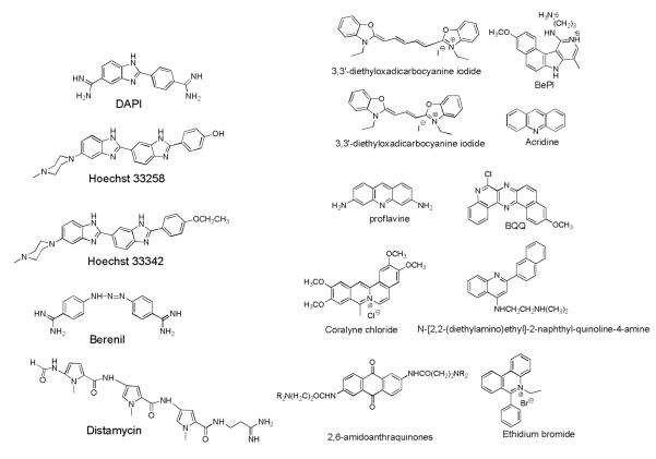
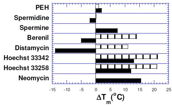
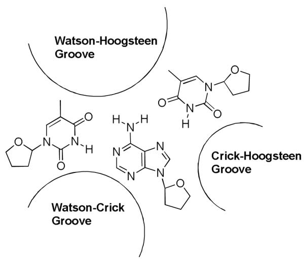
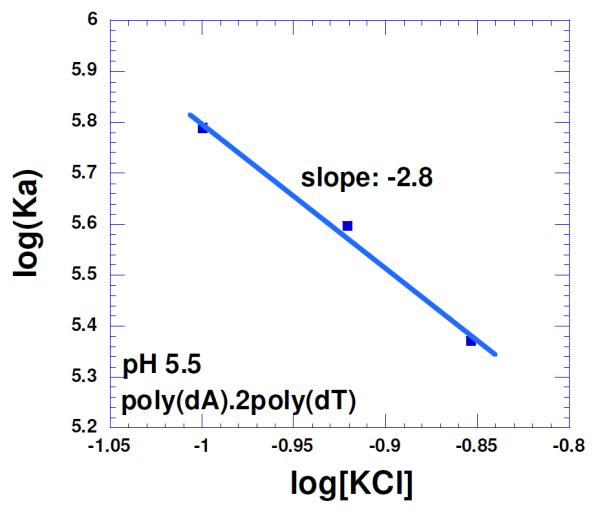
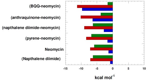
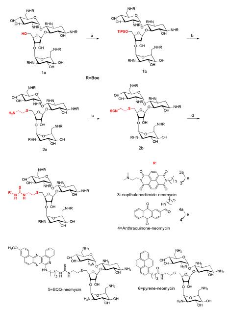
A DNA duplex can be recognized sequence-specifically in the major groove by an oligodeoxynucleotide (ODN). The resulting structure is a DNA triple helix, or triplex. The scientific community has invested significant research capital in the study of DNA triplexes because of their robust potential for providing new applications, including molecular biology tools and therapeutic agents. The triplex structures have inherent instabilities, however, and the recognition of DNA triplexes by small molecules has been attempted as a means of strengthening the three-stranded complex. Over the decades, the majority of work in the field has focused on heterocycles that intercalate between the triplex bases. In this Account, we present an alternate approach to recognition and stabilization of DNA triplexes. We show that groove recognition of nucleic acid triple helices can be achieved with aminosugars. Among these aminosugars, neomycin is the most effective aminoglycoside (groove binder) for stabilizing a DNA triple helix. It stabilizes both the TAT triplex and mixed-base DNA triplexes better than known DNA minor groove binders (which usually destabilize the triplex) and polyamines. Neomycin selectively stabilizes the triplex (TAT and mixed base) without any effect on the DNA duplex. The selectivity of neomycin likely originates from its potential and shape complementarity to the triplex Watson-Hoogsteen groove, making it the first molecule that selectively recognizes a triplex groove over a duplex groove. The groove recognition of aminoglycosides is not limited to DNA triplexes, but also extends to RNA and hybrid triple helical structures. Intercalator-neomycin conjugates are shown to simultaneously probe the base stacking and groove surface in the DNA triplex. Calorimetric and spectrosocopic studies allow the quantification of the effect of surface area of the intercalating moiety on binding to the triplex. These studies outline a novel approach to the recognition of DNA triplexes that incorporates the use of noncompeting binding sites. These principles of dual recognition should be applicable to the design of ligands that can bind any given nucleic acid target with nanomolar affinities and with high selectivity.


Figures

Figure 1
Figure 1
Base interactions in parallel (pyrimidine motif-Top) and antiparallel (Purine motif-Bottom) triple helices. These are defined with respect to the orientation of the TFO and homopurine Watson-Crick (W-C) strand.
Figure 2
Figure 2
Structures of some aminoglycoside antibiotics. Ring numbering scheme is shown for neomycin.
Figure 3
Figure 3
Plots of variation of triplex melting (Tm3→2) and duplex melting (Tm2→1) of poly(dA)•2poly(dT) as a function of increasing neomycin concentration {rdb = drug(neomycin) /base triplet ratio}. 10 mM sodium cacodylate, 150 mM KCl, pH 6.8, [poly(dA)•2poly(dT)] = 15 μM/base triplet.. Reprinted with permission from Bioorg. Med. Chem. Letts 2000,10, 1897-1899.
Figure 4
Figure 4
Effect of aminoglycoside antibiotics on the melting of poly (dA)•2Poly(dT) triplex (rdb = 1.67). Solution conditions: 150 mM KCl, 10 mM sodium cacodylate, pH 6.8, [poly(dA)•2poly(dT)] = 15 μM/base triplet. Number of amines in each antibiotic is shown in parenthesis. Reprinted with permission from J. Amer. Chem. Soc. 2001, 123, 5385-5395.
Figure 5a
Figure 5a
Structures of groove binders (left) and intercalators (right), known to bind to DNA triplexes.
Figure 5b
Figure 5b
Effect of 10 μM (rdb=0.66) groove binders on the DNA triplex melt{poly(dA)•2poly(dT)}(black) and the duplex melt {poly(dA)•poly(dT)}(boxed). Distamycin does not show Tm3→2 transition(<20°C). PEH= pentaethylene hexamine. Solution conditions: 150 mM KCl, 10 mM sodium cacodylate, pH 6.8, DNA = 15 μM/base triplet. Reprinted with permission from J. Amer. Chem. Soc. 2001, 123, 5385-5395.
Figure 6
Figure 6
Structure of a TAT DNA triplex showing different grooves. W-H groove is formed between two pyrimidine strands.-
Figure 7
Figure 7
Salt dependence of the neomycin binding with poly(dA)•2poly(dT) triplex in 10 mM sodium cacodylate, 0.5 mM EDTA and pH 5.5. T= 10 °C. The experimental data were fit with linear regression and the solid line reflects the resulting curve fit. Reprinted with permission from Biochimie, 2010, 92, 5, 514-529.
Figure 8
Figure 8
Electrostatic surface potential maps of (A) neomycin approaching the W-H groove of the triplex, and (B) neomycin buried in the triplex groove. Reprinted with permission from J. Amer. Chem. Soc. 2003, 125, 3733-3744.
Figure 9
Figure 9
Melting curves for Poly (rA)• 2Poly(dT) complex showing ligand (20 μM) induced triplex formation in the presence of berenil and neomycin. Solution conditions: rA: 30 μM/base triplet; 18 mM NaCl; 10 mM sodium cacodylate; 0.1 mM EDTA, pH 6.8. Reprinted with permission from J. Amer. Chem. Soc. 2001, 123, 11093-11094.
Figure 10
Figure 10
Competition dialysis results of pyrene-neomycin and anthraquinone-neomycin as well as their corresponding intercalators pyrene-amine and anthraquinone-amine (1 μM) with various types of nucleic acids (75 μM). Buffer: Na2HPO4 (6 mM), NaH2PO4 (2 mM), Na2EDTA (1 mM), NaCl (185 mM), and pH 7.0. Reprinted with permission from Biochemistry, 2010, 49, 5540-5552. X-axis shows the concentration of bound drug (Cb)-concentration of drug bound to calf thymus DNA (Ccalf).
Figure 10
Figure 10
Competition dialysis results of pyrene-neomycin and anthraquinone-neomycin as well as their corresponding intercalators pyrene-amine and anthraquinone-amine (1 μM) with various types of nucleic acids (75 μM). Buffer: Na2HPO4 (6 mM), NaH2PO4 (2 mM), Na2EDTA (1 mM), NaCl (185 mM), and pH 7.0. Reprinted with permission from Biochemistry, 2010, 49, 5540-5552. X-axis shows the concentration of bound drug (Cb)-concentration of drug bound to calf thymus DNA (Ccalf).
Figure 11
Figure 11
Thermodynamics of binding interactions of ligands with poly(dA)•2poly(dT) triplex at pH 5.5.; red bars represent ΔG, blue bars represent ΔH, and green bars represent TΔS. Experimental condition: sodium cacodylate (10 mM), EDTA (0.5 mM), KCl (150 mM), T=20°C, pH 5.5. Reprinted with permission from Biochemistry, 2010, 49, 5540-5552.
Scheme 1
Scheme 1
a) Triisopropylbenzenesulfonyl chloride, pyridine, r.t. b) 1,2-aminothiolethane, Na, and EtOH c) TCDP, DCM, r.t. d) intercalator-amine, CH2Cl2. e) TFA/CH2Cl2. The site of aminoglycoside modification is shown in red.

Similar articles

Cited by

References

    1. Kool ET. Design of Triplex-forming Oligonucleotides for Binding DNA and RNA: Optimizing Affinity and Selectivity. New J. Chem. 1997;21:33–45.
    1. Dervan PB. Molecular Recognition of DNA by Small Molecules. Bioorg. Med. Chem. 2001;9:2215–2235. - PubMed
    1. Felsenfeld G, Davies D, Rich A. Formation of a Three-Stranded Polynucleotide Molecule. J. Amer. Chem. Soc. 1957;79:2023–2024.
    1. Felsenfeld G, Rich A. Studies on the Formation of Two and Three Stranded Polyribonucleotides. Biochim. Biophys. Acta. 1957;26:457–468. - PubMed
    1. Maher LJ, III, Wold B, Dervan PB. Inhibition of DNA Binding Proteins by Oligonucleotide-Directed Triple Helix Formation. Science. 1989;245:725–730. - PubMed

Publication types