. 2010 Dec 21;49(50):10728-36.
doi: 10.1021/bi1015716. Epub 2010 Nov 22.

Dynamic origins of differential RNA binding function in two dsRBDs from the miRNA "microprocessor" complex

Affiliations

Dynamic origins of differential RNA binding function in two dsRBDs from the miRNA "microprocessor" complex

Christopher Wostenberg et al. Biochemistry. .

Abstract

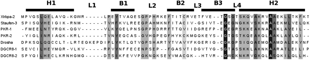
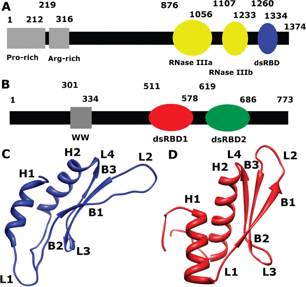
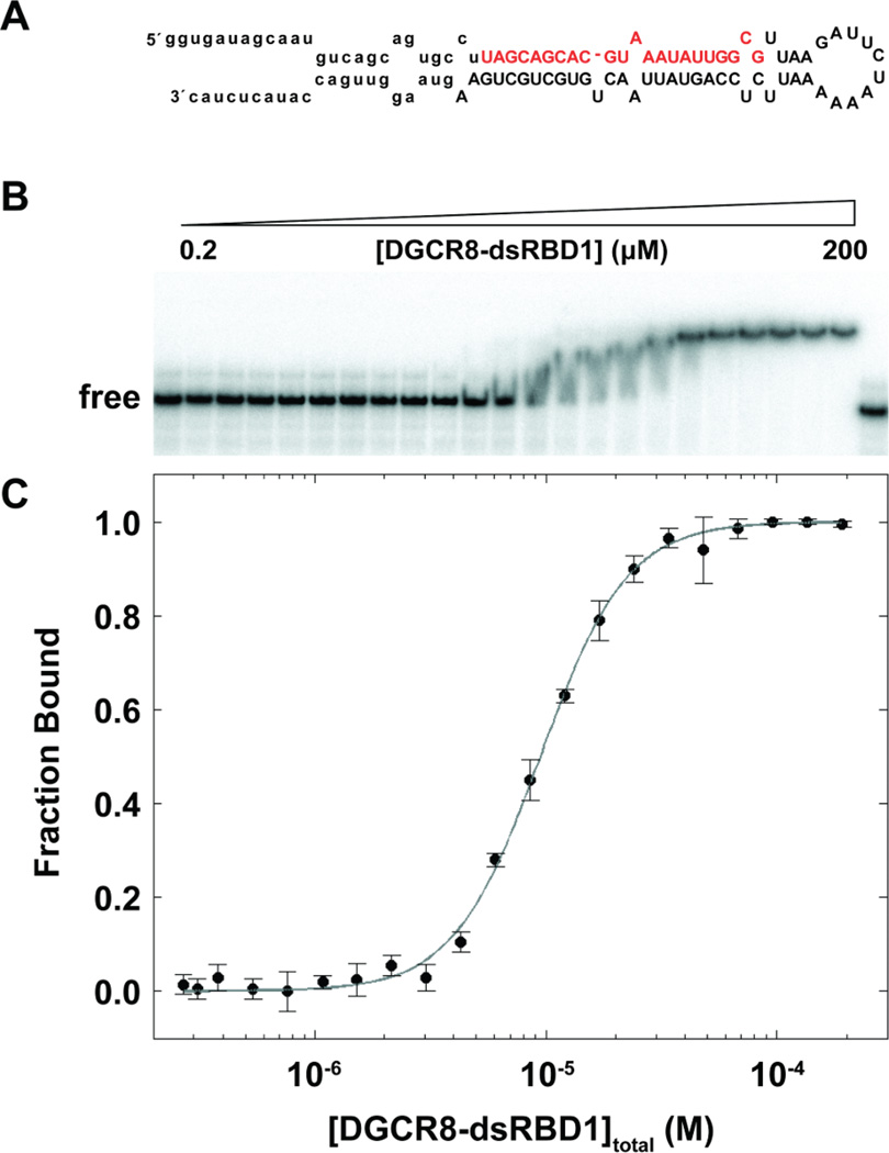
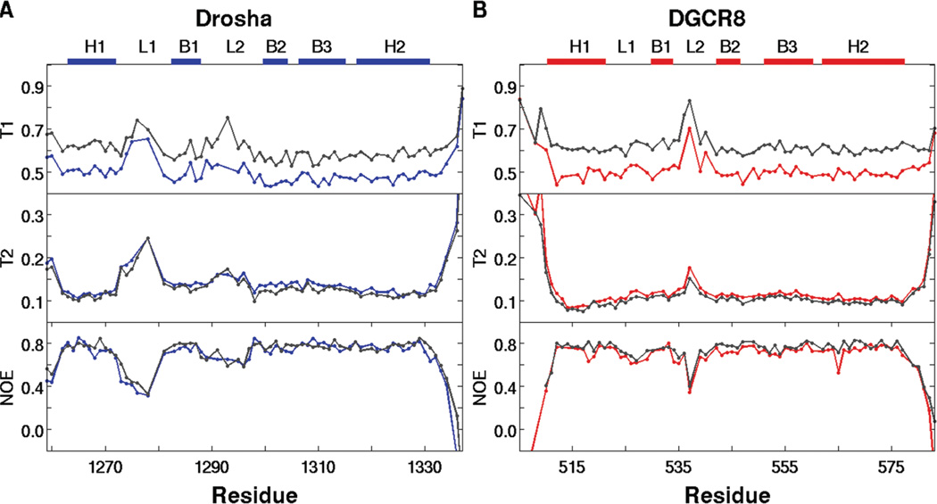
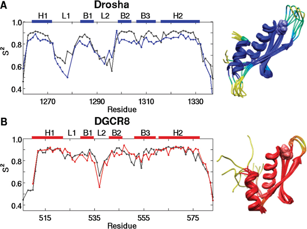
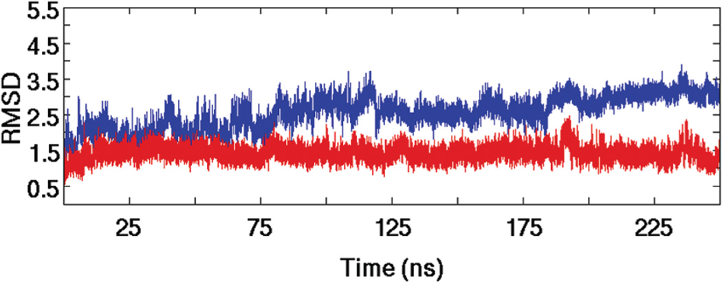
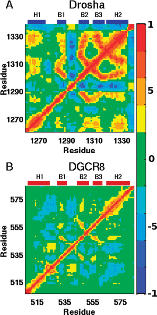
MicroRNAs (miRNAs) affect gene regulation by base pairing with mRNA and contribute to the control of cellular homeostasis. The first step in miRNA maturation is conducted in the nucleus by the "microprocessor" complex made up of an RNase III enzyme, Drosha, that contains one dsRNA binding domain (dsRBD), and DGCR8, that contains two dsRBDs in tandem. The crystal structure of DGCR8-Core (493-720), containing both dsRBDs, and the NMR solution structure of Drosha-dsRBD (1259-1337) have been reported, but the solution dynamics have not been explored for any of these dsRBDs. To better define the mechanism of dsRNA binding and thus the nuclear maturation step of miRNA processing, we report NMR spin relaxation and MD simulations of Drosha-dsRBD (1259-1337) and DGCR8-dsRBD1 (505-583). The study was motivated by electrophoretic mobility shift assays (EMSAs) of the two dsRBDs, which showed that Drosha-dsRBD does not bind a representative miRNA but isolated DGCR8-dsRBD1 does (K(d) = 9.4 ± 0.4 μM). Our results show that loop 2 in both dsRBDs is highly dynamic but the pattern of the correlations observed in MD is different for the two proteins. Additionally, the extended loop 1 of Drosha-dsRBD is more flexible than the corresponding loop in DGCR8-dsRBD1 but shows no correlation with loop 2, which potentially explains the lack of dsRNA binding by Drosha-dsRBD in the absence of the RNase III domains. The results presented in this study provide key structural and dynamic features of dsRBDs that contribute to the binding mechanism of these domains to dsRNA.


Figures

Figure 1
Figure 1
Alignment using Clustal W2 for each of the dsRBDs discussed in this paper showing conserved residues in varying shades of gray, with black corresponding to complete conservation. Approximate location of the secondary elements is shown above the alignment with H representing an α-helix, B representing a β-sheet, and L representing a loop/turn.
Figure 2
Figure 2
Schematic representation of the primary sequence of (A) Drosha and (B) DGCR8. (C) A ribbon diagram representing the solution structure of Drosha-dsRBD (PDB 2KHX, residues 1259–1337) shows the extended loops 1 (L1) and 2 (L2). (D) A ribbon diagram representing the crystal structure of DGCR8-dsRBD1 (PDB 2YT4, residues 505–583) shows a less elongated fold of the dsRBD than Drosha-dsRBD.
Figure 3
Figure 3
EMSA of pri-miR-16-1 binding to DGCR8-dsRBD1. (A) Predicted secondary structure of pri-miR-16-1 with the sequence of the mature miRNA shown in red and the region removed by Drosha cleavage indicated through lower case letters. (B) Representative gel showing addition of DGCR8-dsRBD1 (2–200 µM) to 0.25 nM pri-miR-16-1. (C) Fitted EMSA fraction bound as a function of DGCR8-dsRBD1 concentration with data points and uncertainties represented by filled circles and the best fit to the data (see text) represented as a gray line.
Figure 4
Figure 4
15N spin relaxation data for (A) Drosha-dsRBD and (B) DGCR8-dsRBD1 collected at 500 MHz (blue and red, respectively) and 600MHz (gray). The data show that the extended loop 1 of Drosha-dsRBD is more flexible than that of DGCR8-dsRBD1 on the picosecond to nanosecond time scale. Loop 2 is also seen to be highly dynamic in both proteins. The secondary structure elements for the respective dsRBDs, as well as the positions of loop 1 and loop 2, are represented as colored bars above the plots.
Figure 5
Figure 5
Order parameter (S2) plots for (A) Drosha-dsRBD and (B)DGCR8-dsRBD1 show that loop 2 in both proteins and loop 1 of Drosha-dsRBD are the most dynamic regions of the domains. Experimental data (blue and red lines for Drosha-dsRBD and DGCR8-dsRBD1, respectively) are plotted against MD predicted order parameters (gray). The secondary structure elements for the respective dsRBDs, as well as the positions of loops 1 and 2, are represented as colored bars above the plots. S2 is represented color imetrically in the MD-derived ribbon bundles for (C) Drosha-dsRBD and (D) DGCR8-dsRBD1, with passage from dark blue and dark red toward yellow indicating increased flexibility, based on the experimental order parameters. The conserved aromatic residues (Tyr-1298 of Drosha-dsRBD and Phe-542 of DGCR8-dsRBD1) are shown in space-filling mode to demonstrate how these residues are orientated to preserve the spacing between loop 2 and loop 4. Both bundles are created by taking the structures from the simulation every 50 ns and superimposing them to remove translation and rotation of the center of mass.
Figure 6
Figure 6
The global dynamics of Drosha-dsRBD and DGCR8-dsRBD1 observed in MD simulations indicates qualitative differences between the two domains. The rmsd traces of Drosha-dsRBD (blue line) and DGCR8-dsRBD1 (red line) demonstrate that both proteins are stable over the MD simulations, although Drosha dsRBD is more dynamic overall, as seen in the MD structure bundles found in Figure 5C,D.
Figure 7
Figure 7
Cα correlation matrices reveal the collective backbone motions of (A) Drosha-dsRBD and (B)DGCR8-dsRBD1. The color bar on the right shows the scale indicating strong positive correlation (red), strong negative correlation (blue), and noncorrelated motion (green). Labels above each panel indicate the location of secondary structural elements within the sequence.

References

    1. Bartel DP. MicroRNAs: target recognition and regulatory functions. Cell. 2009;136:215–230. - PMC - PubMed
    1. Zhao Y, Srivastava D. A developmental view of micro-RNA function. Trends Biochem. Sci. 2007;32:189–197. - PubMed
    1. Friedman RC, Farh KKH, Burge CB, Bartel DP. Most mammalian mRNAs are conserved targets of microRNAs. Genome Res. 2009;19:92–105. - PMC - PubMed
    1. Wang Y, Liang Y, Lu Q. MicroRNA epigenetic alterations: predicting biomarkers and therapeutic targets in human diseases. Clin. Genet. 2008;74:307–315. - PubMed
    1. Negrini M, Ferracin M, Sabbioni S, Croce CM. MicroRNAs in human cancer: from research to therapy. J. Cell Sci. 2007;120:1833–1840. - PubMed

Publication types