Review
. 2010 Oct;5(10):1493-506.
doi: 10.2217/fmb.10.107.

Human papillomavirus: gene expression, regulation and prospects for novel diagnostic methods and antiviral therapies

Affiliations
Review

Human papillomavirus: gene expression, regulation and prospects for novel diagnostic methods and antiviral therapies

Sheila V Graham. Future Microbiol. 2010 Oct.

Abstract

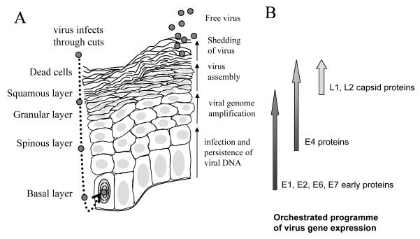
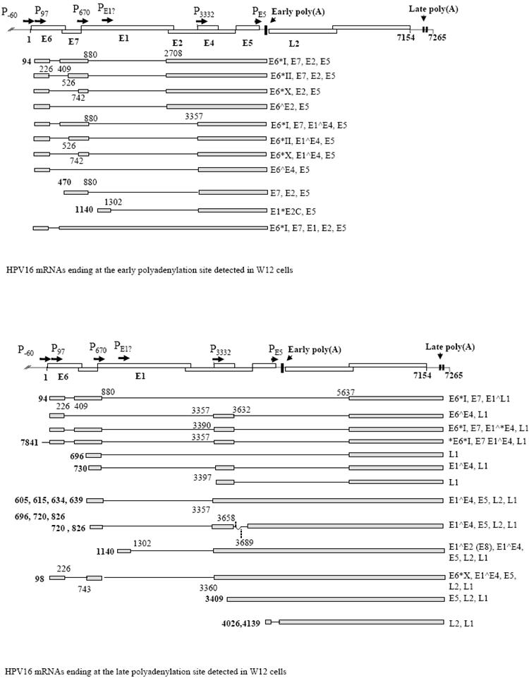
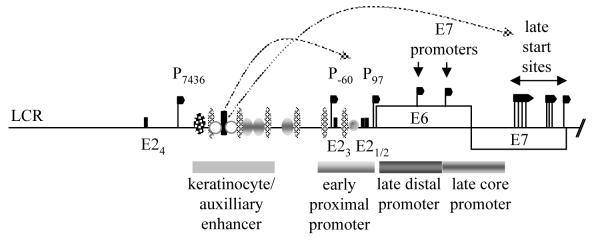
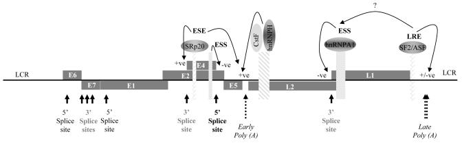
Human papillomaviruses (HPVs) cause diseases ranging from benign warts to invasive tumors. A subset of these viruses termed 'high risk' infect the cervix where persistent infection can lead to cervical cancer. Although many HPV genomes have been sequenced, knowledge of virus gene expression and its regulation is still incomplete. This is due in part to the lack, until recently, of suitable systems for virus propagation in the laboratory. HPV gene expression is polycistronic initiating from multiple promoters. Gene regulation occurs at transcriptional, but particularly post-transcriptional levels, including RNA processing, nuclear export, mRNA stability and translation. A close association between the virus replication cycle and epithelial differentiation adds a further layer of complexity. Understanding HPV mRNA expression and its regulation in the different diseases associated with infection may lead to development of novel diagnostic approaches and will reveal key viral and cellular targets for development of novel antiviral therapies.


Figures

Figure 1
Figure 1. The replication cycle of high risk HPV in a differentiating epithelium
A. The different epithelial layers are indicated on the left hand side of the diagram. Virus is show as small grey circles. The nucleus of the infected, dividing basal epithelial cell is indicated with curved lines representing condensed chromosomes. All nuclei are shaded in light grey. The granular layer is shown with dotted cytoplasm. The key events in the virus replication cycle are indicated to the right hand side of the diagram of the epithelium. B. A schematic diagram of the gene expression program of the virus within the infected epithelium. Shading on the arrows represents the quantity of expression of each protein subset during the virus replication cycle.
Figure 2
Figure 2. The gene expression profile of HPV16 in W12 cervical epithelial cells
A. At the top is shown a diagram of the HPV16 genome linearised at the first nucleotide. Open boxes, open reading frames with proteins encoded indicated with bold text. Promoters are indicated with arrows and “P”. The early and the two late polyadenylation sites are indicated with arrowheads and vertical bars on the genome map. At the bottom is a diagram of the W12 early mRNAs discovered to date [48], (Milligan and Graham, unpublished data). Grey boxes, exons; black lines, introns. Numbers in bold indicate transcription start sites. The remaining numbers locate splice donor and acceptor sites. The coding potential of each mRNA is indicated to the right hand side. B. A diagram of the late mRNAs detected in W12 cells [45] with similar annotation as in part (A).
Figure 3
Figure 3. Regulation of promoters at the start of the HPV16 genome
A schematic diagram of the long control region and the start of the early coding region of the HR HPV genome showing the position of various transcription start sites (black flags), the early proximal promoter region, the keratinocyte/auxiliary enhancer region and the late core and distal promoter regions in the E6/E7 coding region. Data are taken from results with HR HPV16 and 31. LCR, long control region. E2, E2 binding sites indicated by black bars. Ovals and circles indicate approximate regions of transcription factor binding. Only a limited number of binding sites are illustrated. There are very many known binding sites making it impossible to illustrate all transcription factors and their precise binding sites. In addition, most of the transcription factors regulating the E7 promoters and the late promoter have not yet been identified. The box indicates the transcription factors known to bind the HPV16 early region [40].
Figure 4
Figure 4. HPV16 RNA processing regulation; ESE, ESS and polyadenylation control elements and their binding proteins
A schematic diagram of the HPV16 genome showing open reading frames (grey rectangles containing gene names), splice donor (5′) and splice acceptor (3′) splice sites (black arrows), polyadenylation sites (dotted arrows). When transcribe to RNA, cis-acting RNA processing regulatory elements (patterned oblongs) and the proteins these bind (annotated ovals) are indicated. Arrowed lines indicate the target and type of regulation of each RNA/protein interaction. LCR, long control region.

Similar articles

Cited by

References

    1. zur Hausen H. Papillomaviruses in the causation of human cancers - a brief historical account. Virol. 2009;384:260–5. - PubMed
    1. de Villiers EM, Fauquet C, Broker TR, Bernard HU, zur Hausen H. Classification of papillomaviruses. Virol. 2004;324:17–24. - PubMed
    1. Stanley MA. Human papillomavirus vaccines. Rev. Med. Virol. 2006;16:139–49. - PubMed
    1. Doorbar J, Foo C, Coleman N, et al. Characterisation of events during the late stages of HPV16 infection in vivo using high-affinity synthetic Fabs to E4. Virol. 1997;238:40–52. - PubMed
    1. Graham SV. Late events in the life cycle of human papillomaviruses. In: Campo MS, editor. Papillomavirus research: from natural history to vaccines and beyond. Caister Academic Press; Wymondham, Norfolk: 2006. pp. 193–212.

MeSH terms

Substances