. 2010 Nov 12:10:287.
doi: 10.1186/1471-2180-10-287.

Impact of the Staphylococcus epidermidis LytSR two-component regulatory system on murein hydrolase activity, pyruvate utilization and global transcriptional profile

Affiliations

Impact of the Staphylococcus epidermidis LytSR two-component regulatory system on murein hydrolase activity, pyruvate utilization and global transcriptional profile

Tao Zhu et al. BMC Microbiol. .

Abstract

Background: Staphylococcus epidermidis has emerged as one of the most important nosocomial pathogens, mainly because of its ability to colonize implanted biomaterials by forming a biofilm. Extensive studies are focused on the molecular mechanisms involved in biofilm formation. The LytSR two-component regulatory system regulates autolysis and biofilm formation in Staphylococcus aureus. However, the role of LytSR played in S. epidermidis remained unknown.

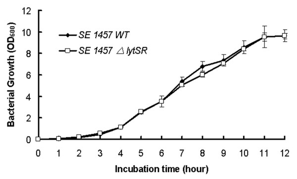
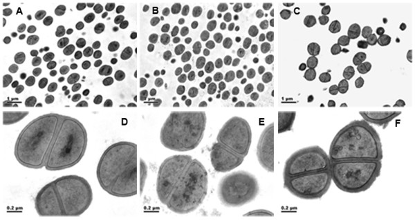
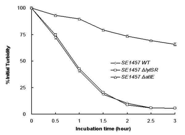
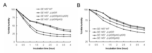
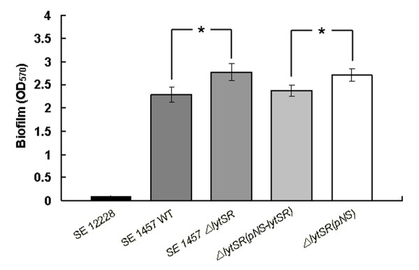
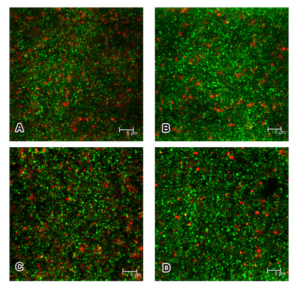
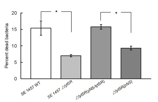
Results: In the present study, we demonstrated that lytSR knock-out in S. epidermidis did not alter susceptibility to Triton X-100 induced autolysis. Quantitative murein hydrolase assay indicated that disruption of lytSR in S. epidermidis resulted in decreased activities of extracellular murein hydrolases, although zymogram showed no apparent differences in murein hydrolase patterns between S. epidermidis strain 1457 and its lytSR mutant. Compared to the wild-type counterpart, 1457ΔlytSR produced slightly more biofilm, with significantly decreased dead cells inside. Microarray analysis showed that lytSR mutation affected the transcription of 164 genes (123 genes were upregulated and 41 genes were downregulated). Specifically, genes encoding proteins responsible for protein synthesis, energy metabolism were downregulated, while genes involved in amino acid and nucleotide biosynthesis, amino acid transporters were upregulated. Impaired ability to utilize pyruvate and reduced activity of arginine deiminase was observed in 1457ΔlytSR, which is consistent with the microarray data.

Conclusions: The preliminary results suggest that in S. epidermidis LytSR two-component system regulates extracellular murein hydrolase activity, bacterial cell death and pyruvate utilization. Based on the microarray data, it appears that lytSR inactivation induces a stringent response. In addition, LytSR may indirectly enhance biofilm formation by altering the metabolic status of the bacteria.


Figures

Figure 1
Figure 1
Physical map of the lytSR operon of S. epidermidis 1457 and construction of lytSR knockout mutant. Arrows depict open reading frames and indicate their orientations. lytSR operon were replaced with the erythromycin resistance gene (ermB) as indicated. The ermB gene and chromosomal regions flanking the corresponding deletions were amplified by PCR and cloned into plasmid pBT2, yielding the integration vectors pBT2-ΔlytSR. The crosses indicate the sites of homologous recombination.
Figure 2
Figure 2
Growth curves of S. epidermidis 1457ΔlytSR. Bacterial cultures were grown in TSB medium at 37 °C, and growth was monitored by measuring the turbidity of the cultures at 600 nm. Data are means ± SD of 3 independent experiments.
Figure 3
Figure 3
Morphology of S. epidermidis 1457ΔlytSR under transmission electron microscope. Strains of S. epidermidis 1457, ΔlytSR and ΔatlE were cultured in TSB till stationary phase, fixed with 2.5% glutaraldehyde in Dulbecco's phosphate-buffered saline (PBS). Thin sections were stained with 1% uranyl acetate-lead acetate and observed under a Philips Tecnai-12 Biotwin transmission electron microscope. A-C ×8,200 magnification of 1457, ΔlytSR and ΔatlE cells respectively; D-F ×43,000 magnification of 1457, ΔlytSR and ΔatlE cells respectively.
Figure 4
Figure 4
Autolysis assay of S. epidermidis 1457ΔlytSR. Bacterial cells were collected from early exponentially growing cultures (OD600 = 0.7) containing 1 M NaCl, washed twice with ice-cold water and resuspended in an equal volume of Tris-HCl(pH 7.2) containing 0.05%(vol/vol) Triton X-100. The rate of autolysis was measured as the decline in optical density. The atlE knockout mutant was used as a negative control. Data are means ± SD of 3 independent experiments.
Figure 5
Figure 5
Zymographic analysis of S. epidermidis 1457ΔlytSR. Extracellular and cell surface proteins were isolated, and 30 μg of each was separated in SDS-polyacrylamide gel electrophoresis gels containing 2.0 mg of M. luteus (A) or S. epidermidis (B) cells/ml. Murein hydrolase activity was detected by incubation overnight at 37 °C in a buffer containing Triton X-100, followed by staining with methylene blue. Lanes: 1 and 6, molecular mass marker; 2 and 7, cell wall protein from 1457ΔlytSR strain; 3 and 8, cell wall protein from wild type strain; 4 and 9, extracellular protein from 1457ΔlytSR strain; 5 and 10, extracellular protein from wild type strain. The results are representative of three independent experiments.
Figure 6
Figure 6
Quantitative murein hydrolase assays of S. epidermidis 1457ΔlytSR. Aliquots (100 μg) of the extracellular proteins concentrated by ultrafiltration from the supernant were added to a 1-mg/ml suspension of M. luteus (A) and S. epidermidis (B) cells separately, and the turbidity at 600 nm was monitored for 4 h. Cell wall hydrolysis was determined by measurement of turbidity every 30 min. Data are means ± SD of 3 independent experiments.
Figure 7
Figure 7
Effect of lytSR gene knocking out on S. epidermidis biofilm formation. The biofilm formation of S. epidermidis ΔlytSR and its parent strain was detected by semi-quantitative microtiter plate assay. Briefly, the overnight bacterial were diluted by 1:200 and cultured in 96-well plate (200 μl/well) at 37 °C for 24 h. The well was washed by PBS for 3 times, fixed by 99% methanol and stained with crystal violet. Data are means ± SD of 3 independent experiments. *P < 0.05; ΔlytSR vs. WT; ΔlytSR(pNS-lytSR) vs. ΔlytSR(pNS-lytSR).
Figure 8
Figure 8
Confocal photomicrographs of 24-hour-old biofilms. Biofilms containing S. epidermidis 1457 strains wild-type (A), ΔlytSR (B), ΔlytSR(pNS-lytSR) (C) and ΔlytSR(pNS) (D) were visualized by using the live/dead viability stain (SYTO9/PI). Green fluorescent cells are viable, whereas red fluorescent cells have a compromised cell membrane, as indicative of dead cells. Scale bars = 5 μm. The result is a stack of images at approximately 0.3 μm depth increments and represents one of the three experiments.
Figure 9
Figure 9
Quantitative analysis of bacteria cell death in 24-hour-old biofilms. Live/dead stained biofilm cells were scraped from the dish and dispersed by pipetting. The integrated intensities of the green (535 nm) and red (625 nm) emission of suspensions excited at 485 nm were measured and the green/red fluorescence ratios (RatioR/G) were calculated. The percentage of dead cells inside biofilm was determined by comparison to the standard curve of RatioR/G versus percentage of dead cells. Data are means ± SEM of 3 independent experiments. *P < 0.05; ΔlytSR vs. WT; ΔlytSR(pNS-lytSR) vs. ΔlytSR(pNS-lytSR).
Figure 10
Figure 10
Pyruvate utilization test of S. epidermidis 1457ΔlytSR. Bacteria were grown in pyruvate fermentation broth at 37 °C, and growth was monitored by measuring the turbidity of the cultures at 600 nm as described in Materials and Methods. Data are means ± SD of 3 independent experiments.

References

    1. Ziebuhr W, Heilmann C, Gotz F, Meyer P, Wilms K, Straube E, Hacker J. Detection of the intercellular adhesion gene cluster (ica) and phase variation in Staphylococcus epidermidis blood culture strains and mucosal isolates. Infection and immunity. 1997;65(3):890–896. - PMC - PubMed
    1. Rupp ME, Archer GL. Coagulase-negative staphylococci: pathogens associated with medical progress. Clin Infect Dis. 1994;19(2):231–243; quiz 244-235. - PubMed
    1. Bowden MG, Chen W, Singvall J, Xu Y, Peacock SJ, Valtulina V, Speziale P, Hook M. Identification and preliminary characterization of cell-wall-anchored proteins of Staphylococcus epidermidis. Microbiology (Reading, England) 2005;151(Pt 5):1453–1464. - PubMed
    1. Vuong C, Kocianova S, Voyich JM, Yao Y, Fischer ER, DeLeo FR, Otto M. A crucial role for exopolysaccharide modification in bacterial biofilm formation, immune evasion, and virulence. The Journal of biological chemistry. 2004;279(52):54881–54886. doi: 10.1074/jbc.M411374200. - DOI - PubMed
    1. Donlan RM, Costerton JW. Biofilms: survival mechanisms of clinically relevant microorganisms. Clinical microbiology reviews. 2002;15(2):167–193. doi: 10.1128/CMR.15.2.167-193.2002. - DOI - PMC - PubMed

Publication types

MeSH terms