. 2010 Nov 12:7:77.
doi: 10.1186/1742-2094-7-77.

Absence of IFNγ expression induces neuronal degeneration in the spinal cord of adult mice

Affiliations

Absence of IFNγ expression induces neuronal degeneration in the spinal cord of adult mice

Sheila C S Victório et al. J Neuroinflammation. .

Abstract

Background: Interferon gamma (IFNγ) is a pro-inflammatory cytokine, which may be up-regulated after trauma to the peripheral or central nervous system. Such changes include reactive gliosis and synaptic plasticity that are considered important responses to the proper regenerative response after injury. Also, IFNγ is involved in the upregulation of the major histocompatibility complex class I (MHC class I), which has recently been shown to play an important role in the synaptic plasticity process following axotomy. There is also evidence that IFNγ may interfere in the differentiation and survival of neuronal cells. However, little is known about the effects of IFNγ absence on spinal cord neurons after injury.

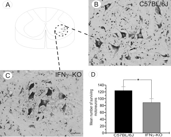
Methods: We performed a unilateral sciatic nerve transection injury in C57BL/6J (wild type) and IFNγ-KO (mutant) mice and studied motoneuron morphology using light and electron microscopy. One week after the lesion, mice from both strains were sacrificed and had their lumbar spinal cords processed for histochemistry (n = 5 each group) and transmission electron microscopy (TEM, n = 5 each group). Spinal cord sections from non-lesioned animals were also used to investigate neuronal survival and the presence of apoptosis with TUNEL and immunohistochemistry.

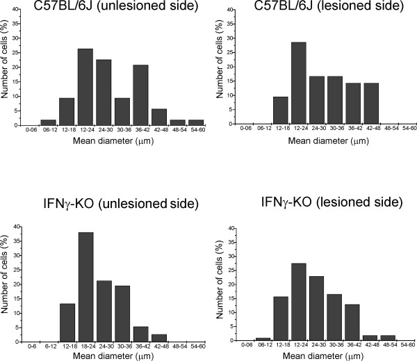
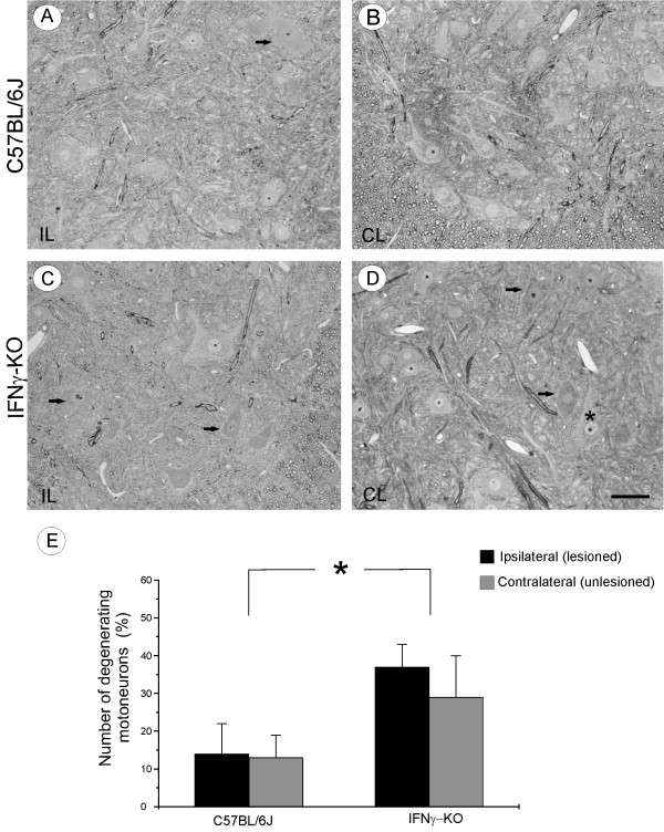
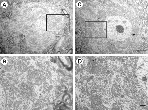
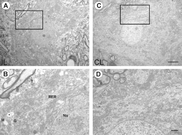
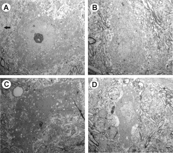
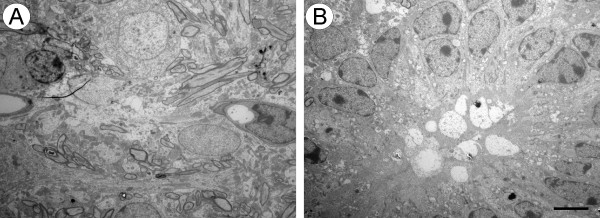
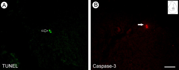
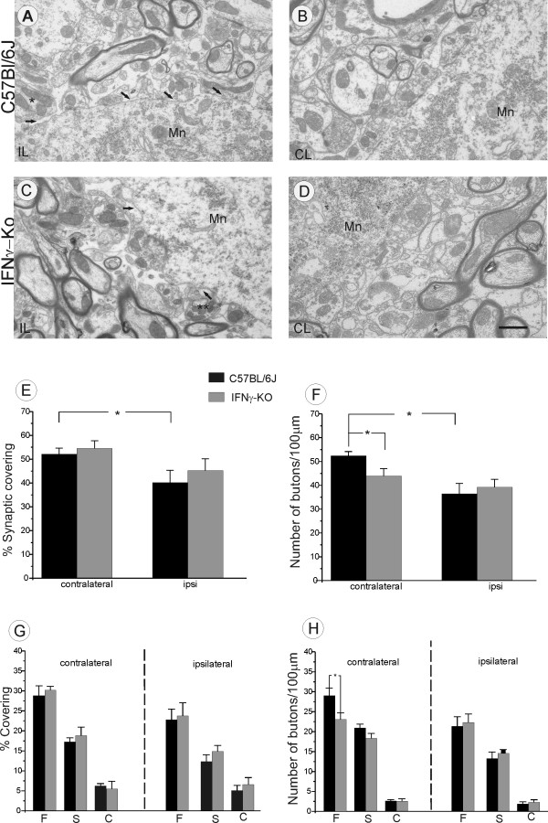
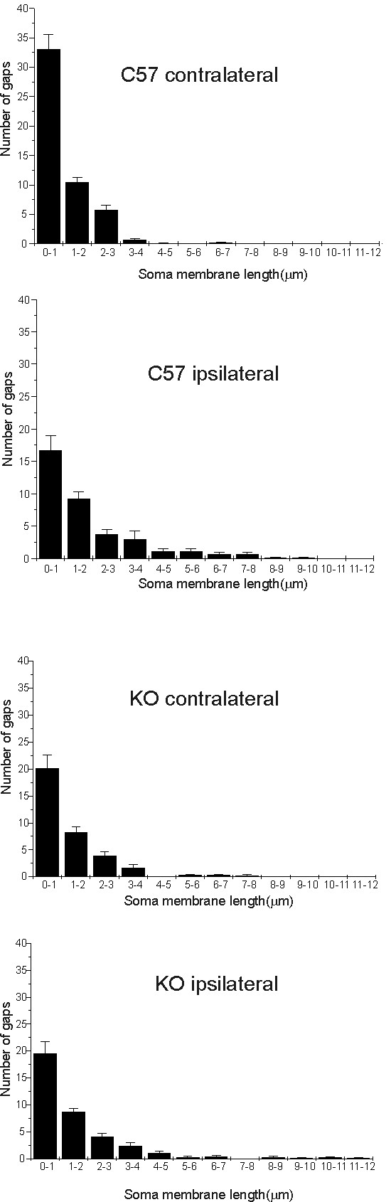
Results: We find that presumed motoneurons in the lower lumbar ventral horn exhibited a smaller soma size in the IFNγ-KO series, regardless of nerve lesion. In plastic embedded sections stained with toluidine blue, the IFNγ-KO mice demonstrated a greater proportion of degenerating neurons in the ventral horn when compared to the control series (p < 0.05). Apoptotic death is suggested based on TUNEL and caspase 3 immunostaining. A sciatic nerve axotomy did not further aggravate the neuronal loss. The cellular changes were supported by electron microscopy, which demonstrated ventral horn neurons exhibiting intracellular vacuoles as well as degenerating nuclei and cytoplasm in the IFNγ-KO mice. Adjacent glial cells showed features suggestive of phagocytosis. Additional ultrastructural studies showed a decreased number of pre-synaptic terminals apposing to motoneurons in mutant mice. Nevertheless, no statistical difference regarding the input covering could be detected among the studied strains.

Conclusion: Altogether, these results suggest that IFNγ may be neuroprotective and its absence results in neuronal death, which is not further increased by peripheral axotomy.


Figures

Figure 1
Figure 1
Motoneuron somata mean diameter (μm), one week after the unilateral transection of the sciatic nerve in C57BL/6J and IFNγ-KO mice. (A) Bimodal distribution in the C57BL/6J on the unlesioned side, with a clear through at 30-36 μm. (B) Lesioned side with non-bimodal size distribution. (C) Size distribution in the IFNγ-KO on the unlesioned side, the cell diameter is especially concentrated at 18-24 μm. (D) Lesioned side of the IFNγ-KO mice also showed a non-bimodal size distribution.
Figure 2
Figure 2
Light photomicrographs of spinal cord ventral horn of C57BL/6J and IFNγ-KO mice from semithin sections stained with toluidine blue. (A) Ipsilateral (lesioned) side and (B) contralateral (unlesioned) side of C57BL/6J mice. (C) IFNγ-KO ipsilateral side shows some motoneurons with hyperchromatic changes in the cytoplasm and nuclei (arrows). (D) IFNγ-KO contralateral side with pyknotic neurons found among at normal neurons (asterisk). (E) The number of degenerated motoneurons in axomized animals. Scale bar: 50 μm
Figure 3
Figure 3
Electron photomicrographs of spinal cord motoneurons of C57BL/6J mice. (A) One α-motoneuron found on the ipsilateral side. The boxed area is shown at higher magnification in (B). Some synaptic terminals are retracted; the cytoplasm and nuclei have a normal appearance. (C) contralateral α-motoneuron. (D) Higher magnification shows cytoplasmatic organelles visible and well organized rough endoplasmic reticulum (RER), Golgi apparatus (GA) and mitochondria. Scale bar: 5 μm (A and C), 1 μm (B-D).
Figure 4
Figure 4
Electron photomicrographs of spinal cord motoneurons of IFNγ-KO mice. (A) α-motoneurons in the lesioned side. Boxed area is shown at higher magnification in (B). The cytoplasm and nuclei are more electron dense and cytoplasmatic organelles are disorganized. Note the presence of numerous vacuoles in the perikaryon (asterisk) and the irregular contour of nuclear membrane. (C) α-motoneurons in the contralateral side. (D) Higher magnification shows the cell less electron dense and with some vacuoles in the cytoplasm. T, synaptic terminal; M, mitochondria; RER, rough endoplasmic reticulum, Nu, nucleus; G, golgi apparatus. Scale bar: 5 μm (A and C), 1 μm (B-D).
Figure 5
Figure 5
Electron photomicrographs showing degenerated neurons in IFNγ-KO mice. (A) One abnormal motoneuron amongst of normal cell (arrow). Cytoplasm and nuclei are electron dense; there are invaginations in the nuclear and cytoplasmic membranes and presence of many vacuoles in the cytoplasm. (B) The cell body appears shrunken and the nuclear envelope fragmented. (C) Darkly stained cytoplasm, condensed nuclear chromatin, and some organelles are hardly visible. (D) Glial cells are in close contact with a degenerating neuron. Note phagocytes of ingested material in the cytoplasm of microglial cells. Scale bar: 2 μm.
Figure 6
Figure 6
Electron micrograph showing two representative dying neurons in layer IX of spinal cord ventral horn of IFNγ-KO mice. (A) Dying neuron and the presence of glial cells in close contact with cell debris. (B) Many microglial cells ingesting a degenerating neuron. Scale bar: 5 μm.
Figure 7
Figure 7
Motoneuron degeneration in IFN-KO mice. (A) The schematic drawing of spinal cord section at the lumbar level demonstrating the sciatic motoneurons pool. (B-C) Representative images showing motoneuron cell bodies in unlesioned C57BL/6J and IFNγ-KO animals. (D) Graphical representation of spinal motoneuron survival. Note a significant reduction in number of motoneurons in IFNγ-KO mice. Scale bar 50 μm
Figure 8
Figure 8
Absence of IFNγ results in neuronal death. (A) TUNEL labeled neuron (arrow) within the motor nucleus indicates the development of apoptosis. (B) Representative immunofluorescence image showing a caspase-3 positive motoneuron in an IFNγ-KO unlesioned mouse. Scale bar 50μm.
Figure 9
Figure 9
Qualitative and quantitative ultrastructural analysis of every input to the surface of α-motoneurons one week after sciatic nerve axotomy. (A) Synaptic retraction following axotomy in the C57BL/6J lesioned (ipsilateral) side. The arrows indicate the retraction of the nerve terminal from the surface of motoneurons. (B) Presynaptic terminals in apposition to motoneuron (Mn) membrane of C57BL/6J contralateral side. (C) Synaptic elimination following axotomy in IFNγ-KO lesioned side. *terminal totally retracted, ** terminal partially retracted. (D) Synaptic covering in the IFNγ-KO contralateral side. (E) Detailed analysis of the covering and number of synaptic boutons in IFNγ-KO mice. Those animals showed a lower detachment of synaptic terminals and a significant reduced number of synaptic terminals in apposition (F), regardless the nerve lesion. (G) Percentage of synaptic covering of F, S and C-terminals on unlesioned and lesioned sides. (H) Graphs of terminal numbers/100 μm of motoneurons membrane. Note a greater loss of F-type of terminals in IFNγ-KO mice. p < 0.05(*). Scale bar 1 μm.
Figure 10
Figure 10
Graphs showing the frequency distribution (in micrometers) of gaps between the terminals along the cell soma membrane of α-motoneurons. (A-B) C57BL/6J animals showed a higher synaptic elimination one week after lesion. The gaps between clusters of boutons are possibly increased due to selective retractions of inputs. (C and D) IFNγ-KO mice showed a lower retraction of terminals after nerve lesion.

Similar articles

Cited by

References

    1. Olsson T, Kristensson K, Ljundahl Å, Maehlen J, Holmdahl R, Klareskog L. Gamma-interferon-like immunireactivity in axotomized rat motor neurons. J Neurosci. 1989;9:3870–3875. - PMC - PubMed
    1. Lindå H, Hammaberg H, Cullheim S, Levinovitz, Khademi M, Olsson T. Expression of MHC class I and β-microglobulin in rat spinal motorneurons: regulatory influences by IFN-gamma and axotomy. Exp Neurol. 1998;150:282–95. doi: 10.1006/exnr.1997.6768. - DOI - PubMed
    1. Renno T, Taupin V, Bourbonnière L, Verge G, Tran E, De Simone R, Krakowski M, Rodriguez M, Peterson A, Owens T. Interferon-gamma in progression to chronic demyelination and neurological deficit following acute EAE. Mol Cell Neurosci. 1998;12:376–89. doi: 10.1006/mcne.1998.0725. - DOI - PubMed
    1. Kerschensteiner M, Meinl E, Hohlfeld R. Neuro-immune crosstalk in CNS diseases. Neuroscience. 2009;158:1122–32. doi: 10.1016/j.neuroscience.2008.09.009. - DOI - PubMed
    1. Hanisch UK. Microglia as a source and target of cytokines. Glia. 2002;40:140–55. doi: 10.1002/glia.10161. - DOI - PubMed

Publication types

MeSH terms