

Leukocyte nucleus segmentation and nucleus lobe counting
- PMID: 21073711
- PMCID: PMC3224570
- DOI: 10.1186/1471-2105-11-558
Leukocyte nucleus segmentation and nucleus lobe counting
Abstract
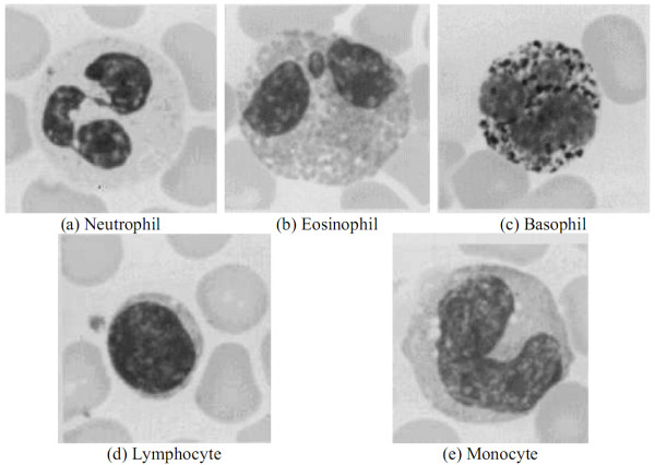
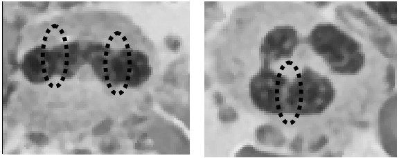
Background: Leukocytes play an important role in the human immune system. The family of leukocytes is comprised of lymphocytes, monocytes, eosinophils, basophils, and neutrophils. Any infection or acute stress may increase or decrease the number of leukocytes. An increased percentage of neutrophils may be caused by an acute infection, while an increased percentage of lymphocytes can be caused by a chronic bacterial infection. It is important to realize an abnormal variation in the leukocytes. The five types of leukocytes can be distinguished by their cytoplasmic granules, staining properties of the granules, size of cell, the proportion of the nuclear to the cytoplasmic material, and the type of nucleolar lobes. The number of lobes increased when leukemia, chronic nephritis, liver disease, cancer, sepsis, and vitamin B12 or folate deficiency occurred. Clinical neutrophil hypersegmentation has been widely used as an indicator of B12 or folate deficiency.Biomedical technologists can currently recognize abnormal leukocytes using human eyes. However, the quality and efficiency of diagnosis may be compromised due to the limitations of the biomedical technologists' eyesight, strength, and medical knowledge. Therefore, the development of an automatic leukocyte recognition system is feasible and necessary. It is essential to extract the leukocyte region from a blood smear image in order to develop an automatic leukocyte recognition system. The number of lobes increased when leukemia, chronic nephritis, liver disease, cancer, sepsis, and vitamin B12 or folate deficiency occurred. Clinical neutrophil hypersegmentation has been widely used as an indicator of B12 or folate deficiency.
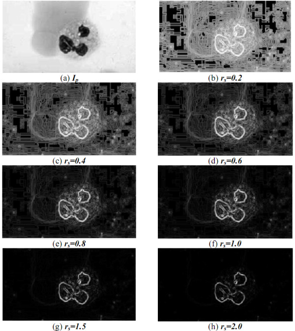
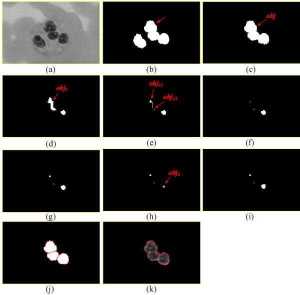
Results: The purpose of this paper is to contribute an automatic leukocyte nuclei image segmentation method for such recognition technology. The other goal of this paper is to develop the method of counting the number of lobes in a cell nucleus. The experimental results demonstrated impressive segmentation accuracy.
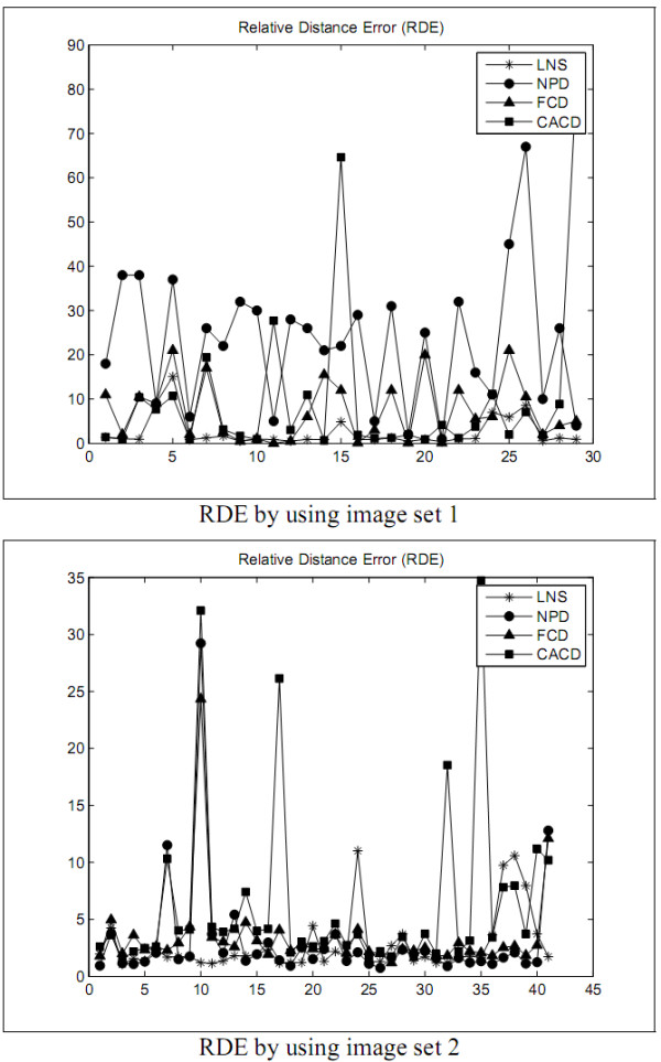
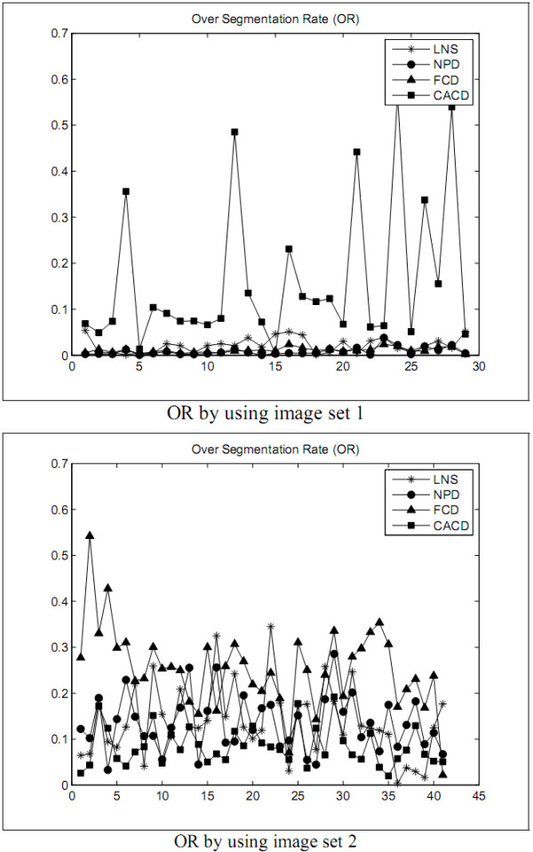
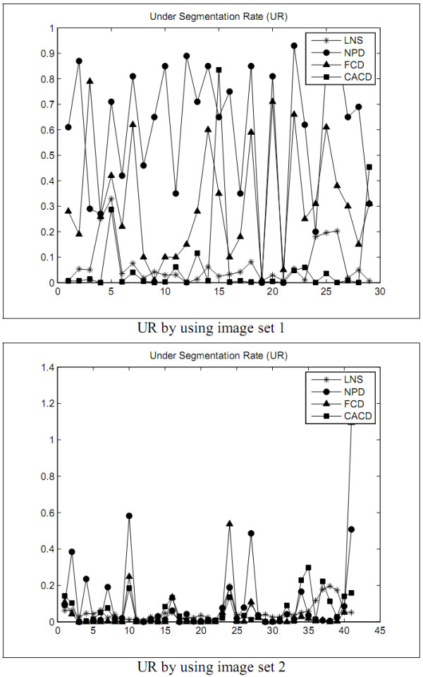
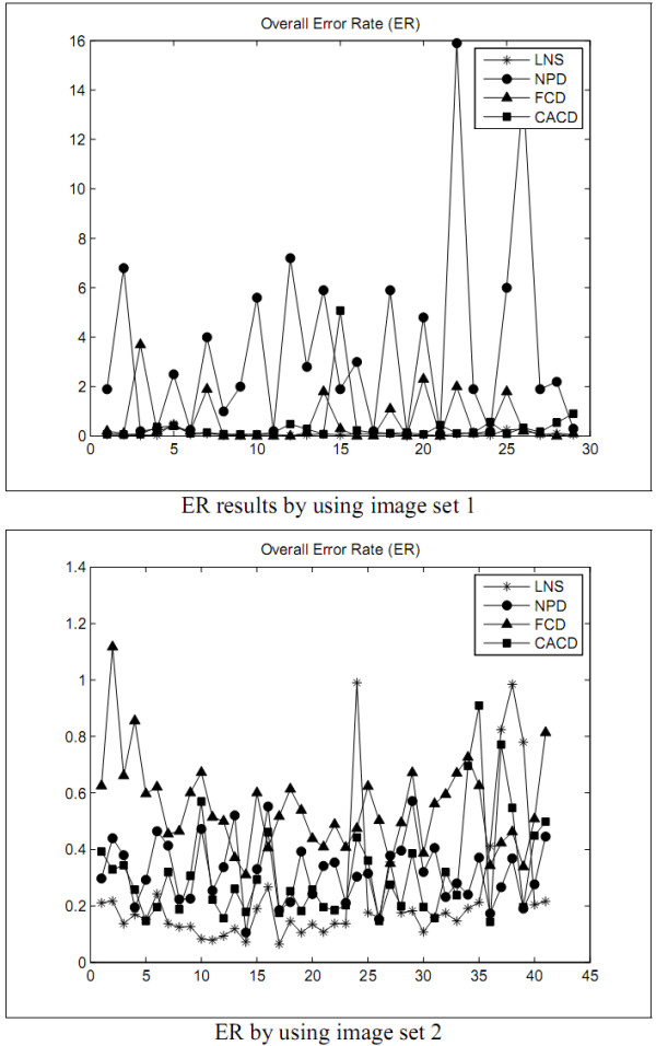
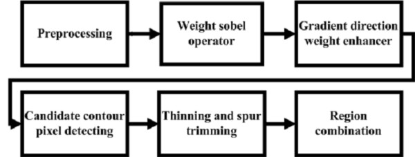
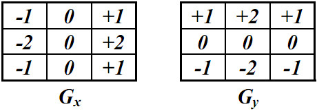
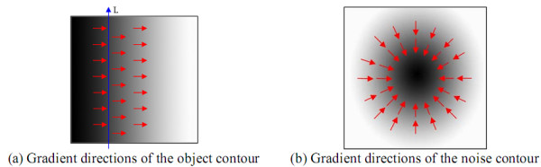
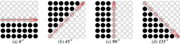
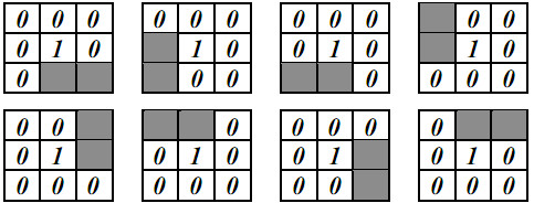
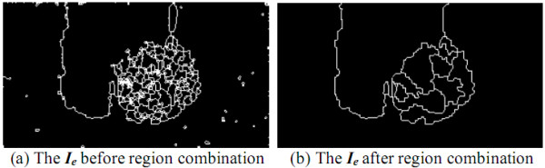
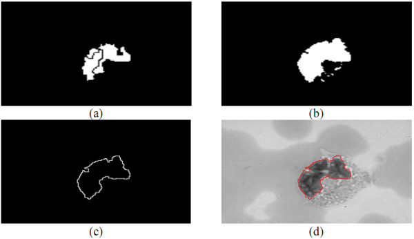
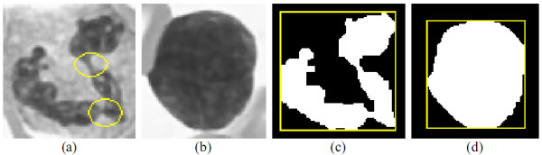
Conclusions: Insensitive to the variance of images, the LNS (Leukocyte Nuclei Segmentation) method functioned well to isolate the leukocyte nuclei from a blood smear image with much better UR (Under Segmentation Rate), ER (Overall Error Rate), and RDE (Relative Distance Error). The presented LC (Lobe Counting) method is capable of splitting leukocyte nuclei into lobes. The experimental results illuminated that both methods can give expressive performances. In addition, three advanced image processing techniques were proposed as weighted Sobel operator, GDW (Gradient Direction Weight), and GBPD (Genetic-based Parameter Detector).
Figures
Similar articles
-
Quick Leukocyte Nucleus Segmentation in Leukocyte Counting.Comput Math Methods Med. 2019 Jun 11;2019:3072498. doi: 10.1155/2019/3072498. eCollection 2019. Comput Math Methods Med. 2019. PMID: 31308855 Free PMC article.
-
A Polylobar Nucleus Identifying and Extracting Method for Leukocyte Counting.Comput Math Methods Med. 2021 Jul 22;2021:5565156. doi: 10.1155/2021/5565156. eCollection 2021. Comput Math Methods Med. 2021. PMID: 34335863 Free PMC article.
-
Semiautomatic white blood cell segmentation based on multiscale analysis.IEEE J Biomed Health Inform. 2013 Jan;17(1):250-6. doi: 10.1109/TITB.2012.2207398. Epub 2012 Jul 27. IEEE J Biomed Health Inform. 2013. PMID: 22855228
-
Recent computational methods for white blood cell nuclei segmentation: A comparative study.Comput Methods Programs Biomed. 2019 May;173:1-14. doi: 10.1016/j.cmpb.2019.03.001. Epub 2019 Mar 6. Comput Methods Programs Biomed. 2019. PMID: 31046984 Review.
-
Automated microscopic image analysis for leukocytes identification: a survey.Micron. 2014 Oct;65:20-33. doi: 10.1016/j.micron.2014.04.001. Epub 2014 Apr 12. Micron. 2014. PMID: 25041828 Review.
Cited by
-
Boundary-Preserved Deep Denoising of Stochastic Resonance Enhanced Multiphoton Images.IEEE J Transl Eng Health Med. 2022 Sep 14;10:1800812. doi: 10.1109/JTEHM.2022.3206488. eCollection 2022. IEEE J Transl Eng Health Med. 2022. PMID: 36304843 Free PMC article.
-
Nuclear morphologies: their diversity and functional relevance.Chromosoma. 2017 Mar;126(2):195-212. doi: 10.1007/s00412-016-0614-5. Epub 2016 Sep 8. Chromosoma. 2017. PMID: 27631793 Free PMC article. Review.
-
Immunosuppressant Drugs Mitigate Immune Responses Generated by Human Mesenchymal Stem Cells Transplanted into the Mouse Parenchyma.Cell Transplant. 2021 Jan-Dec;30:9636897211019025. doi: 10.1177/09636897211019025. Cell Transplant. 2021. PMID: 34044601 Free PMC article.
-
Genetic architecture of band neutrophil fraction in Iceland.Commun Biol. 2022 Jun 1;5(1):525. doi: 10.1038/s42003-022-03462-1. Commun Biol. 2022. PMID: 35650273 Free PMC article.
-
Quick Leukocyte Nucleus Segmentation in Leukocyte Counting.Comput Math Methods Med. 2019 Jun 11;2019:3072498. doi: 10.1155/2019/3072498. eCollection 2019. Comput Math Methods Med. 2019. PMID: 31308855 Free PMC article.
References
-
- Timby BK, Smith NE. Introductory Medical-Surgical Nursing. Nine. Lippincott Williams & Wilkins; 2006.
-
- Human Physiology and Anatomy. Blood Cell Histology. http://www.unomaha.edu/hpa/blood.html
-
- Bagby GC. In: Cecil Medicine. 23. Goldman L, Ausiello D, editor. Philadelphia, Pa: Saunders Elsevier; 2007. Leukopenia and Leukocytosis.
-
- Scientific Psychic. The Hematologist. http://www.scientificpsychic.com/mind/whitecells.html
Publication types
MeSH terms
LinkOut - more resources
Full Text Sources
