. 2010 Nov 13:7:95.
doi: 10.1186/1742-4690-7-95.

The membrane-spanning domain of gp41 plays a critical role in intracellular trafficking of the HIV envelope protein

Affiliations

The membrane-spanning domain of gp41 plays a critical role in intracellular trafficking of the HIV envelope protein

Kosuke Miyauchi et al. Retrovirology. .

Abstract

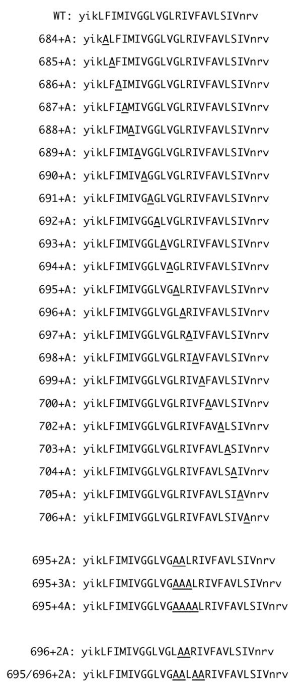
Background: The sequences of membrane-spanning domains (MSDs) on the gp41 subunit are highly conserved among many isolates of HIV-1. The GXXXG motif, a potential helix-helix interaction motif, and an arginine residue (rare in hydrophobic MSDs) are especially well conserved. These two conserved elements are expected to locate on the opposite sides of the MSD, if the MSD takes a α-helical secondary structure. A scanning alanine-insertion mutagenesis was performed to elucidate the structure-function relationship of gp41 MSD.

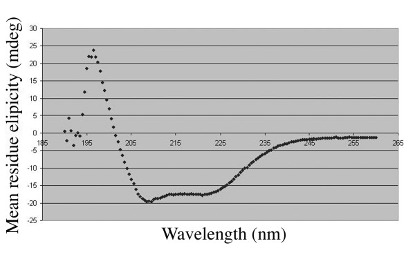
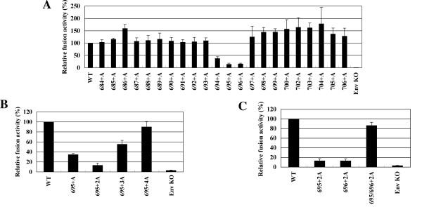
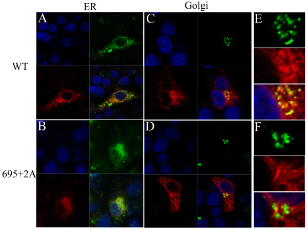
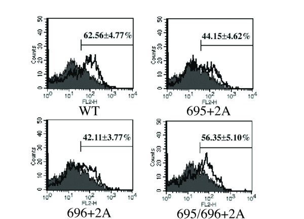
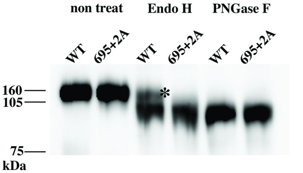
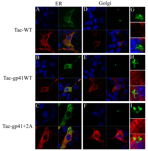
Results: A circular dichroism analysis of a synthetic gp41 MSD peptide determined that the secondary structure of the gp41 MSD was α-helical. We then performed a scanning alanine-insertion mutagenesis of the entire gp41 MSD, progressively shifting the relative positions of MSD segments around the helix axis. Altering the position of Gly694, the last residue of the GXXXG motif, relative to Arg696 (the number indicates the position of the amino acid residues in HXB2 Env) around the axis resulted in defective fusion. These mutants showed impaired processing of the gp160 precursor into gp120 and gp41. Furthermore, these Env mutants manifested inefficient intracellular transport in the endoplasmic reticulum and Golgi regions. Indeed, a transplantation of the gp41 MSD portion into the transmembrane domain of another membrane protein, Tac, altered its intracellular distribution. Our data suggest that the intact MSD α-helix is critical in the intracellular trafficking of HIV-1 Env.

Conclusions: The relative position between the highly conserved GXXXG motif and an arginine residue around the gp41 MSD α-helix is critical for intracellular trafficking of HIV-1 Env. The gp41 MSD region not only modulates membrane fusion but also controls biosynthesis of HIV-1 Env.


Figures

Figure 1
Figure 1
The circular dichroism (CD) profile of the synthetic MSD peptide. The synthetic peptide was dissolved in 15 mM DPC (n-dodecyl pyridinium chloride), 20 mM NaPi, 150 mM NaCl. The spectrum information was collected as described in the materials and methods section. The diagram shown is the average of eight spectra.
Figure 2
Figure 2
Amino acid sequences of the MSD of the wild type (WT) and Ala-insertion mutants used in this study. The predicted MSD portion is indicated in capital letters. The inserted alanine residue is underlined.
Figure 3
Figure 3
The fusion activity of Ala-insertion mutants in the cell-cell fusion assay. COS-7 cells transfected with the T7 RNA polymerase expression vector and the Env expression vector were co-cultured with 293CD4 cells transfected with a plasmid containing T7 promoter-driven renilla luciferase reporter. After a 24-hr co-culture, the renilla luciferase reporter activity was measured and normalized to the firefly activities as described previously [18]. The normalized renilla luciferase activities for (A) single Ala-inserted mutant of Env, (B) the mutant Env with multiple Ala insertion, (C) mutant Env with two alanine residues inserted at positions 695 and 696 are shown. Data are the average of three independent experiments. The error bar indicates a standard error.
Figure 4
Figure 4
The immunoblotting analysis of wild type (WT) and Ala-inserted mutant Env. The envelope proteins expressed in COS-7 cells transfected with the Env expression vector were detected with anti-gp120 antibody (for gp160 and gp120) or with anti-gp41 antibody. The results of single- and multiple-Ala-insertion mutants are shown in (A) and (B), respectively.
Figure 5
Figure 5
The transport defect of alanine insertion mutant Env. Endoplasmic reticulum (ER) (A and B) and Golgi (C to F) regions were visualized by fluorescence protein-conjugated ER or Golgi marker proteins (shown in green). FLAG tagged WT (A, C and E) and 695+2A Env (B, D and F) were stained by anti-FLAG antibody and Alexa Fluor (shown in red). The close-up of the Golgi area was shown in E and F. Nuclei of cells were stained with Hoechst 33258 (shown in blue).
Figure 6
Figure 6
Surface expression level of Env. The cell surface expression level of envelope proteins for WT and Ala-insertion mutants on transfected COS-7 cells was determined by flow cytometry using anti-gp120 antibody.
Figure 7
Figure 7
The analysis of glycosylation of WT and mutant Env. The FLAG-tagged Env purified from transfected COS-7 cells was treated with Endo H or PNGase F glycosidase. The treated protein was separated by SDS-PAGE and detected by immunoblotting analysis using anti-FLAG antibody. The asterisk shows the endo H-resistant fraction of Env.
Figure 8
Figure 8
Intracelluar distribution of Tac-gp41MSD chimera. The influence of MSD in transport of Tac proteines. Endoplasmic reticulum (ER) (A to C) and Golgi (D to I) regions were visualized by fluorescence protein-conjugated ER or Golgi marker proteins (shown in green). Halo tagged Tac-WT (A, D and G), Tac-gp41WT (B, E and H) and Tac-gp41 695+2A Env (C, F and I) were stained by anti-Halo antibody, anti-rabbit Ig and Alexa Fluor (shown in red). The close-up of the Golgi area was shown in G to I. Nuclei of cells were stained with Hoechst 33258 (shown in blue).

Similar articles

Cited by

References

    1. Weiss CD. HIV-1 gp41: mediator of fusion and target for inhibition. AIDS Rev. 2003;5:214–221. - PubMed
    1. Eckert DM, Kim PS. Mechanisms of viral membrane fusion and its inhibition. Annu Rev Biochem. 2001;70:777–810. doi: 10.1146/annurev.biochem.70.1.777. - DOI - PubMed
    1. Freed EO, Martin MA. The role of human immunodeficiency virus type 1 envelope glycoproteins in virus infection. J Biol Chem. 1995;270:23883–23886. doi: 10.1074/jbc.270.41.23883. - DOI - PubMed
    1. Gu M, Rappaport J, Leppla SH. Furin is important but not essential for the proteolytic maturation of gp160 of HIV-1. FEBS Lett. 1995;365:95–97. doi: 10.1016/0014-5793(95)00447-H. - DOI - PubMed
    1. Moulard M, Decroly E. Maturation of HIV envelope glycoprotein precursors by cellular endoproteases. Biochim Biophys Acta. 2000;1469:121–132. - PubMed

Publication types

MeSH terms