. 2011 Mar 1;55(1):153-64.
doi: 10.1016/j.neuroimage.2010.11.013. Epub 2010 Nov 10.

The generation of tetrahedral mesh models for neuroanatomical MRI

Affiliations

The generation of tetrahedral mesh models for neuroanatomical MRI

Carl Lederman et al. Neuroimage. .

Abstract

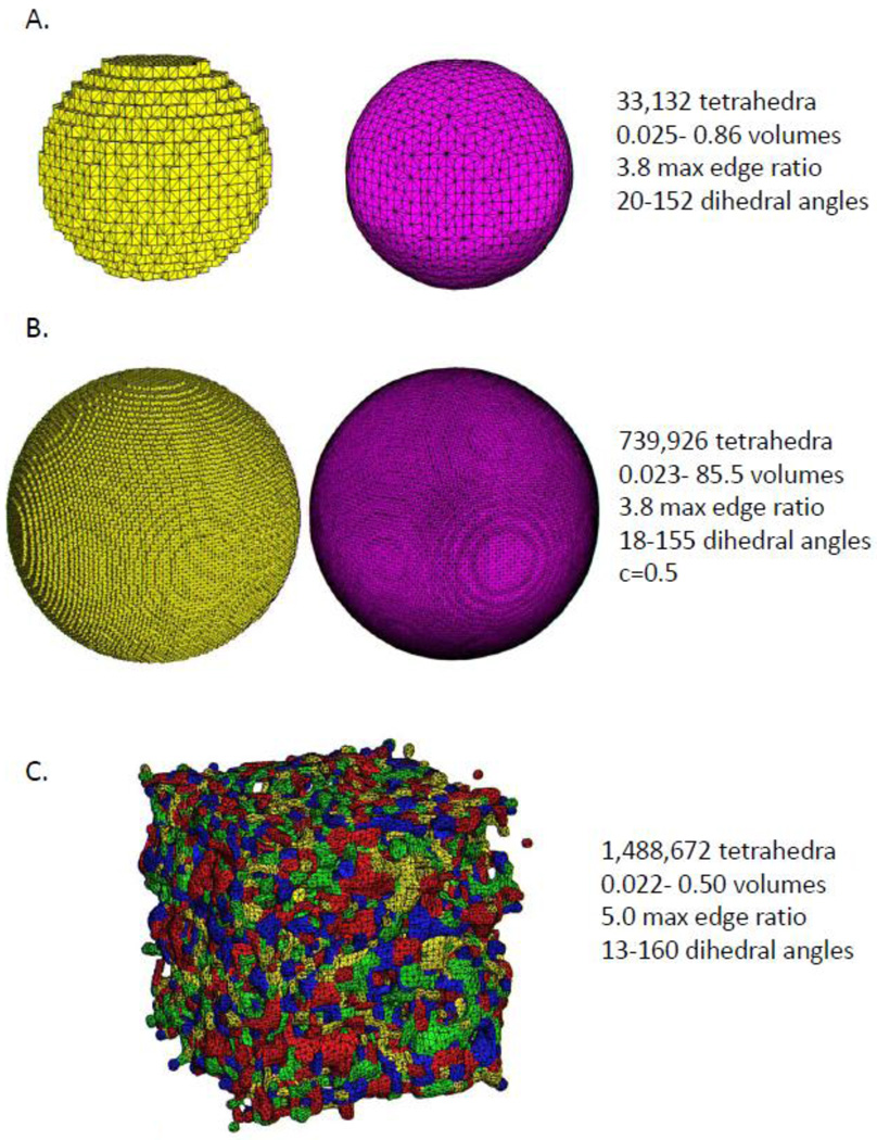
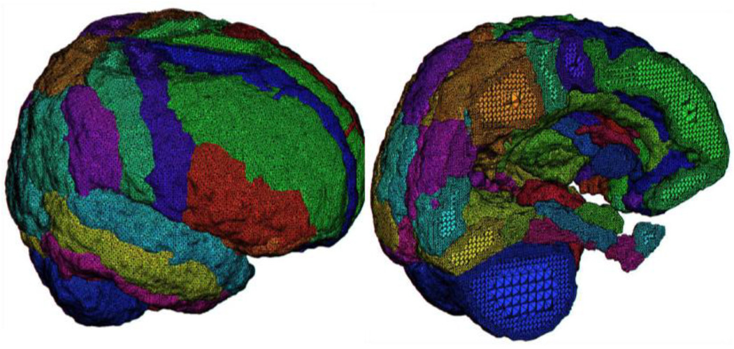
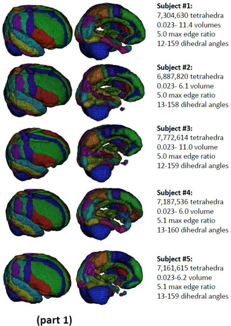
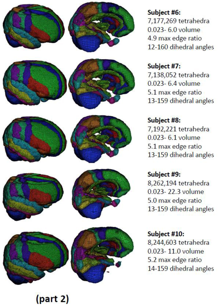
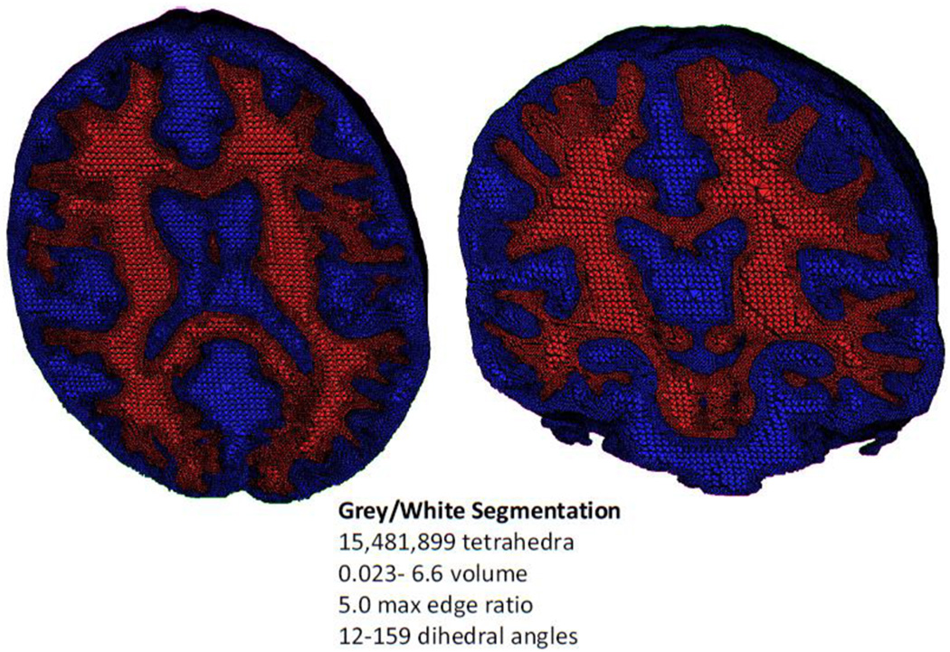
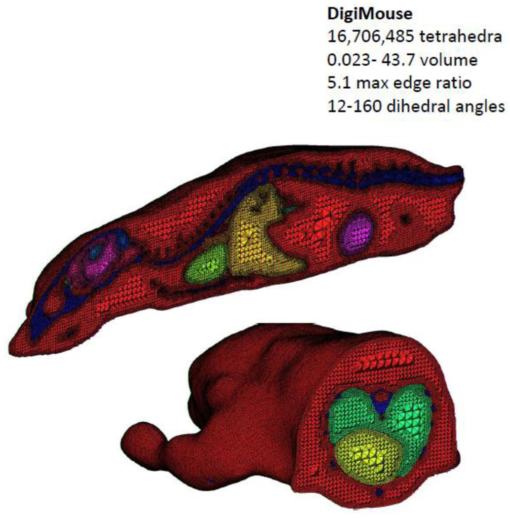
In this article, we describe a detailed method for automatically generating tetrahedral meshes from 3D images having multiple region labels. An adaptively sized tetrahedral mesh modeling approach is described that is capable of producing meshes conforming precisely to the voxelized regions in the image. Efficient tetrahedral mesh improvement is then performed minimizing an energy function containing three terms: a smoothing term to remove the voxelization, a fidelity term to maintain continuity with the image data, and a novel elasticity term to prevent the tetrahedra from becoming flattened or inverted as the mesh deforms while allowing the voxelization to be removed entirely. The meshing algorithm is applied to structural MR image data that has been automatically segmented into 56 neuroanatomical sub-divisions as well as on two other examples. The resulting tetrahedral representation has several desirable properties such as tetrahedra with dihedral angles away from 0 and 180 degrees, smoothness, and a high resolution. Tetrahedral modeling via the approach described here has applications in modeling brain structure in normal as well as diseased brain in human and non-human data and facilitates examination of 3D object deformations resulting from neurological illness (e.g. Alzheimer's disease), development, and/or aging.


Figures

Figure 1
Figure 1
A) This figure illustrates an example of a voxel conforming mesh of the image of a smaller sphere (left). The boundary of the smooth mesh (right) was penalized for any disagreement with the voxelized boundary but still resulted in a much smoother mesh. B) In another illustration, a tetrahedral mesh of the image of a large sphere that conforms exactly to the voxelization (left in yellow) and the recovered smooth sphere (right in fuchsia), resulting in many more tetrahedra being utilized but maintaining dihedral angles between 18–155 degrees. In this example only, the boundary was allowed to move without penalty by up to half a voxel to fully remove the voxelization. C) A tetrahedral mesh generated from an image of random noise. This example demonstrates the robustness of the meshing method, regardless of the number and shape of segmented regions contained in the input image.
Figure 2
Figure 2
A close-up view of the result of tetrahedral mesh generation for an example regionally segmented T1 anatomical image volume comprising 56 distinct cortical and sub-cortical regions. Note the finer tetrahedra promixal to each regional boundary and relatively larger tetrahedra near their centers.
Figure 3
Figure 3
(parts 1 and 2): The results of tetrahedral mesh generation for N=10 example brains drawn from the ADNI normative cohort. Across the subjects, meshes range in size from 6.8 to 8.2 million elements in which the dihedral angles have been constrained to range between 14 and 159 degrees.
Figure 3
Figure 3
(parts 1 and 2): The results of tetrahedral mesh generation for N=10 example brains drawn from the ADNI normative cohort. Across the subjects, meshes range in size from 6.8 to 8.2 million elements in which the dihedral angles have been constrained to range between 14 and 159 degrees.
Figure 4
Figure 4
A nested tetrahedral meshing of a brain segmented into gray (blue) and white (red) matter partitions comprising a total of over 15 million elements.
Figure 5
Figure 5
A tetrahedral representation of the DigiMouse whole body scan. The mesh is comprised of over 16 million tetrahedral elements.

References

    1. Apostolova LG, Thompson PM, Rogers SA, Dinov ID, Zoumalan C, Steiner CA, Siu E, Green AE, Small GW, Toga AW, Cummings JL, Phelps ME, Silverman DH. Surface Feature-Guided Mapping of Cerebral Metabolic Changes in Cognitively Normal and Mildly Impaired Elderly. Mol Imaging Biol. 2009 - PMC - PubMed
    1. Ballmaier M, Schlagenhauf F, Toga AW, Gallinat J, Koslowski M, Zoli M, Hojatkashani C, Narr KL, Heinz A. Regional patterns and clinical correlates of basal ganglia morphology in non-medicated schizophrenia. Schizophr Res. 2008;106(2–3):140–147. - PMC - PubMed
    1. Bank RE. Hierarchical bases and the finite element method. Acta Numerica. 1996;5:1–43.
    1. Bluestone A, Abdoulaev G, Schmitz C, Barbour R, Hielscher A. Three-dimensional optical tomography of hemodynamics in the human head. Opt. Express. 2001;9:272–286. - PubMed
    1. Chan T, Vese L. Active contours without edges. IEEE Transactions on Image Processing. 2001;10(2):266–277. - PubMed

Publication types

MeSH terms