. 2010 Nov 12;143(4):628-38.
doi: 10.1016/j.cell.2010.09.047.

A genome-wide Drosophila screen for heat nociception identifies α2δ3 as an evolutionarily conserved pain gene

Affiliations

A genome-wide Drosophila screen for heat nociception identifies α2δ3 as an evolutionarily conserved pain gene

G Gregory Neely et al. Cell. .

Abstract

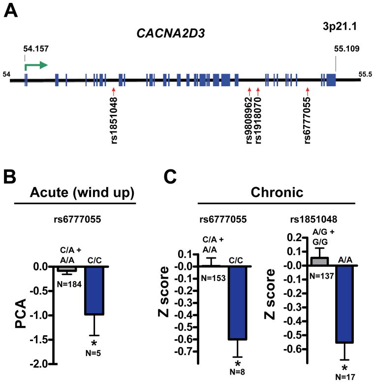
Worldwide, acute, and chronic pain affects 20% of the adult population and represents an enormous financial and emotional burden. Using genome-wide neuronal-specific RNAi knockdown in Drosophila, we report a global screen for an innate behavior and identify hundreds of genes implicated in heat nociception, including the α2δ family calcium channel subunit straightjacket (stj). Mice mutant for the stj ortholog CACNA2D3 (α2δ3) also exhibit impaired behavioral heat pain sensitivity. In addition, in humans, α2δ3 SNP variants associate with reduced sensitivity to acute noxious heat and chronic back pain. Functional imaging in α2δ3 mutant mice revealed impaired transmission of thermal pain-evoked signals from the thalamus to higher-order pain centers. Intriguingly, in α2δ3 mutant mice, thermal pain and tactile stimulation triggered strong cross-activation, or synesthesia, of brain regions involved in vision, olfaction, and hearing.


Figures

Figure 1
Figure 1. Thermal nociception in adult Drosophila
(A) Schematic representation of the thermal nociception assay in adult Drosophila. (B) Avoidance of noxious temperature of 46°C, but not avoidance of “sub-noxious” temperatures (25–35°C), is impaired in painless mutant (Painless(EP(2)2451) flies compared to the control strain Canton S (control). Data are presented as mean values +/− SEM. ~20 flies were tested per group, in replicates of at least four cohorts. Significant differences (P<0.001) were observed for temperature and strain responses. Further post hoc (Tukey’s) analysis showed a significant temperature avoidance response at 46 C for control (*, P<0.05) but not painless flies when compaired to responses at 25°C. (C) To set up the experimental screening system, w1118 (isogenic to the UAS-IR library) × elav-Gal4 flies (Control, grey; n = 1706) and painless mutant flies (painless, blue; n= 1816) were tested for avoidance to noxious heat (46°C). Based on these data a Z-score >= 1.65 was calculated as a specific cutoff to identify lines for further screening. Elav-Gal4 (also containing UAS-Dicer 2 (UAS-DRC2) for more efficient gene silencing) females were crossed to UAS-IR lines to knock-down the target genes in all neurons. All lines that exhibited a thermal avoidance defect (Z-score >= 1.65) were re-rested multiple times. (D) Results of the genome-wide screen. ~ 3% (622 transformants) of total lines tested (16051) exhibited a defect in thermal nociception, resulting in 580 candidate pain genes (622 transgenic lines). (E) Distribution of adult thermal nociception and developmental lethal hits for 16051 Drosophila UAS-IR lines. 1427 elav-Gal4 × UAS-IR lines were developmentally lethal (lethal). Among the 14624 viable lines, 562 lines exhibited defective thermal nociception (pain). Additional 60 lines that exhibited defective nociception as well as a semi-lethal phenotype were labeled as pain & lethal.
Figure 2
Figure 2. Straightjacket controls thermal nociception in adult Drosophila
(A) Diagram of the α2-δ family encoding peripheral subunits of Ca2+ channels. (B) RNAi knock-down of stj impairs noxious thermal avoidance in adult Drosophila (% avoidance of noxious temperature). stj-IR1 = Inverted repeat 1, stj-IR2 = Inverted repeat 2, both crossed to elav-Gal4;UAS-DCR2. (C) Q-PCR for stj-Knock-down efficiency in elav-Gal4>UAS-stj-IR1/2 adult fly brains. (D) Kinetics of temperature-induced paralysis for control and elav-Gal4>UAS-stj flies. (E) stj-Gal4 driving expression of lamin:GFP to label nuclei and cell surface CD8:GFP to visualize axonal projections in the brain of adult flies. The pars intercerebralis is marked with an arrow. (F) Co-localization of anti-STJ immunostaining and stj-Gal4>UAS-lamin:GFP. The pars intercerebralis is marked with an arrow. (G) stj in situ hybridization in the leg of wild type (w1118) flies. Of note, the sense control did not show any signal. DAPI counterstaining is shown as mark nuclei. All data are presented as mean ± sem. * P < 0.05, ** P < 0.01 (Student’s t-test).
Figure 3
Figure 3. Straightjacket controls thermal nociception in Drosophila larvae
(A) stj-GAL4 driven expression of UAS-CD8:GFP in larval body wall sensory neurons co-stained with anti-Futsch as a marker for sensory neurons. CD8:GFP expression co-localizes with sensory neurons (Futsch) in larval abdominal hemi-segments (A3) (top panels), and multidendritic sensory neurons (bottom panels). (B) Pan-neuronal knock-down of stj (UAS-stj-IR x elav), a mutant of stj (stj2), and stj mutant larvae over a corresponding deficiency Df(2R)Exel7128 (stj2/def) show severely impaired thermal nociception responses compared to w1118 x elav controls. painless larvae are shown as a control. The impaired larval thermal responses of a stj/def was rescued by reintroducing a wild type stj allele using the P[acman] system (stj2/def, stj+) (Venken et al., 2009). Percent responses ± sem to a 46°C heated probe are shown for the indicated time points. Mean response latency ± sem. P value was generated using a Kruskal-Wallis non-parametric test for median comparison with the Dunn’s post-hoc test. All P values depicted highlight significance relative to control responses. stj rescue was also significantly difference from stj2 and stj2/def, (P < 0.001). At least 20 animals were tested three times per genotype.
Figure 4
Figure 4. α2δ3 is required for thermal pain responses in mice
(A) Southern blotting of genomic DNA in α2δ3 wild type (+/+) and α2δ3 heterozygous (+/−) ES cells to confirm successful gene targeting. The endogenous wild type and targeted alleles are indicated. A 5′ probe was used on Nhe I digested genomic DNA. (B) α2δ3 and α2δ1 protein expression in brain and isolated DRG lysated from α2δ3+/+ (+/+), α2δ3+/− (+/−), and α2δ3−/− (−/−) mice. Actin is shown as a loading control. (C) Using the hot plate assay, α2δ3 mutant mice (n=16) show a delayed acute thermal nociception response as compared to control α2δ3+/+ mice (n=12). Littermate mice were used as a control. Values represent the latency (seconds) to respond to the indicated temperatures. (D) CFA-induced inflammatory pain behavior. CFA (20 μl) was injected into the hindpaw of α2δ3+/− (+/+, n=10) and α2δ3−/− (−/−, n=21) littermates and mice were tested for thermal pain (54°C) using the hot plate assay on the indicated days. Days 1, 3, 5, and 7 indicate days after CFA injection. All data are presented as mean values ± sem. *p < 0.05; **p < 0.01; *** P < 0.001 comparing mutant versus control mice. # P< 0.05 comparing sensitization to baseline (day -2) of the same genotype (Student’s t-test).
Figure 5
Figure 5. Polymorphisms in CACNA2D3 (α2δ3) associate with decreased acute and chronic pain in humans
(A) Schematic representation of the human CACNA2D3 gene locus on chromosome 3p21.1. The positions of the SNPs assayed are indicated. Blue boxes represent exons. The relative gene position is given in megabases (Mb). (B) Homozygous carriers of the rs6777055 minor allele (C/C) were significantly less sensitive to heat wind-up induced sensitivity relative to the other genotypes (C/A or A/A). (C) Of 169 lumbar chronic root pain patients 1 year post discectomy those homozygous for the minor allele C/C at SNP rs6777055 and A/A at SNP rs1851048 were less sensitive than the other allele combinations. In each case, the homozygous minor allele is associated with significantly less pain. Note that genotyping was not always successful for every individual, hence, the slightly different total numbers in the chronic pain group. All data are presented as mean values ± sem. *p < 0.05 (Student’s t-test).
Figure 6
Figure 6. α2δ3 is expressed in the brain and relays the pain signal to higher order brain centers
(A) β-Gal staining of whole brain slices from α2δ3+/− mice that carry the LacZ cassette. Different brain regions that are positive for LacZ expression are indicated. White lines indicate the brain slices displayed in Fig. 7A. (B–C) Quantification of % BOLD change and mean activation volume (in voxels) for (B) the thalamus and (C) the S1 somato-sensory cortex of α2δ3+/+ and α2δ3−/− mice. Of note, it has been proposed that the S1 cortical region is involved in the localisation of nociception (Treede et al., 1999). The different stimulation temperatures are indicated. Data are presented as mean +/− sem. *p < 0.05, **p < 0.01 (Student’s t-test comparing the respective control and α2δ3−/− groups). (D) Cross-correlation matrix of time profiles. Whereas the pain signal spreads from the thalamus to other higher order pain centers in α2δ3+/+ mice (red areas), in α2δ3−/− mice correlated activation can be only observed up to the level of the thalamus. Very weak activity is found in somato-sensory cortex (SC) for α2δ3−/− mice (green stripes). Data from the structures of left side of the brain are shown following challenge with noxious heat (55°C) at the right hindpaw. SI: sensory input; Th: thalamus; SC: somato-sensory cortex; AC: association cortex; LL: link to limbic system; LS: limbic system; HT: hypothalamus; BG: basal ganglia; C. cerebellum; M: motor cortex, P: periaquaeductal gray. Correlation-coefficients (cc) are given in the range from 0 (green), to +1 (red). n=20 for α2δ3+/+; n = 18 for α2δ3−/−.
Figure 7
Figure 7. α2δ3 mutant mice exhibit sensory cross-activation in response to thermal and tactile stimuli
(A) Second order statistical parameters maps showing only the significant differences of heat (55°C) and tactile (vibrissae) stimulation induced brain activation between α2δ3+/+ and α2δ3−/− mutant mice. Activation was assessed by BOLD-fMRI. The three planes correspond to the white lines shown in Fig. 6A. The green/blue scale indicates increased peak activation (55°C) in α2δ3+/+ control mice compared to α2δ3−/− mutant mice. The yellow/red scale indicates increased activation in α2δ3−/− mutant mice compared to α2δ3+/+ control mice. Images depict significant differences of second order group statistics corrected for multiple comparisons over all mice tested (n = 20 for α2δ3+/+ mice, n = 18 for α2δ3−/− mice). Arrows point to activated regions; note that for heat stimulation the S1/S2 somato-sensory cortex, the cingulate (Cg) cortex and the motor (M) cortex show significantly higher activity in α2δ3+/+ controls. In α2δ3−/− mice, heat stimulation leads to significantly higher activity auditory cortex (AC), the visual cortex (VC), and the olfactory tubercle (OT), as well as the amygdala (Amd) and the hypothalamus (HT). For tactile stimulation, only one small region in the S1 somatosensory cortex, ipsilateral to the side of stimulation (right), showed significantly higher activity in α2δ3+/+ controls, whereas α2δ3−/− mice again exhibited increased activation of the VC, AC, and OT, in addition to the caudate putamen (Cpu), S1 and S2 regions of the somato-sensory cortex, and the superior colliculus (SC). (B,C) % BOLD changes in the auditory cortex (AC), olfactory tubercle (OT), and visual cortex (VC) in control and α2 δ3 −/− mice following (B) heat (55°C) and (C) tactile vibrissal stimulation. Data is presented as mean values ± sem. *p < 0.05; **p < 0.01 (Student’s t-test).

Comment in

  • A straightjacket for pain?
    Beggs S, Salter MW. Beggs S, et al. Cell. 2010 Nov 12;143(4):505-7. doi: 10.1016/j.cell.2010.10.038. Cell. 2010. PMID: 21074043

References

    1. Aleman A, Rutten GJ, Sitskoorn MM, Dautzenberg G, Ramsey NF. Activation of striate cortex in the absence of visual stimulation: an fMRI study of synesthesia. Neuroreport. 2001;12:2827–2830. - PubMed
    1. Arthurs OJ, Boniface S. How well do we understand the neural origins of the fMRI BOLD signal? Trends in neurosciences. 2002;25:27–31. - PubMed
    1. Atlas SJ, Keller RB, Chang Y, Deyo RA, Singer DE. Surgical and nonsurgical management of sciatica secondary to a lumbar disc herniation: five-year outcomes from the Maine Lumbar Spine Study. Spine. 2001;26:1179–1187. - PubMed
    1. Basbaum AI, Bautista DM, Scherrer G, Julius D. Cellular and molecular mechanisms of pain. Cell. 2009;139:267–284. - PMC - PubMed
    1. Beauchamp MS, Ro T. Neural substrates of sound-touch synesthesia after a thalamic lesion. J Neurosci. 2008;28:13696–13702. - PMC - PubMed

Publication types