

Protein composition of microparticles shed from human placenta during placental perfusion: Potential role in angiogenesis and fibrinolysis in preeclampsia
- PMID: 21074265
- PMCID: PMC3762591
- DOI: 10.1016/j.placenta.2010.10.011
Protein composition of microparticles shed from human placenta during placental perfusion: Potential role in angiogenesis and fibrinolysis in preeclampsia
Abstract
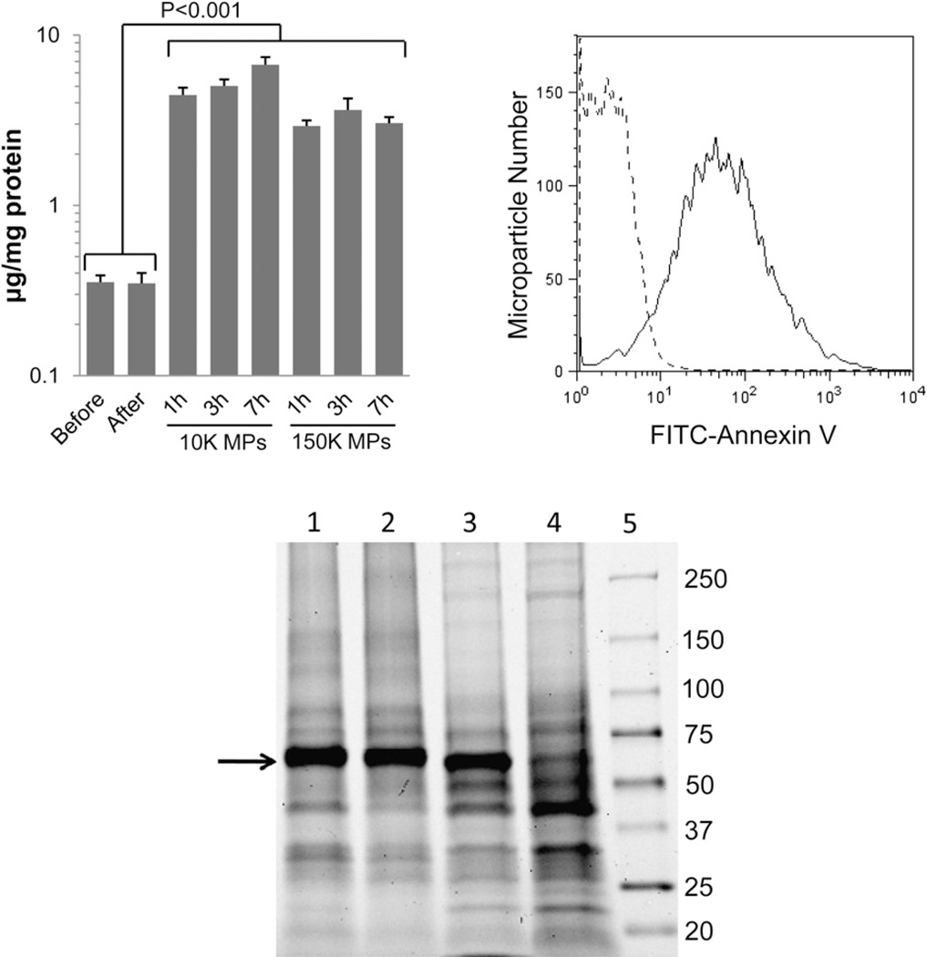
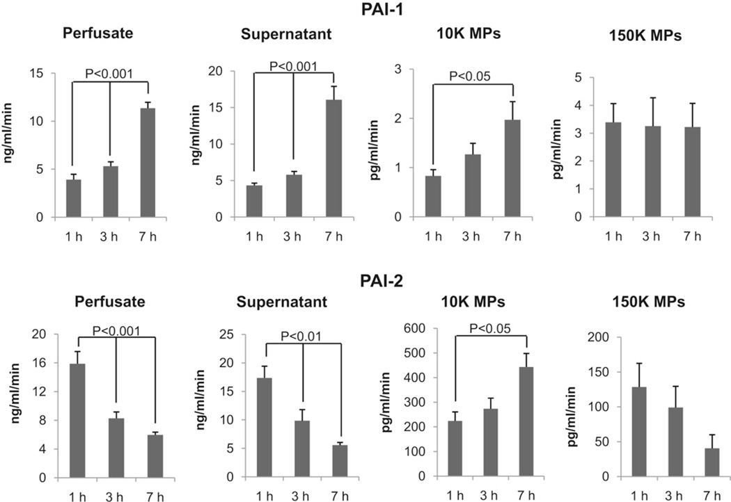
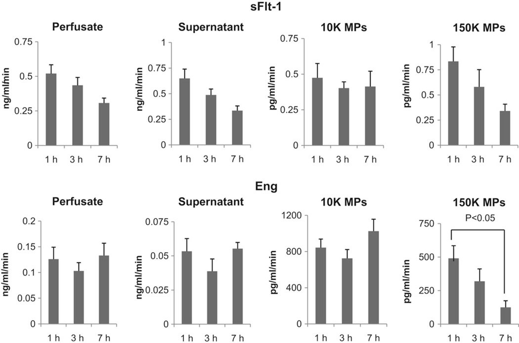
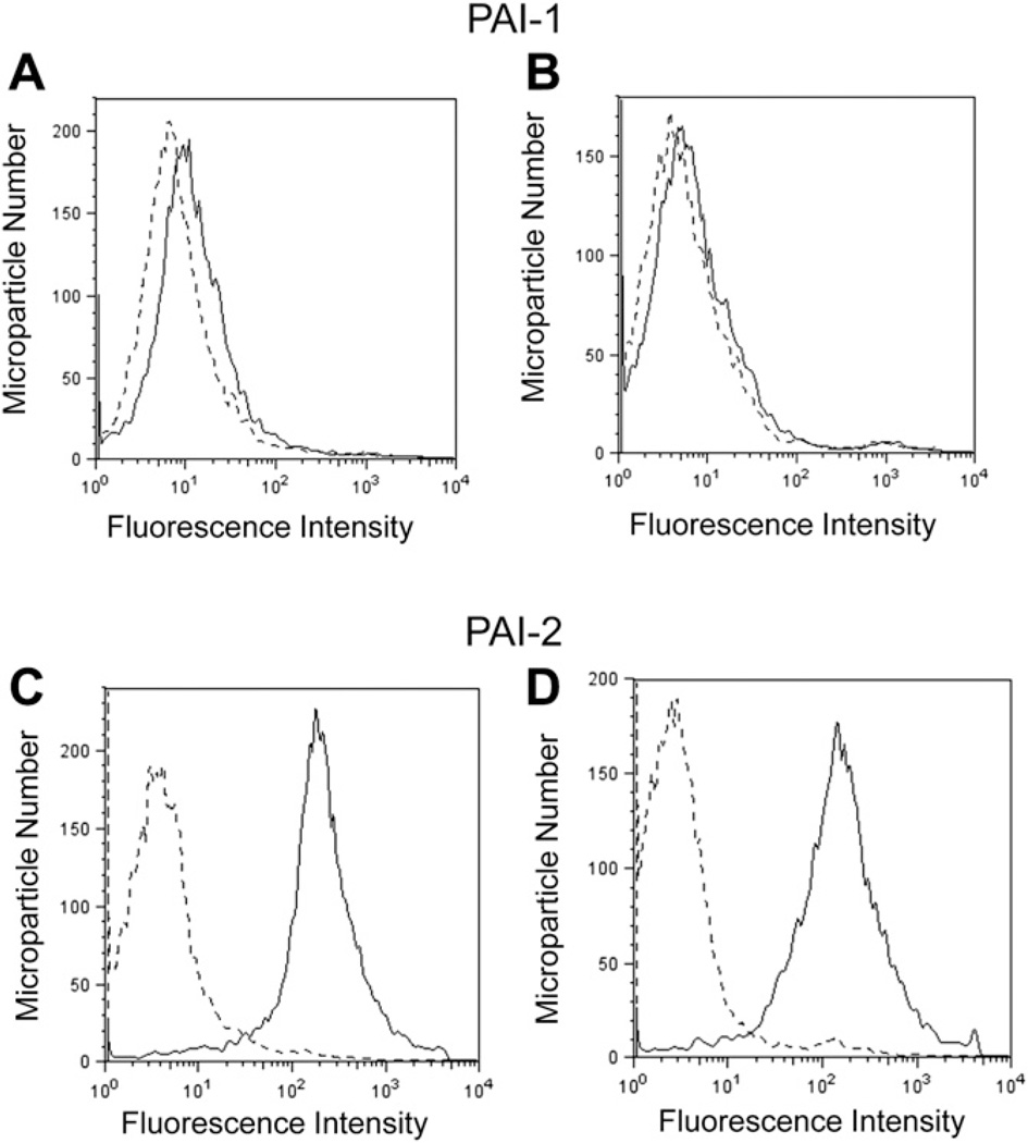
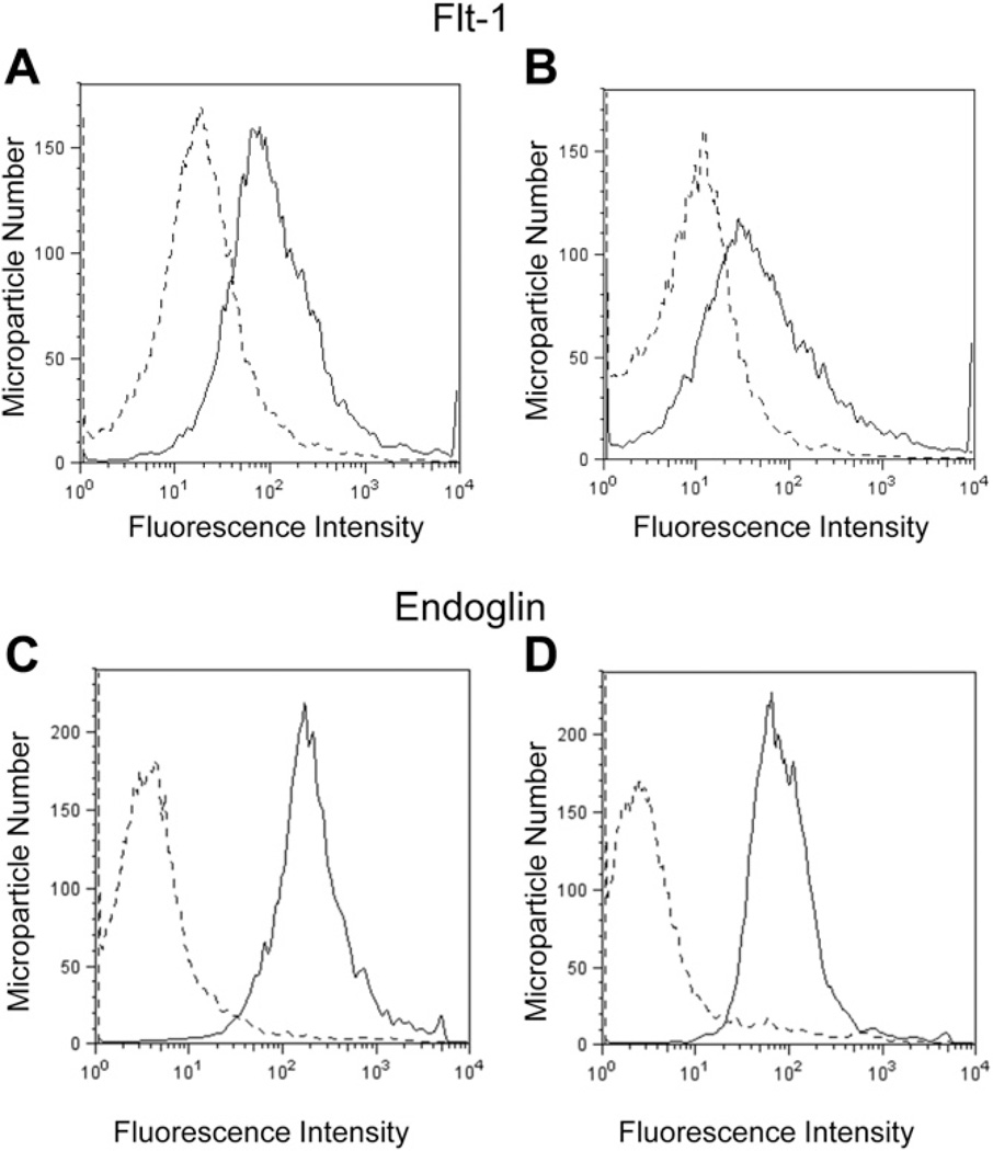
Shedding of syncytiotrophoblast microparticles (MPs) from placenta to maternal blood occurs in normal pregnancy and is enhanced during preeclampsia (PE). The syncytiotrophoblast synthesizes plasminogen activator inhibitors (PAIs) which regulate fibrinolysis, as well as soluble forms of the fms-like tyrosine kinase (sFlt-1) and endoglin, which exert anti-angiogenic actions. An increase in the ratio of PAI-1/PAI-2 and elevated levels of sFlt-1 and sEng in maternal serum are linked to placental damage and maternal endothelial cell dysfunction in PE. The goal of the current study was to determine whether MPs released to maternal perfusate during dual perfusion contain these factors associated with placental pathophysiology in PE. Initially, high levels of alkaline phosphatase activity and Annexin V binding were found in MPs isolated by sequential centrifugation of maternal perfusates at 10,000 and 150,000×g(10 K and 150 K MPs), indicating their plasma membrane origin. ELISA revealed the presence of these factors at the following relative levels: Eng>PAI-2⋙PAI-1>sFlt-1. Based on comparisons of their concentration in perfusates, MPs, and MP-free 150 K supernatants, we determined that MPs constitute a significant portion of Eng released by placenta. Flow cytometric analysis of 10 K MPs supported the levels of expression found by ELISA and indicated that Eng and PAI-2 were almost exclusively localized to the surface of MPs, a site with biological potential. These results indicate that MPs shed from the syncytial surface express factors which may alter the fibrinolytic and angiogenic balance at the maternal-fetal interface and play a role in the pathophysiology of PE.
Copyright © 2010 Elsevier Ltd. All rights reserved.
Figures
References
-
- Aly AS, Khandelwal M, Zhao J, Mehmet AH, Sammel MD, Parry S. Neutrophils are stimulated by syncytiotrophoblast microvillous membranes to generate superoxide radicals in women with preeclampsia. Am J Obstet Gynecol. 2004;190:252–258. - PubMed
-
- Goswami D, Tannetta DS, Magee LA, Fuchisawa A, Redman CW, Sargent IL, et al. Excess syncytiotrophoblast microparticle shedding is a feature of early-onset pre-eclampsia, but not normotensive intrauterine growth restriction. Placenta. 2006;27:56–61. - PubMed
-
- Gupta AK, Rusterholz C, Huppertz B, Malek A, Schneider H, Holzgreve W, et al. A comparative study of the effect of three different syncytiotrophoblast micro-particles preparations on endothelial cells. Placenta. 2005;26:59–66. - PubMed
-
- Knight M, Redman CW, Linton EA, Sargent IL. Shedding of syncytiotrophoblast microvilli into the maternal circulation in pre-eclamptic pregnancies. Br J Obstet Gynaecol. 1998;105:632–640. - PubMed
-
- Huppertz B, Frank HG, Kingdom JC, Reister F, Kaufmann P. Villous cytotrophoblast regulation of the syncytial apoptotic cascade in the human placenta. Histochem Cell Biol. 1998;110:495–508. - PubMed
Publication types
MeSH terms
Substances
Grants and funding
LinkOut - more resources
Full Text Sources
Other Literature Sources
Medical
Miscellaneous
