. 2011 Jan;50(1):239-47.
doi: 10.1016/j.yjmcc.2010.11.002. Epub 2010 Nov 11.

Thioredoxin 1 enhances neovascularization and reduces ventricular remodeling during chronic myocardial infarction: a study using thioredoxin 1 transgenic mice

Affiliations

Thioredoxin 1 enhances neovascularization and reduces ventricular remodeling during chronic myocardial infarction: a study using thioredoxin 1 transgenic mice

Ram Sudheer Adluri et al. J Mol Cell Cardiol. 2011 Jan.

Abstract

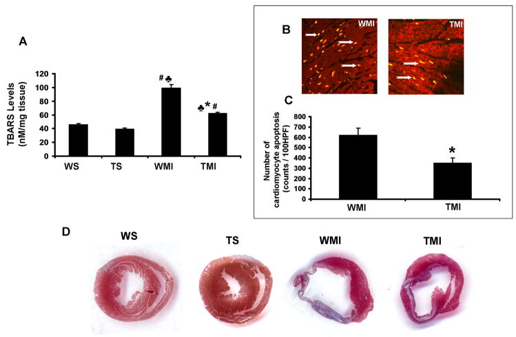
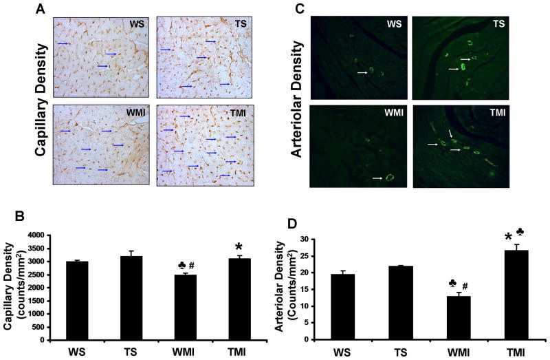
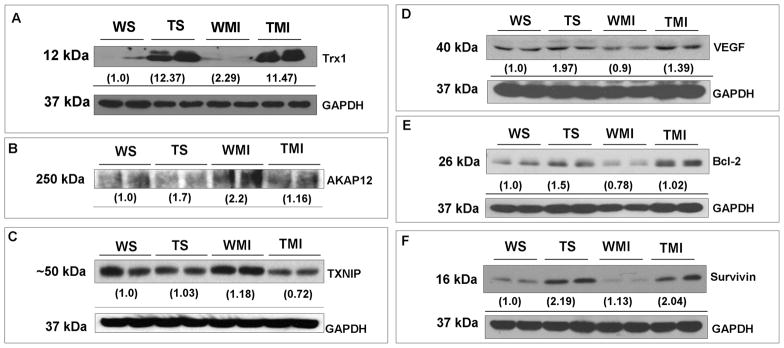
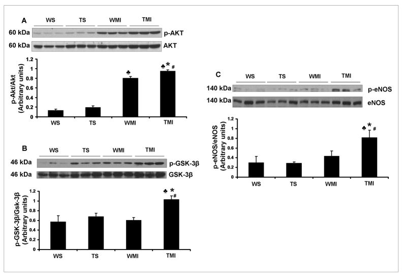
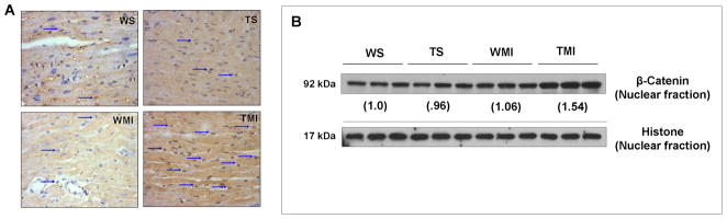
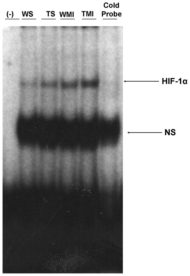
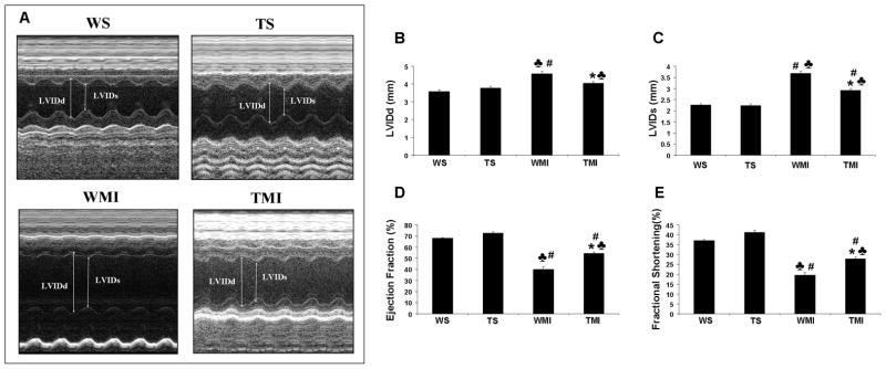
Oxidative stress plays a crucial role in disruption of neovascularization by alterations in thioredoxin 1 (Trx1) expression and its interaction with other proteins after myocardial infarction (MI). We previously showed that Trx1 has angiogenic properties, but the possible therapeutic significance of overexpressing Trx1 in chronic MI has not been elucidated. Therefore, we explored the angiogenic and cardioprotective potential of Trx1 in an in vivo MI model using transgenic mice overexpressing Trx1. Wild-type (W) and Trx1 transgenic (Trx1(Tg/+)) mice were randomized into W sham (WS), Trx1(Tg/+) sham (TS), WMI, and TMI. MI was induced by permanent occlusion of LAD coronary artery. Hearts from mice overexpressing Trx1 exhibited reduced fibrosis and oxidative stress and attenuated cardiomyocyte apoptosis along with increased vessel formation compared to WMI. We found significant inhibition of Trx1 regulating proteins, TXNIP and AKAP 12, and increased p-Akt, p-eNOS, p-GSK-3β, HIF-1α, β-catenin, VEGF, Bcl-2, and survivin expression in TMI compared to WMI. Echocardiography performed 30days after MI revealed significant improvement in myocardial functions in TMI compared to WMI. Our study identifies a potential role for Trx1 overexpression and its association with its regulatory proteins TXNIP, AKAP12, and subsequent activation of Akt/GSK-3β/β-catenin/HIF-1α-mediated VEGF and eNOS expression in inducing angiogenesis and reduced ventricular remodeling. Hence, Trx1 and other proteins identified in our study may prove to be potential therapeutic targets in the treatment of ischemic heart disease.


Figures

Fig. 1
Fig. 1. Trx1 overexpression attenuated ischemic stress-induced oxidative stress and myocardial fibrosis after MI
A, Shows the quantitative analysis of TBARS concentration after surgical intervention. Trx1 transgenic animals resulted in significant reduction in oxidative stress after MI compared to WT animals (n=4). B, Representative digital micrographs showing cardiomyocyte apoptosis in the border zone of TMI and WMI groups. C, Quantitative analysis of cardiomyocyte apoptosis after MI from 3–5 animals, in counts/100 high-power field (HPF). The apoptotic cardiomyocytes are significantly reduced in TMI compared to WMI. D, Shows the representative picture of myocardial fibrosis (Masson’s trichrome staining) from different groups after surgical intervention. A significant increase in fibrosis (collagen staining in blue colour and thinner infarct) is evident in WMI compared to WS and TS. Transgenic animals subjected to MI resulted in a thicker infarct and less fibrosis (blue colour) with the islands of viable cardiac tissue with markedly less scar extension (n=3–4). WS indicates wild type sham; TS, Trx1 transgenic sham; WMI, wild type animals subjected to MI; TMI, Trx1 transgenic animals subjected to MI. ♣, P≤0.05 vs. WS; #, P≤0.05 vs. TS; *, P≤0.05 vs. WMI.
Fig. 2
Fig. 2. Trx1 overexpression enhanced the neovascularization after MI
A, Representative digital micrographs showing capillary density/CD31 immuno-staining after 7 days of surgical intervention in different experimental groups. B, Quantitative analysis of capillary density in counts/mm2. The values are mean±SEM of 3–4 animals per group. C, Representative digital micrographs showing arteriolar density/α-smooth muscle actin immuno-staining in different experimental groups after 7 days of surgical intervention. D, Quantitative analysis of arteriolar density, in counts/mm2. The values are mean±SEM of 3–4 animals per group. Both capillary and arteriolar density was found to be significantly increased in transgenic animals subjected to MI compared to WT animals. Abbreviations are as in Fig. 1. ♣, P≤0.05 vs. WS; #, P≤0.05 vs. TS; *, P≤0.05 vs. WMI.
Fig. 3
Fig. 3. Trx1 overexpression altered the expression of TXNIP, AKAP12, VEGF, Bcl-2 and survivin during ischemic stress
Representative Western blots show the expression of Trx1, TXNIP, AKAP12, VEGF, Bcl-2 and survivin, and their corresponding loading control-GAPDH. Western blot analysis revealed downregulation of AKAP12 (Fig. 3B) and TXNIP (Fig. 3C), whereas increased VEGF (Fig. 3D), Bcl-2 (Fig. 3E) and survivin (Fig. 3F) expression in TMI compared to the WMI. Numbers below the bands represent the average-fold change compared to WS from 3–5 independent experiments. Trx1 representative blot (Fig. 3A) is given for the validation of overexpression of Trx1 in transgenic animals. Abbreviations are as given in Fig. 1.
Fig. 4
Fig. 4. Trx1 overexpression influenced the expression of p-Akt, p-GSK-3β and p-eNOS after MI
Representative Western blots show the expression of p-Akt (Fig. 4A), p-GSK-3β (Fig. 4B) and p-eNOS (Fig. 4C). Bar graphs in A, B and C represent the quantitative analysis and difference in the expression of p-Akt, p-GSK-3β and p-eNOS in between different groups, after they were normalized with corresponding non-phosphorylated protein controls, respectively in arbitrary units. The values are mean±SEM (n=3–5 from each group). There was a significant increase in the expression of p-Akt, p-GSK-3β and p-eNOS in TMI compared with the WMI. Abbreviations are as given in Fig. 1. ♣, P≤0.05 vs. WS; #, P≤0.05 vs. TS; *, P≤0.05 vs. WMI.
Fig. 5
Fig. 5. Trx1 overexpression increased the nuclear translocation of β-catenin in ischemic stress
A, Representative digital micrographs (immunohistochemical analysis by DAB staining and counter nuclear staining by haematoxylin) showing β-catenin nuclear translocation (indicated by arrows) after surgical intervention in different experimental groups; n=3 per group. B, Representative Western blots show the expression of β-catenin in nuclear fractions and its corresponding loading control. There was a significant increase in the translocation of β-catenin into the nucleus, evaluated by both immunohistochemistry and Western blot analysis, in TMI compared to WMI. Numbers below the bands represent the average-fold change compared to WS. The values are mean±SEM of 3–5 animals from each group. Abbreviations are as given in Fig. 1.
Fig. 6
Fig. 6. Trx1 overexpression increased the HIF-1α DNA binding activity after MI
EMSA analysis revealed increased nuclear translocation and DNA binding activity of HIF-1α in TMI compared to WMI (n=3 from each group). The position of the HIF-1α band was confirmed by the disappearance of the corresponding band in the last lane from right side when we used unlabelled probe (Cold probe). WS indicates wild type sham; TS, Trx1 transgenic sham; WMI, wild type animals subjected to MI; TMI, Trx1 transgenic animals subjected to MI; NS, non-specific.
Fig. 7
Fig. 7. Trx1 overexpression prevented post-infarcted ventricular remodeling (by echocardiography)
A. Representative echocardiograph pictures of parasternal short-axis M mode images 30 days after surgical intervention from different groups. B, The quantitative data of left ventricular internal diameter in diastole (LVIDd); C, The quantitative data of left ventricular internal diameter in systole (LVIDs); D, ejection fraction; and E, fractional shortening. These results demonstrate that there was significant functional disorder in the WMI compared to WS and TS. Trx1 overexpression significantly improved functional parameters compared to the wild type animals after MI. Values are mean±SEM (n=4–6 per group). WS indicates wild type sham; TS, Trx1 transgenic sham; WMI, wild type animals subjected to MI; TMI, Trx1 transgenic animals subjected to MI. ♣, P≤0.05 vs. WS; #, P≤0.05 vs. TS; *, P0.05 vs. WMI.
Fig. 8
Fig. 8
Schematic diagram shows the possible molecular mechanism of Trx1-induced neovascularization and reduced ventricular remodeling in ischemic myocardium.

Similar articles

Cited by

References

    1. Molin D, Post MJ. Therapeutic angiogenesis in the heart: protect and serve. Curr Opin Pharmacol. 2007;7:158–63. - PubMed
    1. Losordo DW, Dimmeler S. Therapeutic angiogenesis and vasculogenesis for ischemic disease. Part I: angiogenic cytokines. Circulation. 2004;109:2487–91. - PubMed
    1. Samuel SM, Thirunavukkarasu M, Penumathsa SV, Koneru S, Zhan L, Maulik G, et al. Thioredoxin-1 gene therapy enhances angiogenic signaling and reduces ventricular remodeling in infarcted myocardium of diabetic rats. Circulation. 2010;121:1244–55. - PMC - PubMed
    1. Billiet L, Rouis M. Thioredoxin-1 is a novel and attractive therapeutic approach for various diseases including cardiovascular disorders. Cardiovasc Hematol Disord Drug Targets. 2008;8:293–6. - PubMed
    1. Ago T, Sadoshima J. Thioredoxin and ventricular remodeling. J Mol Cell Cardiol. 2006;41:762–73. - PMC - PubMed

Publication types

MeSH terms