. 2011 Mar;8(3):448-54.
doi: 10.1016/j.hrthm.2010.11.019. Epub 2010 Nov 12.

Morphologic pattern of the intrinsic ganglionated nerve plexus in mouse heart

Affiliations

Morphologic pattern of the intrinsic ganglionated nerve plexus in mouse heart

Kristina Rysevaite et al. Heart Rhythm. 2011 Mar.

Abstract

Background: Both normal and genetically modified mice are excellent models for investigating molecular mechanisms of arrhythmogenic cardiac diseases that may be associated with an imbalance between sympathetic and parasympathetic nervous input to the heart.

Objective: The purpose of this study was to (1) determine the structural organization of the mouse cardiac neural plexus, (2) identify extrinsic neural sources and their relationship with the cardiac plexus, and (3) reveal any anatomic differences in the cardiac plexus between mouse and other species.

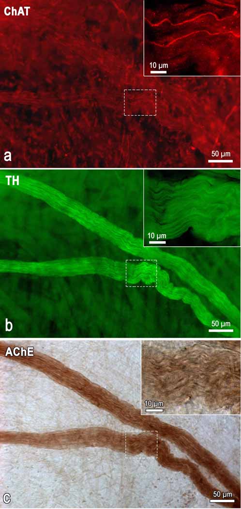
Methods: Cardiac nerve structures were visualized using histochemical staining for acetylcholinesterase (AChE) on whole heart and thorax-dissected preparations derived from 25 mice. To confirm the reliability of staining parasympathetic and sympathetic neural components in the mouse heart, we applied a histochemical method for AChE and immunohistochemistry for tyrosine hydroxylase (TH) and/or choline acetyltransferase (ChAT) on whole mounts preparations from six mice.

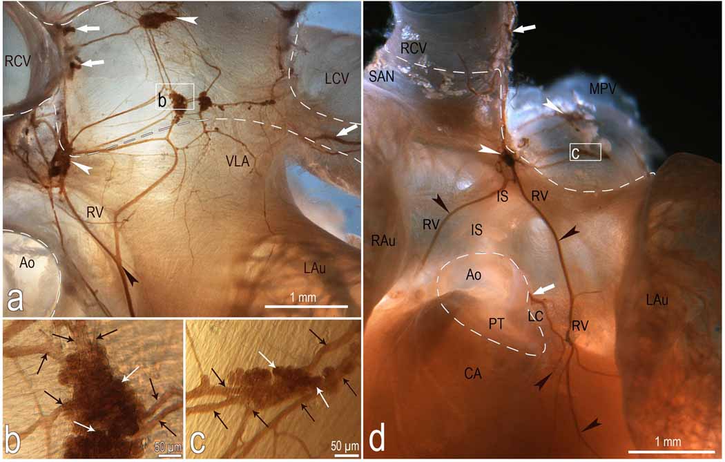
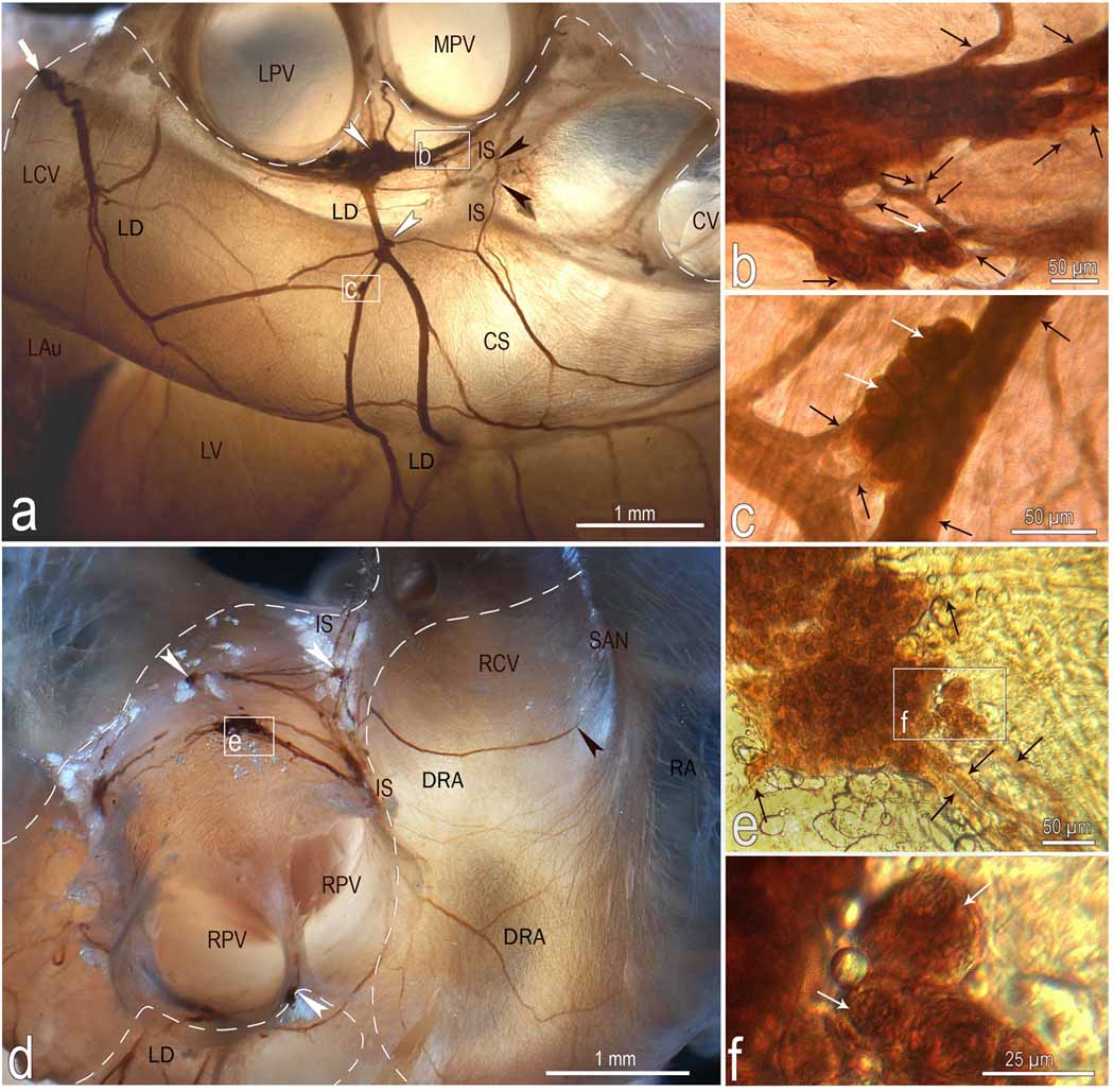
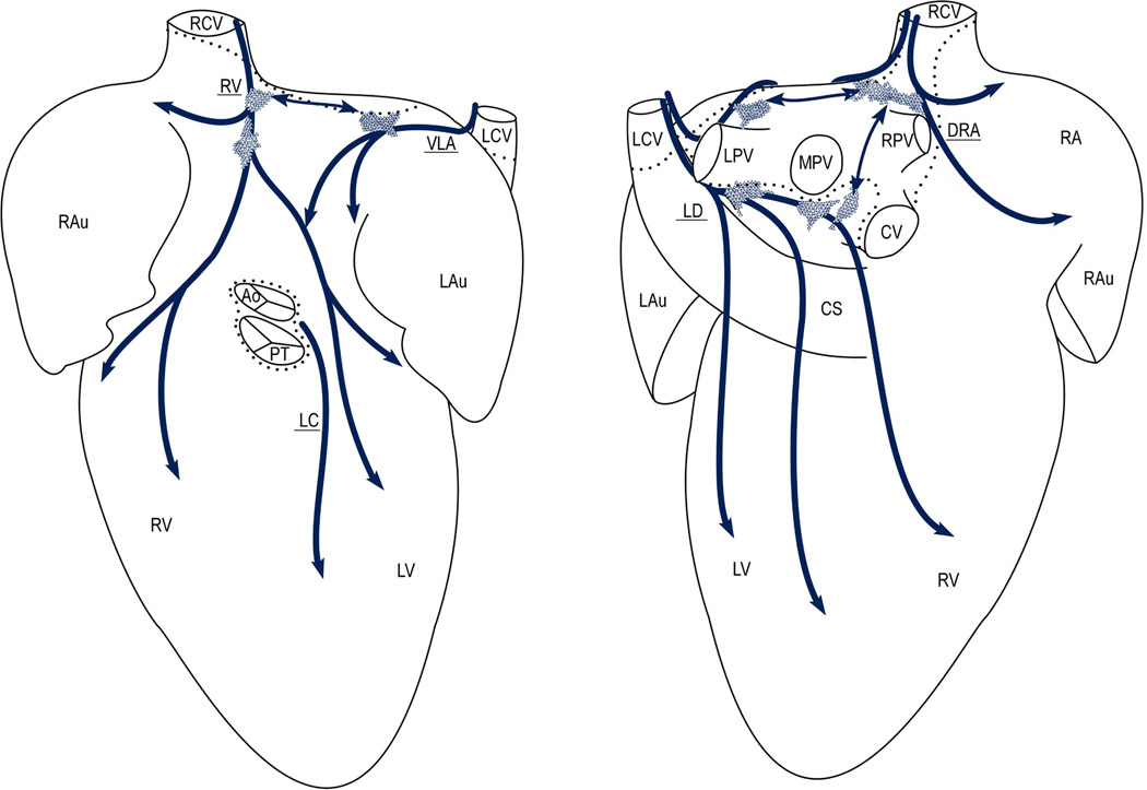
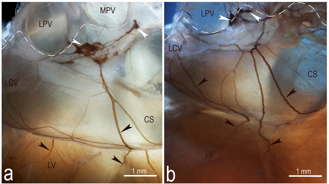
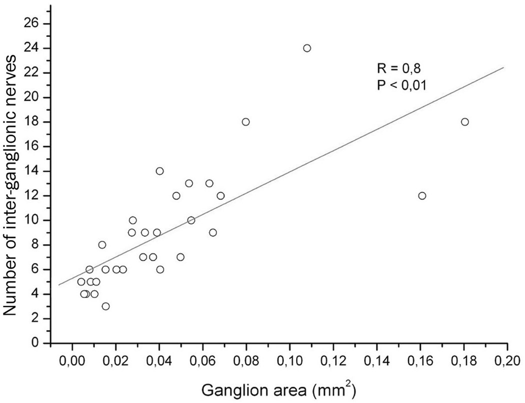
Results: Double immunohistochemical labeling of TH and ChAT on AChE-positive neural elements in mouse whole mounts demonstrated equal staining of nerves and ganglia for AChE that were positive for both TH and ChAT. The extrinsic cardiac nerves access the mouse heart at the right and left cranial veins and interblend within the ganglionated nerve plexus of the heart hilum that is persistently localized on the heart base. Nerves and bundles of nerve fibers extend epicardially from this plexus to atria and ventricles by left dorsal, dorsal right atrial, right ventral, and ventral left atrial routes or subplexuses. The right cranial vein receives extrinsic nerves that mainly originate from the right cervicothoracic ganglion and a branch of the right vagus nerve, whereas the left cranial vein is supplied by extrinsic nerves from the left cervicothoracic ganglion and the left vagus nerve. The majority of intrinsic cardiac ganglia are localized on the heart base at the roots of the pulmonary veins. These ganglia are interlinked by interganglionic nerves into the above mentioned nerve plexus of the heart hilum. In general, the examined hearts contained 19 ± 3 ganglia, giving a cumulative ganglion area of 0.4 ± 0.1 mm(2).

Conclusion: Despite substantial anatomic differences in ganglion number and distribution, the structural organization of the intrinsic ganglionated plexus in the mouse heart corresponds in general to that of other mammalian species, including human.


Conflict of interest statement

No Conflicts

Figures

Fig 1
Fig 1
Microphotograph demonstrating the presence of AChE within intrinsic cardiac nerves composed predominantly by TH-positive (adrenergic) and a few of ChAT-positive (cholinergic) nerve fibers. The image was taken from a whole mount heart preparation that was immunohistochemically double labeled for ChAT (a) and TH (b) and subsequently stained histochemically for AChE (c). Note the sharp contrast of the AChE staining compared with the ChAT labeling. Boxed areas within images were enlarged as the right upper insets to demonstrate the appearance of cholinergic and adrenergic nerve fibers within AChE positive nerve of mouse heart.
Fig 2
Fig 2
Macrophotographs to illustrate the location and morphologic pattern of the right ventral (a, d), ventral left atrial (a) and left coronary (d) nerve subplexuses in a mouse heart stained histochemically for AChE. Boxed areas in a and d were enlarged employing a contact microscope and shown as b and c insets. Note the right ganglia situated at the root of right cranial vein and the left ganglia on the cranial-dorsal aspect of left atrium. Black arrowheads, some cardiac nerves; white arrowheads, ganglia. White solid arrows, nerves accessing the heart hilum and originating the right ventral (RV) and left coronary (LC) neural subplexuses; black thin arrows, interganglionic nerves, white thin arrows, some neurons located inside ganglia. Dashed line shows the limits of heart hilum. Abbreviations: Ao – aorta; CA – conus arteriosus; IS – ventral interatrial groove; LAu – left auricle; LCV – left cranial vein; MPV – middle pulmonary vein; PT – pulmonary trunk; RAu – right auricle; RCV – right cranial vein; SAN – sinuatrial nodal zone; neural subplexuses: RV – right ventral; LC – left coronary, VLA – ventral left atrial.
Fig 3
Fig 3
Location, course and structure of left dorsal (LD) and dorsal right atrial (DRA) neural subplexuses in a mouse heart stained histochemically for acetylcholinesterase. Boxed areas b and c in a, e in d and f in e were enlarged using a contact microscope and shown respectively as b, c, e, and f insets. Black arrowheads in a point to the nerves that penetrate into interatrial septum, while in d one that proceeds toward the region of the sinuatrial node (SAN). White arrowheads, some ganglia; white solid arrows, nerves entering the ganglionated nerve plexus of the heart hilum; Black thin arrows, interganglionic nerves, white thin arrows, some neurons. Dashed line limits the heart hilum. In d, note the right ganglia situated at the root of right cranial vein on the interatrial groove. Other abbreviations: CS – coronary sinus; CV – orifice of caudal (inferior caval) vein; IS – interatrial groove; LAu – inferior surface of left auricle; LCV – left cranial vein; LPV – orifice of left pulmonary vein; LV – left ventricle; RA – right atrial wall; MPV – orifice of middle pulmonary vein; RPV – orifice of right pulmonary vein; RCV – root of right cranial (superior caval) vein.
Fig 4
Fig 4
Diagram summarizing the distribution and morphology of distinct intrinsic ganglionated nerve subplexuses in 15 mouse hearts as they were seen from the ventral (left) and dorsal (right) sides on a pressure-inflated heart stained histochemically for AChE. Dotted lines limit the heart hilum; thick arched arrows show the course of neural subplexuses; polygonal areas show the main locations of intrinsic ganglia in mouse heart. Note the left ganglia at the root of the left pulmonary vein and the right ganglia distributed above interatrial septum at the roots of right cranial (RCV) and middle pulmonary (MPV) veins. Abbreviations: Ao – aorta; PT – pulmonary trunk; CS – coronary sinus; veins: CV – caudal (inferior caval), LCV – left cranial (left azygos), RCV – right cranial (superior caval), LPV – left pulmonary, RPV – right pulmonary, and MPV – middle pulmonary; LAu – left auricle; LV – left ventricle; RA – right atrium; RAu – right auricle; RV – right ventricle; neural subplexuses: VLA – ventral left atrial, DRA – dorsal right atrial, LC – left coronary, LD – left dorsal, and RV – right ventral.
Fig 5
Fig 5
Macrophotographs illustrating the structural variability of the left dorsal neural subplexus in two mouse hearts stained histochemically for AChE. White arrowheads, some ganglia, black arrowheads, topographically comparable nerves at coronary sinus. Dashed lines demarcate the limits of heart hilum. Abbreviations: CS – coronary sinus; LCV – left cranial (left azygos) vein; LPV – left pulmonary vein; LV – left ventricle; MPV – middle pulmonary vein. Note the persistent location of ganglia specified by white arrowheads.
Fig 6
Fig 6
The number of interganglionic nerves plotted against areas of 31 ganglia that were derived from ten mouse hearts. The straight line indicates the linear regression of the plotted data. Each point corresponds to one of the analyzed ganglion. R, correlation coefficient at P < 0,05.

Similar articles

Cited by

References

    1. Gorman MW, Tune JD, Richmond KN, Feigl EO. Quantitative analysis of feedforward sympathetic coronary vasodilation in exercising dogs. J Appl Physiol. 2000;89:1903–1911. - PubMed
    1. Tsuboi M, Furukawa Y, Nakajima K, Kurogouchi F, Chiba S. Inotropic, chronotropic, and dromotropic effects mediated via parasympathetic ganglia in the dog heart. Am J Physiol Heart Circ Physiol. 2000;279:H1201–H1207. - PubMed
    1. Cifelli C, Rose RA, Zhang H, et al. RGS4 regulates parasympathetic signaling and heart rate control in the sinoatrial node. Circ Res. 2008;103:527–535. - PubMed
    1. Gatti PJ, Johnson TA, Phan P, Jordan IK, 3rd, Coleman W, Massari VJ. The physiological and anatomical demonstration of functionally selective parasympathetic ganglia located in discrete fat pads on the feline myocardium. J Auton Nerv Syst. 1995;51:255–259. - PubMed
    1. Armour JA. Potential clinical relevance of the 'little brain' on the mammalian heart. Exp Physiol. 2008;93:165–176. - PubMed

Publication types

Substances