Comment
. 2010 Nov 16;18(5):411-22.
doi: 10.1016/j.ccr.2010.10.024.

A stapled p53 helix overcomes HDMX-mediated suppression of p53

Affiliations
Comment

A stapled p53 helix overcomes HDMX-mediated suppression of p53

Federico Bernal et al. Cancer Cell. .

Abstract

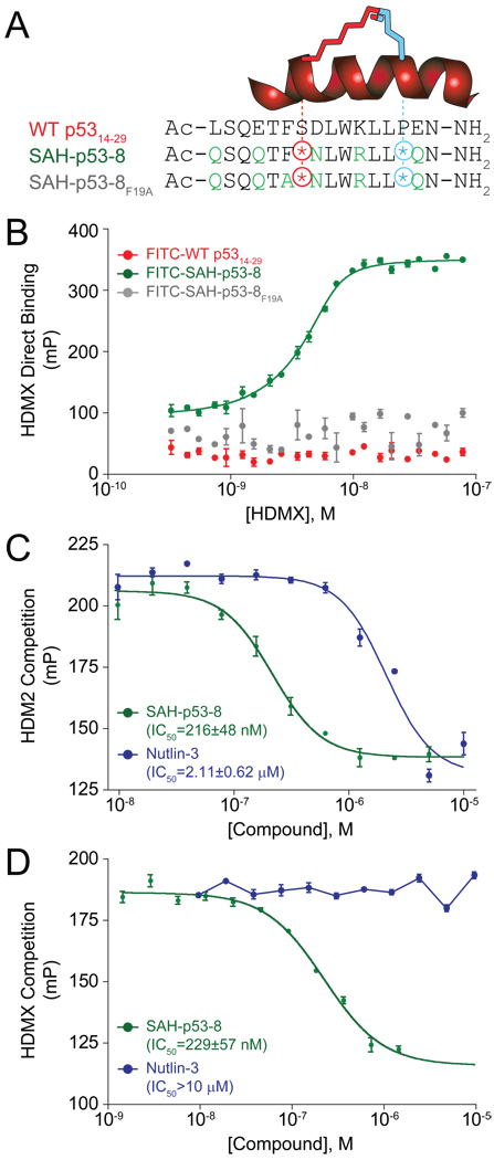
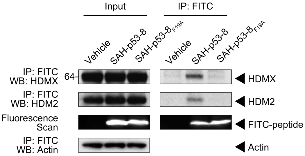
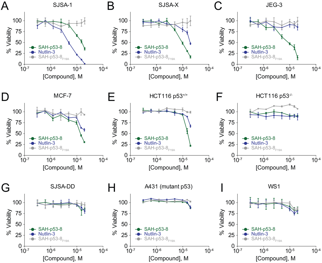
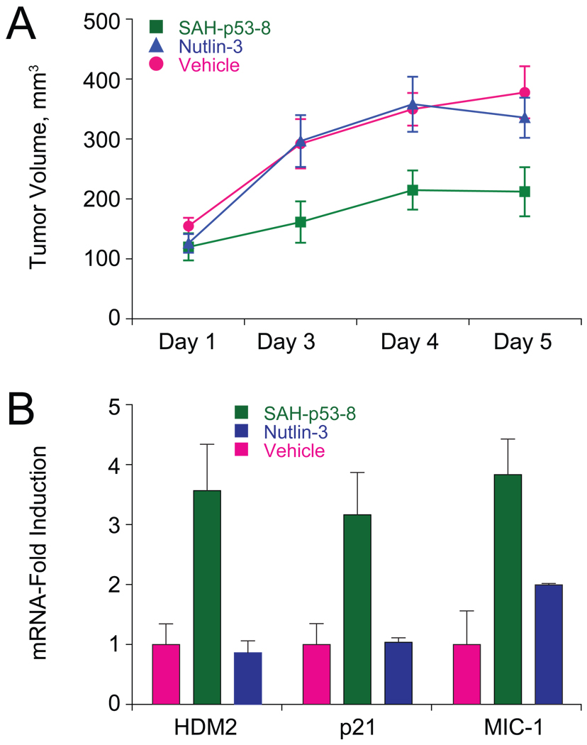
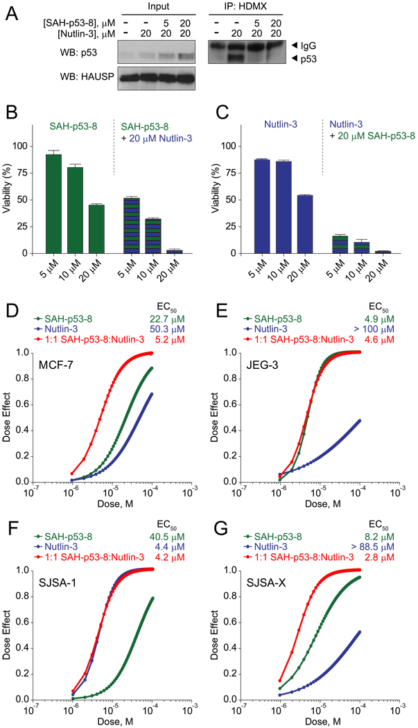
Cancer cells neutralize p53 by deletion, mutation, proteasomal degradation, or sequestration to achieve a pathologic survival advantage. Targeting the E3 ubiquitin ligase HDM2 can lead to a therapeutic surge in p53 levels. However, the efficacy of HDM2 inhibition can be compromised by overexpression of HDMX, an HDM2 homolog that binds and sequesters p53. Here, we report that a stapled p53 helix preferentially targets HDMX, blocks the formation of inhibitory p53-HDMX complexes, induces p53-dependent transcriptional upregulation, and thereby overcomes HDMX-mediated cancer resistance in vitro and in vivo. Importantly, our analysis of p53 interaction dynamics provides a blueprint for reactivating the p53 pathway in cancer by matching HDM2, HDMX, or dual inhibitors to the appropriate cellular context.


Figures

Figure 1
Figure 1. Primary sequence and HDM2/HDMX binding activity of SAH-p53-8
(A) Composition of wild-type p5314–29, SAH-p53-8, and SAH-p53-8F19A peptides. (B) Direct binding of FITC-peptides to recombinant HDMX as measured by fluorescence polarization. Competition of SAH-p53-8 and Nutlin-3 with FITC-SAH-p53-8 for binding to HDM2 (C) and HDMX (D). mP: units of milli-polarization. Data are mean +/− s.e.m. for experiments performed in at least triplicate.
Figure 2
Figure 2. SAH-p53-8 targets both HDM2 and HDMX in situ
SJSA-X cells were treated for 12 hours with either vehicle, FITC-SAH-p53-8 (15 µM), or FITC-SAH-p53-8F19A (15 µM). Anti-FITC immunoprecipitates from cellular extracts were subjected to HDM2 and HDMX western analyses, with β-actin used as a loading control. The presence of FITC-labeled peptide in the extracts was detected by a fluorescence scan of the immunoblot.
Figure 3
Figure 3. Viability of cancer cells exposed to HDM2/HDMX inhibitors
Cancer cell lines with differential expression levels of HDM2 and HDMX (A–E), deficient or dysfunctional p53 (F–H), and a non-tumor cell control (I) were treated with 0.3–20 µM Nutlin-3, SAH-p53-8, or SAH-p53-8F19A for 24 h. The cells were exposed to CellTiter-Glo™ reagent (Promega) and viability was assessed by ATP-induced chemiluminescence. Data are mean +/− s.e.m. for experiments performed in at least triplicate.
Figure 4
Figure 4. SAH-p53-8 blocks the formation of p53-HDMX complexes
(A) JEG-3 choriocarcinoma cells were exposed to vehicle, 20 µM SAH-p53-8, or 20 µM Nutlin-3 for 6 h. Cellular extracts and anti-HDMX immunoprecipitates were subjected to SDS-PAGE and analyzed by western blotting for p53, HDMX, and HAUSP. (B) JEG-3 cells were treated with 0–20 µM SAH-p53-8 in the presence of 10 µM MG-132 for 6 h. Cellular extracts and anti-HDMX immunoprecipitates were analyzed by western blotting for p53, HDMX, and HAUSP. (C) Doxycycline-inducible U2OS cells were treated with or without doxycycline in the presence of Nutlin-3 (10 µM), SAH-p53-8 (10 µM), or both, and induction of HA-HDMX and p53 was detected and quantitated by immunofluorescence. Scale bar, 10 µm (D) Cultured U2OS cells were treated with or without doxycycline in the presence of Nutlin-3, SAH-p53-8, or both and then processed for P-LISA. Scale bar, 10 µm (E) Quantitation of p53-HDMX complexes as detected by P-LISA. Data are mean +/− s.d. *p=0.0003, **p=0.0001, unpaired t-test with Welch’s correction.
Figure 5
Figure 5. Reactivation of p53-dependent Transcription and Apoptosis by SAH-p53-8
(A) JEG-3 cells were treated with vehicle, SAH-p53-8 (20 µM), or Nutlin-3 (20 µM) and transcriptional upregulation of HDM2, p21, and MIC-1 was evaluated by qPCR analysis. (B) JEG-3 cells were treated with vehicle, or 0.3–10 µM SAH-p53-8 or Nutlin-3 for 12 hours, followed by exposure to Caspase-3/7-Glo™ reagent (Promega). Caspase-3/7 activation was assessed by monitoring the cleavage of proluminescent caspase-3/7 substrate. Data are mean +/− s.e.m. for experiments performed in at least triplicate.
Figure 6
Figure 6. SAH-p53-8 Overcomes HDMX-mediated p53 Suppression and Blocks Tumor Growth in Vivo
(A) Cohorts (n=7) of JEG-3 xenograft mice were treated with vehicle (5% DMSO in D5W) or 10 mg kg−1 of SAH-p53-8 or Nutlin-3 by intravenous injection daily for 4 days and tumor volume was monitored by caliper measurement on days 1, 3, 4 and 5. Data are mean +/− s.d. (D3: SAH/Veh, p=0.032; SAH/Nut, p=0.032, Nut/Veh, p=0.94; D4: SAH/Veh, p=0.008; SAH/Nut, p=0.026, Nut/Veh, p=0.88; D5: SAH/Veh, p=0.017; SAH/Nut, p=0.037, Nut/Veh, p=0.46). (B) RNA was prepared from the excised JEG-3 tumors and qPCR analysis was performed to measure levels of the p53 transcriptional targets HDM2, p21, and MIC-1. Data are mean +/− s.e.m.
Figure 7
Figure 7. Pharmacologic Induction of p53-HDMX Complexes Sensitizes Nutlin-3-Resistant Cancer Cells to HDMX Inhibition
(A) MCF-7 breast adenocarcinoma cells were exposed to vehicle, Nutlin-3 (20 µM), or Nutlin-3 in combination with 5 or 20 µM SAH-p53-8 in the presence of 10 µM MG-132 for 6 h. Cellular extracts and anti-HDMX immunoprecipitates were subjected to SDS-PAGE and analyzed by western blotting for p53, HDMX, and HAUSP. For synergy studies, MCF-7 cells were treated with 5–20 µM SAH-p53-8 with or without 20 µM Nutlin-3 (B) or 5–20 µM Nutlin-3 with or without 20 µM SAH-p53-8 (C), and cell viability measured at 24 hours by CellTiter-Glo™ assay. Data are mean +/− s.e.m. for experiments performed in at least triplicate. (D–G) Dose-effect synergy analyses of MCF-7 (D), JEG-3 (E), SJSA-1 (F), and SJSA-X (G) cells treated with 0.5–20 µM SAH-p53-8, Nutlin-3, or an equimolar combination.
Figure 8
Figure 8. Molecular Determinants of Cancer Cell Susceptibility to HDM2, HDMX, and Dual Inhibition
In the context of HDM2-driven p53 suppression, HDM2 inhibition triggers a surge in p53 levels and induces cancer cell death (A). However, if HDMX is present, p53 can become sequestered in p53-HDMX complexes, limiting the cellular response to HDM2 inhibition (B). When p53-HDMX complex levels are elevated, HDMX inhibition reactivates the p53 pathway and induces cell death (C). However, if cellular levels of p53 are low, targeting HDMX has little to no effect on cell viability (D). When p53 levels are suppressed by HDM2, and HDMX is also expressed, maximal reactivation of the p53 pathway is achieved by combined blockade of HDM2 and HDMX, which elevates p53 levels and blocks formation of inhibitory p53-HDMX complexes, respectively (E). Thus, cancer cell susceptibility to pharmacologic inhibition of HDM2, HDMX, or both targets is determined by the respective levels and interactions of p53, HDM2, and HDMX.

Comment on

References

    1. Baker SJ, Fearon ER, Nigro JM, Hamilton SR, Preisinger AC, Jessup JM, vanTuinen P, Ledbetter DH, Barker DF, Nakamura Y, et al. Chromosome 17 deletions and p53 gene mutations in colorectal carcinomas. Science. 1989;244:217–221. - PubMed
    1. Bernal F, Tyler AF, Korsmeyer SJ, Walensky LD, Verdine GL. Reactivation of the p53 tumor suppressor pathway by a stapled p53 peptide. J Am Chem Soc. 2007;129:2456–2457. - PMC - PubMed
    1. Bird GH, Bernal F, Pitter K, Walensky LD. Synthesis and biophysical characterization of stabilized alpha-helices of BCL-2 domains. Methods Enzymol. 2008;446:369–386. - PMC - PubMed
    1. Bird GH, Madani N, Perry AF, Princiotto AM, Supko JG, He X, Gavathiotis E, Sodroski JG, Walensky LD. Hydrocarbon double-stapling remedies the proteolytic instability of a lengthy peptide therapeutic. Proc Natl Acad Sci U S A. 2010;107:14093–14098. - PMC - PubMed
    1. Böttger A, Böttger V, Garcia-Echeverria C, Chène P, Hochkeppel HK, Sampson W, Ang K, Howard SF, Picksley SM, Lane DP. Molecular characterization of the hdm2-p53 interaction. J Mol Biol. 1997;269:744–756. - PubMed

Publication types

MeSH terms