. 2010 Nov 16;18(5):448-58.
doi: 10.1016/j.ccr.2010.10.020.

Oncogenic KRas suppresses inflammation-associated senescence of pancreatic ductal cells

Affiliations

Oncogenic KRas suppresses inflammation-associated senescence of pancreatic ductal cells

Kyoung Eun Lee et al. Cancer Cell. .

Abstract

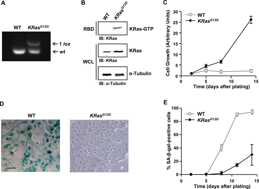
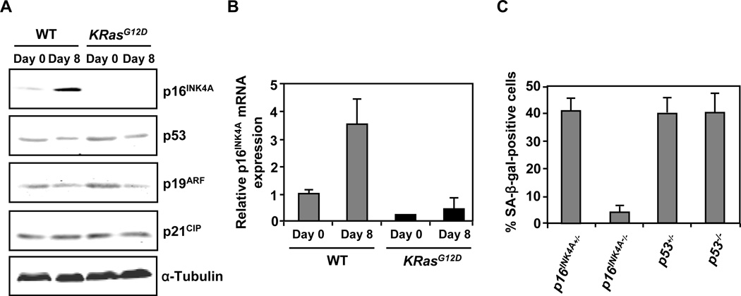
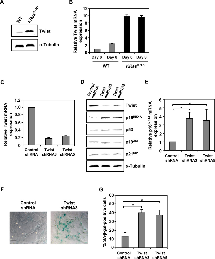
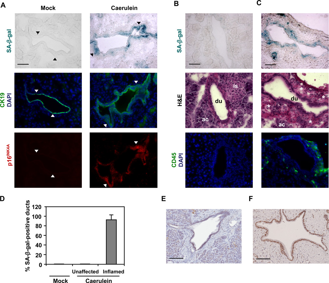
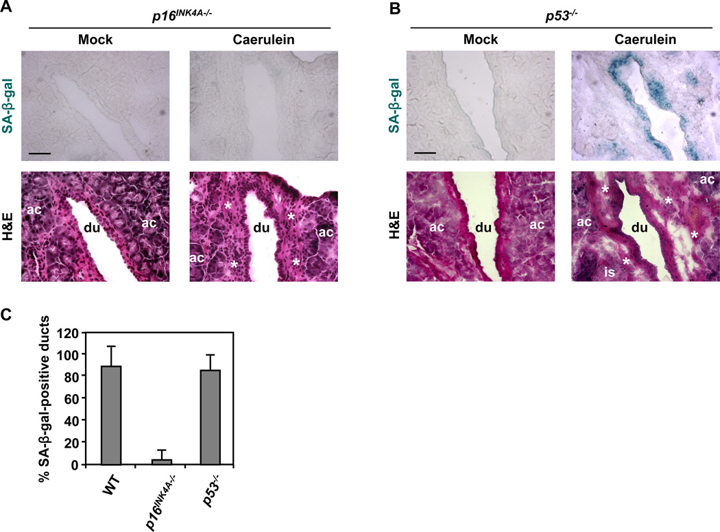
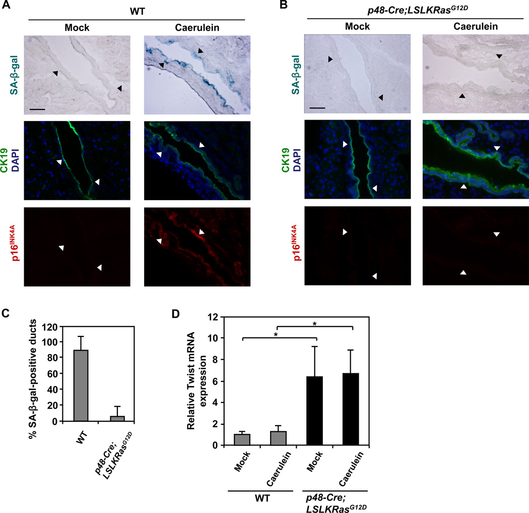
Mutational activation of KRas is the first and most frequently detected genetic lesion in pancreatic ductal adenocarcinoma (PDAC). However, the precise role of oncogenic KRas in the pathogenesis of PDAC is not fully understood. Here, we report that the endogenous expression of oncogenic KRas suppresses premature senescence in primary pancreatic duct epithelial cells (PDEC). Oncogenic KRas-mediated senescence bypass is conferred by the upregulation of the basic helix-loop-helix transcription factor Twist that in turn abrogates p16(INK4A) induction. Moreover, the KRas-Twist-p16(INK4A) senescence bypass pathway is employed in vivo to prevent inflammation-associated senescence of pancreatic ductal epithelium. Our findings indicate that oncogenic KRas could contribute to PDAC initiation by protecting cells from entering a state of permanent growth arrest.


Figures

Figure 1
Figure 1. Oncogenic KRas protects PDEC from undergoing premature senescence
(A) PCR analysis of genomic DNA prepared from LSL-KRasG12D PDEC infected with adenoviral-GFP (WT) or adenoviral-Cre (KRasG12D). The excision-recombination event of the LSL cassette leaves behind a single LoxP (1 lox) site. (B) Measurement of Ras activation in WT and KRasG12D PDEC by GST-RBD pull-down assay. α-Tubulin serves as a loading control. IB, immunoblot; WCL, whole cell lysates. (C) Growth analysis of WT and KRasG12D PDEC. The number of DAPI-stained nuclei was counted in 9 random fields of view (FOV) each containing at least 50 cells at day 2, 5, 8, and 14. The average number of nuclei present in the FOV at each time point was then normalized to the average number of nuclei per FOV at day 2. Error bars indicate standard deviation (SD). Data are representative of five independent experiments. (D) SA-β-gal staining of WT and KRasG12D PDEC cultures at day 8. Scale bar: 100 µm. (E) Quantification of SA-β-gal staining in WT and KRasG12D PDEC cultures at day 2, 5, 8, 11, and 14. Cells were counterstained with Hoechst 33342 for β-gal quantification. Error bars indicate SD (n = 6 FOV). Data are representative of five independent experiments. See also Figure S1.
Figure 2
Figure 2. Oncogenic KRas confers bypass of premature senescence in PDEC through the suppression of p16INK4A induction
(A) Western blot analysis for senescence effectors in WT and KRasG12D PDEC cultures at day 0 and day 8. Equal loading was verified with anti-α-Tubulin. (B) Quantitative RT-PCR analysis of p16INK4A in WT and KRasG12D PDEC cultures at day 0 and day 8. Error bars indicate SD (n = 3). (C) Quantification of SA-β-gal staining in p16INK4A+/−, p16INK4A−/−, p53+/−, p53−/− PDEC cultures at day 8. Error bars indicate SD (n = 6 FOV). Data are representative of three independent experiments.
Figure 3
Figure 3. Oncogenic KRas-mediated bypass of premature senescence in PDEC is dependent on Twist
(A) Western blot analysis for Twist in WT and KRasG12D PDEC cultures at day 8. α-Tubulin serves as a loading control. (B) Quantitative RT-PCR analysis of Twist in WT and KRasG12D PDEC cultures at day 0 and day 8. Error bars indicate SD (n = 3). (C) Quantitative RT-PCR analysis of Twist in KRasG12D PDEC cultures 8 days after infection with recombinant lentiviruses encoding shRNA targeted against GFP (control shRNA) or Twist (Twist shRNA3 or Twist shRNA5). Error bars indicate SD (n = 3). (D) Western blot analysis for Twist and senescence effectors in KRasG12D PDEC cultures 8 days after infection with recombinant lentiviruses encoding control shRNA, Twist shRNA3, or Twist shRNA5. Equal loading was verified with anti-α-Tubulin. (E) Quantitative RT-PCR analysis of p16INK4A in KRasG12D PDEC cultures 8 days after infection with recombinant lentiviruses encoding control shRNA, Twist shRNA3, or Twist shRNA5. Error bars indicate SD (n = 3). *, p-value < 0.05. (F) SA-β-gal staining of KRasG12D PDEC cultures 8 days after infection with recombinant lentiviruses encoding control shRNA or Twist shRNA3. Scale bar: 100 µm. (G) Quantification of SA-β-gal staining in KRasG12D PDEC cultures 8 days after infection with recombinant lentiviruses encoding control shRNA, Twist shRNA3, or Twist shRNA5. Error bars indicate SD (n = 6 FOV). *, p-value < 0.05. Data are representative of three independent experiments. See also Figure S2.
Figure 4
Figure 4. Inflammatory insult triggers premature senescence in pancreatic ductal epithelium in vivo
(A–D) Caerulein was administered to 1-month-old mice as 8 hourly intraperitoneal injections (50 ng/g of body weight/injection) for two days. Three days after the last caerulein injection, pancreata were harvested. At least 3 sections were analyzed per animal (n = 5 per genotype per treatment). (A) SA-β-gal staining and immunofluorescence staining for p16INK4A and CK19 on consecutive sections of pancreata from WT mice treated with caerulein or saline (mock). CK19 was used to identify ductal cells. Nuclei were counterstained with DAPI. Arrowheads mark corresponding areas. (B and C) SA-β-gal staining, hematoxylin and eosin (H&E) staining, and immunofluorescence staining for CD45 on consecutive sections of pancreata from WT mice treated with caerulein. Pancreatic ducts in unaffected (B) or inflamed (C) areas are from the same tissue section. CD45 was used to identify leukocytes. Nuclei were counterstained with DAPI. ac, acinus; du, duct; is, islet; asterisk, immune infiltrate. (D) Quantification of SA-β-gal staining in pancreata from WT mice treated with caerulein or saline (mock). Pancreatic ducts bearing 10% or more β-gal-positive cells in each duct were scored as positive. At least four pancreatic ducts were scored in each mouse. Error bars indicate SD (n = 5). (E and F) Immunohistochemical analysis of p16INK4A in human chronic pancreatitis tissue samples. Representative areas of normal pancreatic (E) and pancreatitis (F) tissues are shown. Scale bars: 50 µm (A, B, and C), 100 µm (E and F). See also Figure S3.
Figure 5
Figure 5. Inflammation-induced premature senescence in pancreatic ductal epithelium depends on p16INK4A upregulation
Caerulein was administered as described in Figure 4. At least 3 sections were analyzed per animal (n = 5 per genotype per treatment). (A and B) SA-β-gal staining and H&E staining on consecutive sections of pancreata from p16INK4A−/− (A) and p53−/− mice (B) treated with caerulein or saline (mock). Ac, acinus; du, duct; is, islet; asterisk, immune infiltrate. Scale bars: 50 µm. (C) Quantification of SA-β-gal staining in pancreata from WT, p16INK4A−/−, and p53−/− mice treated with caerulein. Only pancreatic ducts in inflamed areas were counted. At least four pancreatic ducts were scored in each mouse. Error bars indicate SD (n = 5). See also Figure S4.
Figure 6
Figure 6. Inflammation-associated premature senescence of pancreatic ductal epithelium in vivo can be blocked by oncogenic KRas through the suppression of p16INK4A
Caerulein was administered as described in Figure 4. At least 3 sections were analyzed per animal (n = 5 per genotype per treatment). (A and B) SA-β-gal staining and immunofluorescence staining for p16INK4A and CK19 on consecutive sections of pancreata from WT (A) and p48-Cre;LSL-KRasG12D (B) mice treated with caerulein or saline (mock). CK19 was used to identify ductal cells. Nuclei were counterstained with DAPI. Arrowheads mark corresponding areas. Scale bars: 50 µm. (C) Quantification of SA-β-gal staining in pancreata from WT and p48-Cre;LSL-KRasG12D mice treated with caerulein. Only pancreatic ducts in inflamed areas were counted. At least four pancreatic ducts were scored in each mouse. Error bars indicate SD (n = 5). (D) Quantitative RT-PCR analysis of Twist in pancreata from WT and p48-Cre;LSL-KRasG12D mice treated with caerulein or saline (mock). Error bars indicate SD (n = 5 per genotype). *, p-value < 0.05. See also Figure S5.

References

    1. Acosta JC, O'Loghlen A, Banito A, Guijarro MV, Augert A, Raguz S, Fumagalli M, Da Costa M, Brown C, Popov N, et al. Chemokine signaling via the CXCR2 receptor reinforces senescence. Cell. 2008;133:1006–1018. - PubMed
    1. Agbunag C, Bar-Sagi D. Oncogenic K-ras drives cell cycle progression and phenotypic conversion of primary pancreatic duct epithelial cells. Cancer Res. 2004;64:5659–5663. - PubMed
    1. Agbunag C, Lee KE, Buontempo S, Bar-Sagi D. Pancreatic duct epithelial cell isolation and cultivation in two-dimensional and three-dimensional culture systems. Methods Enzymol. 2006;407:703–710. - PubMed
    1. Almoguera C, Shibata D, Forrester K, Martin J, Arnheim N, Perucho M. Most human carcinomas of the exocrine pancreas contain mutant c-K-ras genes. Cell. 1988;53:549–554. - PubMed
    1. Ansieau S, Bastid J, Doreau A, Morel AP, Bouchet BP, Thomas C, Fauvet F, Puisieux I, Doglioni C, Piccinin S, et al. Induction of EMT by twist proteins as a collateral effect of tumor-promoting inactivation of premature senescence. Cancer Cell. 2008;14:79–89. - PubMed

Publication types

MeSH terms