. 2010 Nov 18;8(5):445-54.
doi: 10.1016/j.chom.2010.10.005.

Statins enhance formation of phagocyte extracellular traps

Affiliations

Statins enhance formation of phagocyte extracellular traps

Ohn A Chow et al. Cell Host Microbe. .

Abstract

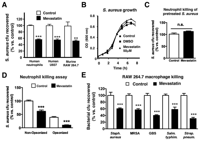
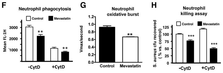
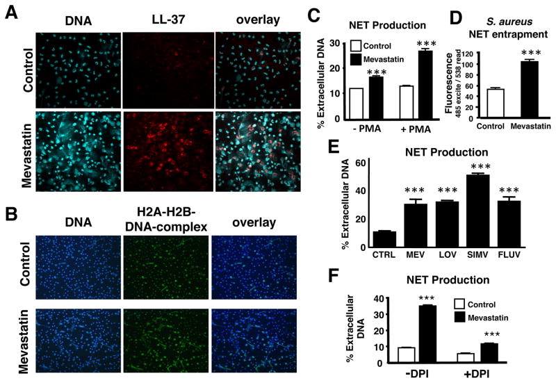
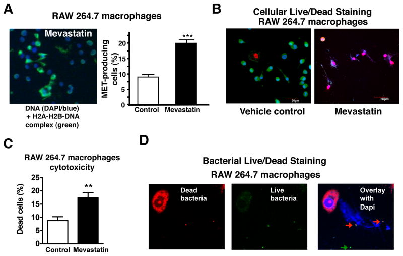
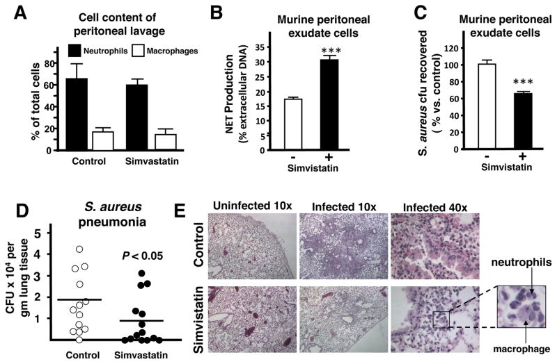
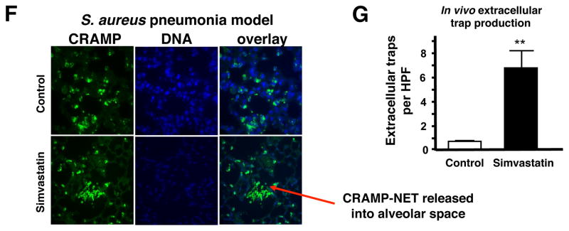
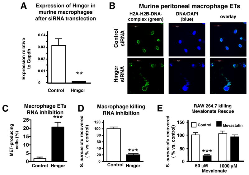
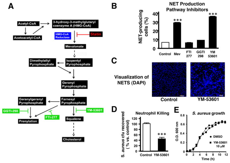
Statins are inhibitors of 3-hydroxy 3-methylglutaryl coenzyme A (HMG-CoA) reductase, the rate-limiting enzyme in cholesterol biosynthesis. Recent clinico-epidemiologic studies correlate patients receiving statin therapy with having reduced mortality associated with severe bacterial infection. Investigating the effect of statins on the innate immune capacity of phagocytic cells against the human pathogen Staphylococcus aureus, we uncovered a beneficial effect of statins on bacterial clearance by phagocytes, although, paradoxically, both phagocytosis and oxidative burst were inhibited. Probing instead for an extracellular mechanism of killing, we found that statins boosted the production of antibacterial DNA-based extracellular traps (ETs) by human and murine neutrophils and also monocytes/macrophages. The effect of statins to induce phagocyte ETs was linked to sterol pathway inhibition. We conclude that a drug therapy taken chronically by millions alters the functional behavior of phagocytic cells, which could have ramifications for susceptibility and response to bacterial infections in these patients.


Figures

Figure 1
Figure 1. Statin Induction of Phagocyte Antimicrobial Activity In Vitro
(A) In vitro killing of S. aureus by primary human neutrophils or RAW 264.7 and human U937 cells treated with mevastatin or vehicle control. (B). Growth of S. aureus strain Newman in RPMI with or without mevastatin (50 μM) or DMSO vehicle control. (C) In vitro killing of S. aureus pretreated with mevastatin or vehicle control by primary human neutrophils. (D) In vitro killing of opsonized versus non-opsonized S. aureus by primary human neutrophils treated with mevastatin or vehicle control. (E) In vitro killing of S. aureus strain Newman, S. aureus strain Sanger (MRSA), S. agalactiae strain COH1 (GBS), S. typhimurium and Streptococcus pneumoniae strain D39 by RAW 264.7 cells treated with mevastatin or vehicle control. (F) Mean fluorescence intensity as parameter for phagocytosis of neutrophils after infection with FITC-labelled S. aureus Wood strain bioparticles measured by flow cytometry. As a control, 10 μg/ml of cytochalasin D was added to the samples 10 min prior to infection to prevent phagocytosis. (G) Oxidative burst of primary human neutrophils stimulated with mevastatin or vehicle control measured by flow cytometry after 30 min incubation in the presence of 2′,7′ dichlorofluorescein. (H) Extracellular killing of S. aureus by primary human neutrophils treated with mevastatin or vehicle control. To prevent phagocytosis, 10 μg/ml of cytochalasin D was added to the samples 10 min prior to infection. Experiments performed 3–4 times with similar results, representative experiment shown ± standard deviation. ** P < 0.01, *** P < 0.005, n.s. = not significant by two-tailed Student’s t-test comparing control versus statin-treated group.
Figure 2
Figure 2. Statin Induction of Neutrophil Extracellular Traps (NETs)
(A) Representative fluorescent images of neutrophils stimulated with mevastatin or vehicle control and PMA to induce NETs. NET-formation was visualized in blue (Dapi) and LL-37 expression was visualized by Alexa red-immunostaining. (B) NET-formation was visualized in blue (Dapi) and H2A-H2B-DNA-complexes were visualized by Alexa green-immunostaining. (C) Quantification of NET-production by primary human neutrophils ± stimulation with PMA and treatment with mevastatin or vehicle control. (D) Entrapment of fluorescently labeled S. aureus by PMA-stimulated human neutrophils treated with mevastatin or vehicle control. In (C-D) *** P < 0.005 by two-tailed Student’s t-test comparing control versus statin-treated group. (E) Quantification of NET-production by primary human neutrophils ± stimulation with PMA and treatment with different statins (MEV = mevastatin, LOV = lovastatin, SIMV = simvastatin, FLUV = fluvastatin) or vehicle control (CTRL). *** P < 0.005 by one-way ANOVA with Dunnet’s post-test vs. control group. (F) NET production of PMA-stimulated human neutrophils treated with 10 μg/ml DPI or vehicle control to inhibit the NADPH-oxidase-dependent ROS production. *** P < 0.005 or n.s. = not significant by two-tailed Student’s t-test comparing control versus statin-treated group. Experiments performed 3–4 times with similar results, representative experiment shown ± standard deviation. (See also the Supplemental Figures S1 and S2)
Figure 3
Figure 3. Statin-Induced Formation of Macrophage Extracellular Traps (METs)
(A) MET formation by RAW 264.7 cells. Representative fluorescent image of MET formation visualized using blue (Dapi) and Alexa green-immunostaining for H2A-H2B-DNA-complexes; quantification of MET production by RAW 264.7 cells after overnight treatment with mevastatin or vehicle control and subsequent stimulation with PMA. Experiment was performed at 3 independent occasions with similar results, representative experiment shown ± standard deviation. *** P < 0.005 by t-test; (B) Live/Dead viability/cytotoxicity staining to determine viability of MET-producing cells or (C) Live/Dead BacLight Bacterial Viability staining to determine viability of FITC-labelled S. aureus entrapped in the METs produced by RAW cells after overnight treatment with mevastatin and subsequent stimulation with PMA. Note that all trap-forming macrophages as well as entrapped bacteria are dead as shown by the red dye (arrows); non-entrapped bacteria are not stained by the red dye (green arrow) indicating that they are alive. (See also the Supplemental Figures S3 and S4).
Figure 4
Figure 4. Statin Therapy in Mice Boosts Extracellular Trap Formation Ex Vivo and In Vivo
(A) Cell content (percentage of total cells) of thioglycolate-induced peritoneal cells extracted from mice fed with standard chow or standard chow + simvastatin. (B) Quantification of NET production by peritoneal cells. (C) Ex vivo killing of S. aureus strain Newman by thioglycolate-induced peritoneal cells extracted from mice fed with standard chow or standard chow + simvastatin. In (B–C): *** P < 0.005 by two-tailed Student’st-test comparing control versus statin-treated group. Experiments performed 3–4 times with similar results, representative experiment shown ± standard deviation. (D) Recovered bacteria from lungs of mice pre-fed for with standard chow or standard chow supplemented with simvastatin and infected intranasally with 2 × 10⁸ CFU S. aureus strain Newman for 48 h. Data shown are pooled from 2 independent experiments with each n = 10 or n = 7 mice, respectively. * P < 0.05 by two-tailed Mann Whitney test. (E) Representative light micrograph (HE-stained) of lung tissue sections of infected mice pre-fed for with standard chow (control, upper panel) or standard chow supplemented with simvastatin (lower panel). (F) Representative fluorescent images of extracellular trap formation (visualized by Alexa 488 (green)-labelled CRAMP production and counterstained with Dapi) in paraffin-embedded lung sections of mice pre-fed for with standard chow or standard chow supplemented with simvastatin and intranasally infected with 2 × 10⁸ cfu of S. aureus strain Newman for 48 h. (G) Quantification of in vivo extracellular trap production visualized in (F). Data are shown as average of 6 high-power field (HPF) obtained with a 10x/0.3 UPlanFCN objective. ** P < 0.01 by two-tailed Student’s t-test. (See also the Supplemental Figure S5).
Figure 5
Figure 5. Statin Induction of Extracellular Traps Involves HMG-CoA Reductase Inhibition
(A) Hmgcr transcript expression in thioglycolate-induced macrophages transfected with Hmgcr siRNA. (B) Representative fluorescent images of murine thioglycolate-induced peritoneal macrophages following transfection with Hmgcr or control siRNA forming extracellular traps. MET-formation was visualized in blue (Dapi) and H2A-H2B-DNA-complexes were visualized by Alexa green-immunostaining. (C) Quantification of MET production by murine thioglycolate-induced peritoneal macrophages following transfection with Hmgcr or control siRNA. (D) Killing of S. aureus by murine thioglycolate-induced peritoneal macrophages following transfection with Hmgcr or control siRNA. (E) Killing of S. aureus by RAW 264.7 cells following treatment with mevastatin or vehicle control ± mevalonate. * P < 0.05, ** P < 0.01, *** P < 0.005 by two-tailed Student’s t-test comparing control versus statin-treated group. Experiments performed 3–4 times with similar results, representative experiment shown ± standard deviation. (See also the Supplemental Figures S6).
Figure 6
Figure 6. Neutrophil Extracellular Trap Induction Involves the Sterol Synthesis Pathway
(A) Diagram of the mevalonate pathway. (B) Neutrophil extracellular trap (NET) production following treatment of primary human neutrophils with inhibitors of the mevalonate pathway. *** P < 0.005 by one-way ANOVA with Dunnet post-test versus control group. (C) Representative fluorescent images of NET formation by human primary neutrophils in response to YM-53601 or vehicle control treatment visualized by DAPI staining. (D) Killing of S. aureus by human neutrophils following treatment with YM-53601 or vehicle control. *** P < 0.005 by two-tailed Student’s t-test comparing control versus statin-treated group. (E) Growth of S. aureus strain Newman in RPMI containing YM-53601 (10 μM) or DMSO vehicle control. Experiments performed 3–4 times with similar results, representative experiment shown ± standard deviation.

References

    1. Almog Y, Novack V, Eisinger M, Porath A, Novack L, Gilutz H. The effect of statin therapy on infection-related mortality in patients with atherosclerotic diseases. Crit Care Med. 2007;35:372–378. - PubMed
    1. Ando H, Takamura T, Ota T, Nagai Y, Kobayashi K. Cerivastatin improves survival of mice with lipopolysaccharide-induced sepsis. J Pharmacol Exp Ther. 2000;294:1043–1046. - PubMed
    1. Bartlett AH, Foster TJ, Hayashida A, Park PW. Alpha-toxin facilitates the generation of CXC chemokine gradients and stimulates neutrophil homing in Staphylococcus aureus pneumonia. J Infect Dis. 2008;198:1529–1535. - PubMed
    1. Beiter K, Wartha F, Albiger B, Normark S, Zychlinsky A, Henriques-Normark B. An endonuclease allows Streptococcus pneumoniae to escape from neutrophil extracellular traps. Curr Biol. 2006;16:401–407. - PubMed
    1. Benati D, Ferro M, Savino MT, Ulivieri C, Schiavo E, Nuccitelli A, Pasini FL, Baldari CT. Opposite effects of simvastatin on the bactericidal and inflammatory response of macrophages to opsonized S. aureus. J Leukoc Biol 2009 - PubMed

Publication types

MeSH terms

Substances