. 2010 Dec;48(6):315-23.
doi: 10.1016/j.ceca.2010.09.005. Epub 2010 Nov 13.

The type III inositol 1,4,5-trisphosphate receptor is associated with aggressiveness of colorectal carcinoma

Affiliations

The type III inositol 1,4,5-trisphosphate receptor is associated with aggressiveness of colorectal carcinoma

Kazunori Shibao et al. Cell Calcium. 2010 Dec.

Abstract

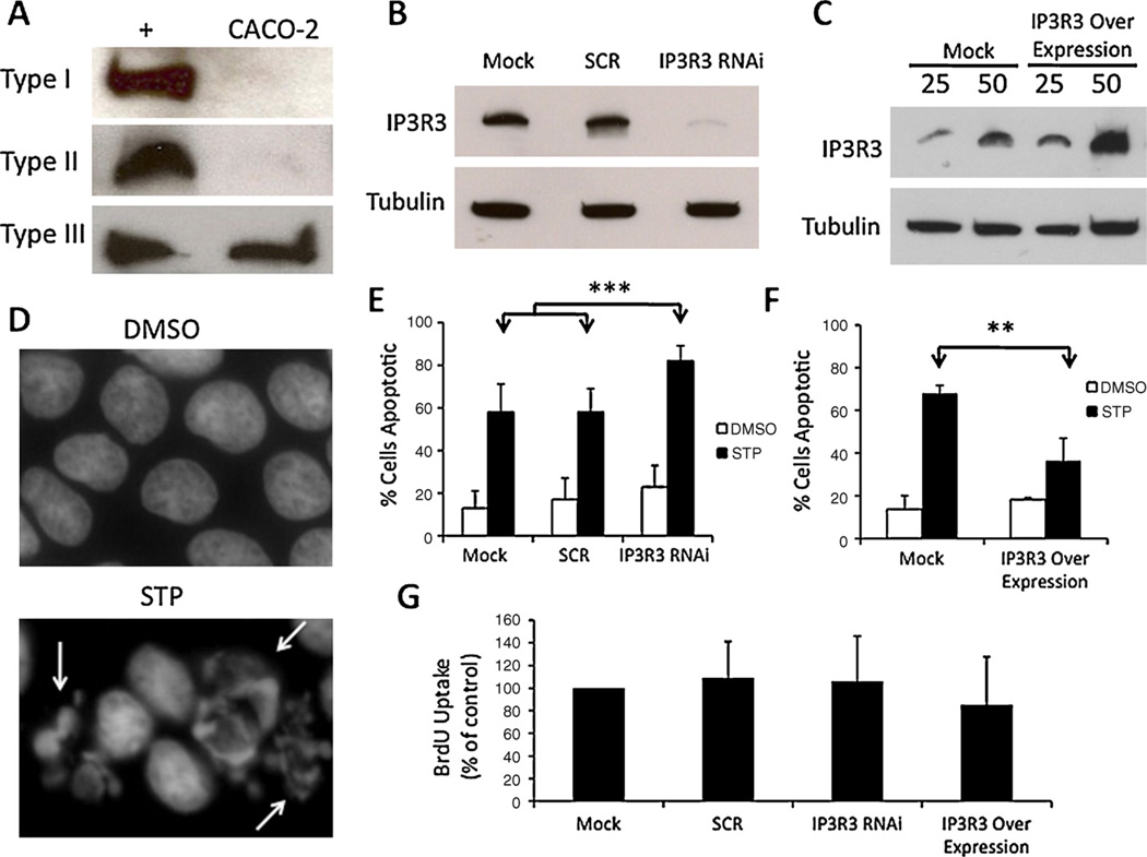
The inositol 1,4,5-trisphosphate receptor (InsP3R) mediates Ca(2+) signaling in epithelia and regulates cellular functions such as secretion, apoptosis and cell proliferation. Loss of one or more InsP3R isoform has been implicated in disease processes such as cholestasis. Here we examined whether gain of expression of InsP3R isoforms also may be associated with development of disease. Expression of all three InsP3R isoforms was evaluated in tissue from colorectal carcinomas surgically resected from 116 patients. Type I and II InsP3Rs were seen in both normal colorectal mucosa and colorectal cancer, while type III InsP3R was observed only in colorectal cancer. Type III InsP3R expression in the advancing margins of tumors correlated with depth of invasion, lymph node metastasis, liver metastasis, and TNM stage. Heavier expression of type III InsP3R also was associated with decreased 5-year survival. shRNA knockdown of type III InsP3R in CACO-2 colon cancer cells enhanced apoptosis, while over-expression of the receptor decreased apoptosis. Thus, type III InsP3R becomes expressed in colon cancer, and its expression level is directly related to aggressiveness of the tumor, which may reflect inhibition of apoptosis by the receptor. These findings suggest a previously unrecognized role for Ca(2+) signaling via this InsP3R isoform in colon cancer.


Conflict of interest statement

Conflict of interest

There are no conflict of interest to disclose for all authors.

Figures

Fig. 1
Fig. 1
Human colorectal mucosa expresses type I and type II but not type III InsP3 receptors. Immunohistochemistry was performed on formalin-fixed, paraffin-embedded tissue specimens in colorectal mucosa. (A) Type I InsP3 receptor expression in normal mucosa. Strong type I InsP3 receptor expression is seen in a granular pattern, diffusely throughout the cytoplasm (brown). (B) Type II InsP3 receptor expression in normal mucosa. Strong type II InsP3 receptor expression is seen in the region near the apical membrane. (C) Normal mucosa does not express the type III InsP3 receptor. Original magnification A, B, and C: 200×; insets of A and B: 1000×. Nuclei were counterstained with haematoxylin (blue). (For interpretation of the references to color in this figure legend, the reader is referred to the web version of the article.)
Fig. 2
Fig. 2
Colorectal cancer expresses various levels of all InsP3 receptor isoforms. Immunostaining of colorectal cancers variably reveals strong (A, C, and E) or weak (B, D, and F) labeling of each isoform of the InsP3 receptor. (A) Strong type I InsP3 receptor expression in a granular pattern in the cytoplasm. (B) Weak expression of the type I InsP3 receptor in colorectal cancer. (C) Strong type II InsP3 receptor expression in the apical region of a colorectal cancer. (D) A representative case of no type II InsP3 receptor expression in colorectal cancer. (E) Strong luminal expression of type III InsP3 receptor. (F) A representative case of no type III InsP3 receptor expression. Original magnification (A–F): 400×. Nuclei were counterstained with haematoxylin (blue). (For interpretation of the references to color in this figure legend, the reader is referred to the web version of the article.)
Fig. 3
Fig. 3
Colorectal cancer in the invasive margin showed strong expression of type III InsP3 receptor. (A) Tumor center and invasive margin of colorectal cancer are shown. (B) Higher magnification of the rectangular area in (A). Although type III InsP3 receptor expression was weak in the center of the tumor (A, asterisk), stronger labeling was evident in the invasive margin of the tumor (B, arrow heads). Original magnification A: 200×, B: 400×. Nuclei were counterstained with haematoxylin (blue). (For interpretation of the references to color in this figure legend, the reader is referred to the web version of the article.)
Fig. 4
Fig. 4
Enhanced expression of type III InsP3 receptor is associated with poorer survival rate in colorectal cancer patients. Kaplan-Meier curve is shown for InsP3 receptor expression levels (weak vs. strong) in the invasive margin of colorectal carcinoma and the corresponding survival rate of patients over 5 years. (A) Weak (n = 41) and strong (n = 75) type I InsP3 receptor expression, p = 0.66. (B) Weak (n = 33) and strong type II InsP3 receptor expression (n = 83), p = 0.26. (C) Weak (n = 82) and strong type III InsP3 receptor expression (n = 34). The 5-year survival rate of patients with strong type III InsP3 receptor expression (61.5%) was significantly lower than that of patients with weak expression (82.7%). p = 0.033.
Fig. 5
Fig. 5
Type III InsP3 receptor expression is protective against apoptosis in a human colorectal cancer cell line. (A) Western blot of type I, II, III InsP3R in positive control (+) and CACO-2 cells shows nearly all InsP3R is type III in CACO-2 cells. Controls for type I, II, and III were mouse cortex, Hepatocyte, and CHO lysates, respectively. (B) An isoform-specific shRNA resulted in ~90% reduction in type III InsP3R in CACO-2 cells and was used in subsequent studies. The same concentration (5 nM) of a non-specific scrambled (SCR) shRNA was used as a control in all RNAi experiments. Alpha-tubulin was used as a loading control, and 50 µg of protein was used for Western Blot. Mock corresponds to a mock transfection control that contains transfection reagent but no RNAi material. (C) Type III InsP3R3 was over-expressed in CACO-2 cells via transient transfection of type III InsP3R cDNA. “25” and “50” correspond to 25 and 50 µg protein loaded, respectively. Tubulin used as loading control. Expression was increased by ~100%. (D) Hoescht stain of CACO-2 nuclei under DMSO (vehicle) and STP (1 µM) treatments (24 h). Arrows indicate fragmented nuclei, which were scored as apoptotic. (E) Percentage of cells scored as apoptotic under RNAi conditions. For mock transfected cells, 13 ± 8% of cells underwent apoptosis in the presence of DMSO and 58 ± 13% were apoptotic in the presence of STP. For Scrambled transfected cells, 17 ± 10% of cells were apoptotic in the presence of DMSO as compared to 58 ± 11% in the presence of STP. For shIP3R3 transfected cells, 23 ± 10% of cells were apoptotic in the presence of DMSO as compared to 82 ± 7% in the presence of STP. Over 300 cells were analyzed under each condition and in each experiment. Values are mean ± S.D. of triplicate measurements (***p < 0.0001). (F) For mock transfected cells, 13 ± 6% of cells underwent apoptosis in the presence of DMSO and 67 ± 3% were apoptotic in the presence of STP. In cells transfected with type III InsP3R over expressed, 18 ± 1% of cells were apoptotic in the presence of DMSO and 36 ± 10% were apoptotic in the presence of STP (**p < 0.01). (G) BrdU uptake was used to measure proliferation of cells with either reduced or enhanced expression of InsP3R3. No statistically significant differences relative to control (Mock transfection) were observed. (For interpretation of the references to color in this figure legend, the reader is referred to the web version of the article.)

References

    1. Stewart BK, Kleihues P. World Cancer Report, World Health Organization, International Agency of Research on Cancer. Lyon: IARC Press; 2003. Colorectal Cancer.
    1. Grady WM, Carethers JM. Genomic and epigenetic instability in colorectal cancer pathogenesis. Gastroenterology. 2008;135:1079–1099. - PMC - PubMed
    1. Taketo MM, Edelmann W. Mouse models of colon cancer. Gastroenterology. 2009;136:780–798. - PubMed
    1. Rodrigues MA, Gomes DA, Leite MF, et al. Nucleoplasmic calcium is required for cell proliferation. J. Biol. Chem. 2007;282:17061–17068. - PMC - PubMed
    1. Mendes CC, Gomes DA, Thompson M, et al. The type III inositol 1, 4,5-trisphosphate receptor preferentially transmits apoptotic Ca2+ signals into mitochondria. J. Biol. Chem. 2005;280:40892–40900. - PubMed

Publication types

Substances