

A phase II comparative study of gross tumor volume definition with or without PET/CT fusion in dosimetric planning for non-small-cell lung cancer (NSCLC): primary analysis of Radiation Therapy Oncology Group (RTOG) 0515
- PMID: 21075551
- PMCID: PMC3117034
- DOI: 10.1016/j.ijrobp.2010.09.033
A phase II comparative study of gross tumor volume definition with or without PET/CT fusion in dosimetric planning for non-small-cell lung cancer (NSCLC): primary analysis of Radiation Therapy Oncology Group (RTOG) 0515
Abstract
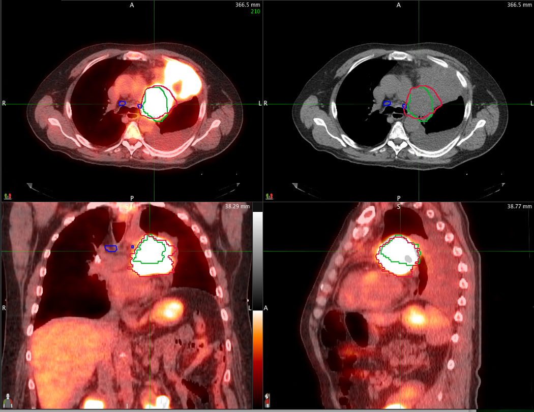
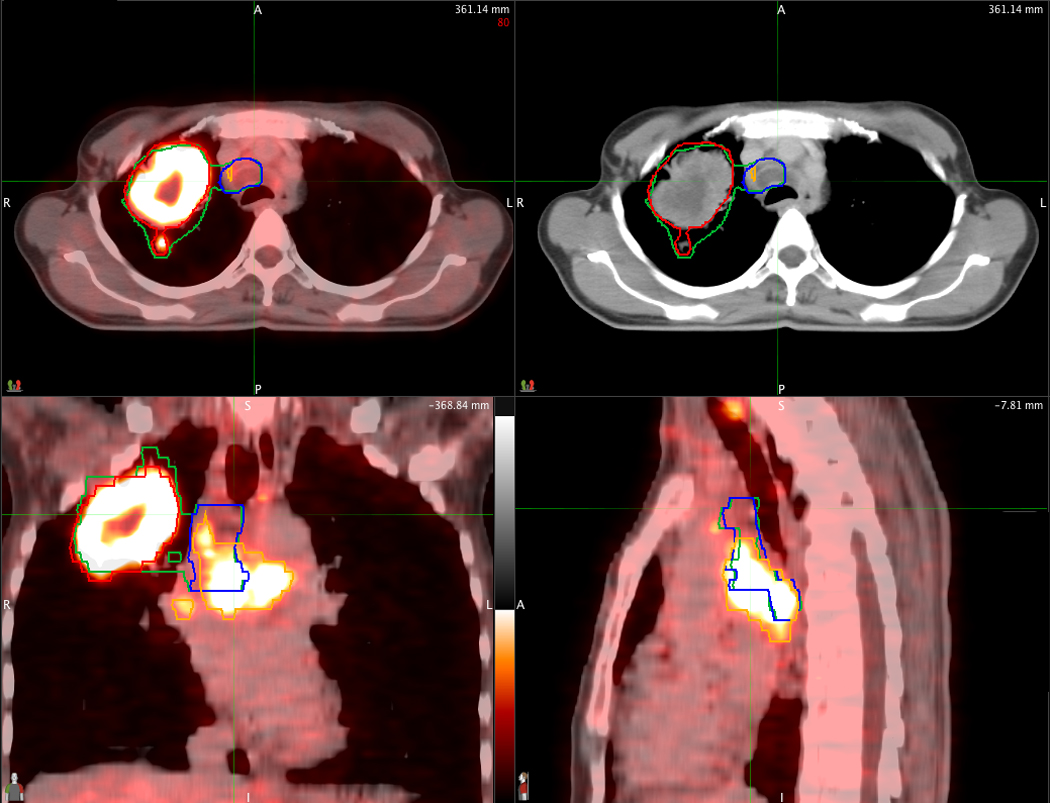
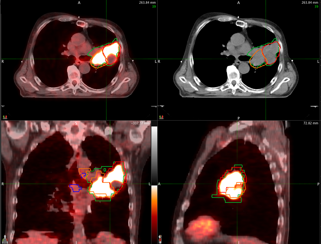
Background: Radiation Therapy Oncology Group (RTOG) 0515 is a Phase II prospective trial designed to quantify the impact of positron emission tomography (PET)/computed tomography (CT) compared with CT alone on radiation treatment plans (RTPs) and to determine the rate of elective nodal failure for PET/CT-derived volumes.
Methods: Each enrolled patient underwent definitive radiation therapy for non-small-cell lung cancer (≥ 60 Gy) and had two RTP datasets generated: gross tumor volume (GTV) derived with CT alone and with PET/CT. Patients received treatment using the PET/CT-derived plan. The primary end point, the impact of PET/CT fusion on treatment plans was measured by differences of the following variables for each patient: GTV, number of involved nodes, nodal station, mean lung dose (MLD), volume of lung exceeding 20 Gy (V20), and mean esophageal dose (MED). Regional failure rate was a secondary end point. The nonparametric Wilcoxon matched-pairs signed-ranks test was used with Bonferroni adjustment for an overall significance level of 0.05.
Results: RTOG 0515 accrued 52 patients, 47 of whom are evaluable. The follow-up time for all patients is 12.9 months (2.7-22.2). Tumor staging was as follows: II = 6%; IIIA = 40%; and IIIB = 54%. The GTV was statistically significantly smaller for PET/CT-derived volumes (98.7 vs. 86.2 mL; p < 0.0001). MLDs for PET/CT plans were slightly lower (19 vs. 17.8 Gy; p = 0.06). There was no significant difference in the number of involved nodes (2.1 vs. 2.4), V20 (32% vs. 30.8%), or MED (28.7 vs. 27.1 Gy). Nodal contours were altered by PET/CT for 51% of patients. One patient (2%) has developed an elective nodal failure.
Conclusions: PET/CT-derived tumor volumes were smaller than those derived by CT alone. PET/CT changed nodal GTV contours in 51% of patients. The elective nodal failure rate for GTVs derived by PET/CT is quite low, supporting the RTOG standard of limiting the target volume to the primary tumor and involved nodes.
Copyright © 2012 Elsevier Inc. All rights reserved.
Conflict of interest statement
Figures
Similar articles
-
Impact of [18F]fluorodeoxyglucose PET-CT staging on treatment planning in radiotherapy incorporating elective nodal irradiation for non-small-cell lung cancer: a prospective study.Int J Radiat Oncol Biol Phys. 2011 Jul 15;80(4):1008-14. doi: 10.1016/j.ijrobp.2010.04.018. Epub 2010 Jul 23. Int J Radiat Oncol Biol Phys. 2011. PMID: 20656419
-
Impact of FDG-PET on radiation therapy volume delineation in non-small-cell lung cancer.Int J Radiat Oncol Biol Phys. 2004 May 1;59(1):78-86. doi: 10.1016/j.ijrobp.2003.10.044. Int J Radiat Oncol Biol Phys. 2004. PMID: 15093902
-
Role of computed tomography and [18F] fluorodeoxyglucose positron emission tomography image fusion in conformal radiotherapy of non-small cell lung cancer: a comparison with standard techniques with and without elective nodal irradiation.Tumori. 2007 Jan-Feb;93(1):88-96. doi: 10.1177/030089160709300116. Tumori. 2007. PMID: 17455878 Clinical Trial.
-
Clinical implications of defining the gross tumor volume with combination of CT and 18FDG-positron emission tomography in non-small-cell lung cancer.Int J Radiat Oncol Biol Phys. 2007 Mar 1;67(3):709-19. doi: 10.1016/j.ijrobp.2006.09.046. Epub 2006 Dec 29. Int J Radiat Oncol Biol Phys. 2007. PMID: 17197120 Review.
-
Current status of PET/CT for tumour volume definition in radiotherapy treatment planning for non-small cell lung cancer (NSCLC).Lung Cancer. 2007 Aug;57(2):125-34. doi: 10.1016/j.lungcan.2007.03.020. Epub 2007 May 2. Lung Cancer. 2007. PMID: 17478008 Review.
Cited by
-
Pancreatic cancer: early detection, diagnosis, and screening.Clin J Gastroenterol. 2012 Oct;5(5):322-6. doi: 10.1007/s12328-012-0327-0. Epub 2012 Aug 10. Clin J Gastroenterol. 2012. PMID: 26181069
-
Comparison of different registration landmarks for MRI-CT fusion in radiotherapy for lung cancer with post-obstructive lobar collapse.J Appl Clin Med Phys. 2019 Jan;20(1):50-54. doi: 10.1002/acm2.12495. Epub 2018 Nov 22. J Appl Clin Med Phys. 2019. PMID: 30565844 Free PMC article.
-
Magnetic resonance (MR) imaging for tumor staging and definition of tumor volumes on radiation treatment planning in nonsmall cell lung cancer: A prospective radiographic cohort study of single center clinical outcome.Medicine (Baltimore). 2017 Feb;96(8):e5943. doi: 10.1097/MD.0000000000005943. Medicine (Baltimore). 2017. PMID: 28225485 Free PMC article.
-
Pretreatment FDG-PET metrics in stage III non-small cell lung cancer: ACRIN 6668/RTOG 0235.J Natl Cancer Inst. 2015 Feb 16;107(4):djv004. doi: 10.1093/jnci/djv004. Print 2015 Apr. J Natl Cancer Inst. 2015. PMID: 25688115 Free PMC article.
-
A Prospective Study Comparing Dosimetry between Computed Tomography (CT) based Radiation Planning and Positron Emission Computed Tomography (PET-CT) based Radiation Planning in Treatment of Non-Metastatic Non Small Cell Lung Carcinoma.Asian Pac J Cancer Prev. 2023 Jul 1;24(7):2543-2550. doi: 10.31557/APJCP.2023.24.7.2543. Asian Pac J Cancer Prev. 2023. PMID: 37505789 Free PMC article.
References
-
- Morgensztern D, Goodgame B, Baggstrom MQ, et al. The effect of FDG-PET on the stage distribution of non-small cell lung cancer. J Thorac Oncol. 2008;3:135–139. - PubMed
-
- MacManus M, Nestle U, Rosenzweig KE, et al. Use of PET and PET/CT for radiation therapy planning: IAEA expert report 2006–2007. Radiother Oncol. 2009;91:85–94. - PubMed
-
- Greene FL, Page DL, Fleming ID, et al. AJCC Cancer Staging Manual. 6th Edition. New York: Springer-Verlag; 2002.
-
- Eisenhauer EA, Therasse P, Bogaerts J, et al. New response evaluation criteria in solid tumours: revised RECIST guideline (version 1.1) Eur J Cancer. 2009;45:228–247. - PubMed
-
- Toloza EM, Harpole L, McCrory DC. Noninvasive staging of non-small cell lung cancer: a review of the current evidence. Chest. 2003;123:137S–146S. - PubMed
