. 2011 Jan 28;286(4):2728-38.
doi: 10.1074/jbc.M110.192344. Epub 2010 Nov 12.

The proprotein convertase PC7: unique zymogen activation and trafficking pathways

Affiliations

The proprotein convertase PC7: unique zymogen activation and trafficking pathways

Estelle Rousselet et al. J Biol Chem. .

Abstract

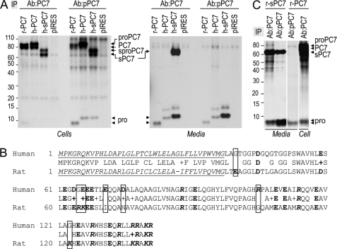
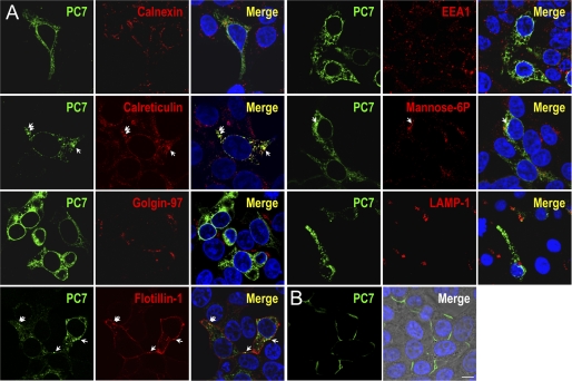
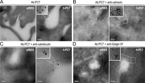
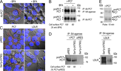
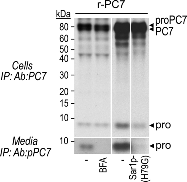
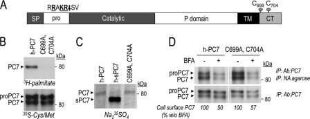
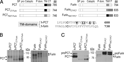
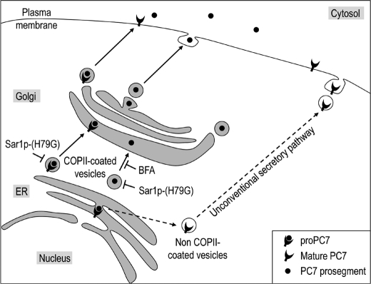
The zymogen activation mechanism and physiological functions of the most ancient and highly conserved basic amino acid-specific proprotein convertase 7 (PC7) are not known. Herein, we characterized the biosynthesis, subcellular localization, and trafficking of the membrane-bound full-length rat and human PC7. The prosegment of PC7 is primarily secreted alone as a non-inhibitory protein via the conventional, Golgi-dependent, secretory pathway. Mature PC7 is partially sulfated and thus reaches the cell surface via the conventional route. However, a fraction of PC7 reaches the cell surface through a brefeldin A- and COPII-independent unconventional secretory pathway. The latter trafficking may explain the rapid (<10 min) transit of a fraction of PC7 from the ER to the cell surface. Electron microscopy further confirmed the localization of PC7 to the cell surface of HEK293 cells. Within the cytosolic tail, only two cysteines (Cys(699) and Cys(704)) are palmitoylated, but this modification does not affect the choice of trafficking pathway. Swapping the transmembrane-cytosolic tail (TMCT) sequences of the convertases Furin and PC7 revealed that PC7(TMCT-Furin) is much more sulfated and hence traffics more efficiently through the conventional secretory pathway. In contrast, the Furin(TMCT-PC7) is no longer sulfated and thus reaches the cell surface by the unconventional pathway. Because trafficking of PC7(CT-Furin) and Furin(CT-PC7) resemble their wild type counterparts, we deduce that the transmembrane domain of PC7 regulates the sorting of PC7 toward the unconventional secretory pathway. In conclusion, PC7 is distinct from other proprotein convertases in its zymogen activation, subcellular localization, and trafficking.


Figures

FIGURE 1.
FIGURE 1.
Zymogen activation of PC7. A, autoradiographs of labeled PC7 and its prosegment in cell lysates (left panel) and media (right panel) of HEK293 cells expressing r-PC7, h-PC7, or h-sPC7 and pulse-labeled with [3H]Leu for 4 h. Proteins were immunoprecipitated with the Ab:PC7 (recognizing both the prosegment and mature PC7) or Ab:pPC7 (recognizing only the prosegment and the zymogen form of PC7) and analyzed by SDS-PAGE separation. B, alignment of the prosegment of h-PC7 and r-PC7. The signal peptide appears in italic and is underlined; bold residues emphasize negative charges, and bold and italic residues, the positive charges. Framed boxes emphasize differences in charge between the two species. C, autoradiographs of labeled PC7 and its prosegment in the media and cell lysates of HEK293 cells expressing soluble or full-length rat PC7 (r-sPC7, r-PC7) and pulse-labeled with [3H]Leu for 4 h. Proteins were immunoprecipitated with either the Ab:PC7 or Ab:pPC7 and analyzed by SDS-PAGE separation.
FIGURE 2.
FIGURE 2.
Subcellular localization of PC7. A, immunofluorescence of PC7 (green labeling) on permeabilized HEK293 cells expressing r-PC7. Cell compartment markers are labeled in red. PC7 co-localizes with calreticulin in the ER, but not with calnexin, another ER marker. There is also some co-localization in clathrin-independent vesicles with GFP-Flotillin-1 and in late endosomes with the mannose-6-phosphate. In contrast, no co-localization is observed in the TGN with Golgin 97, in early endosomes (EEA1), or in lysosomes (LAMP-1). B, PC7 cell surface immunofluorescence of non-permeabilized HEK293 cells expressing r-PC7 shows that PC7 is present at the cell surface (green labeling). Cell nuclei are marked by Hoescht 33258 staining (blue labeling). Bar, 10 μm.
FIGURE 3.
FIGURE 3.
Electron microscopy. Immunolabeling of cryo-sections of HEK293 cells transfected with pIRES-2-EGFP or h-PC7. PC7 is labeled with the Ab:PC7 conjugated to 10 nm colloidal gold particles (arrowheads). Cell compartment markers are visualized by secondary antibodies conjugated to 18 nm colloidal gold particles (arrows). A, PC7 labeling was abundant at and below the cell surface of cells expressing h-PC7. Co-localization was observed with calreticulin (C), but not with calnexin (B) and to a lesser extent with Golgin 97 (D). PC7 labeling was absent in cells transfected with the control empty vector (pIRES). Bars, 200 nm (A), 100 nm (B–D).
FIGURE 4.
FIGURE 4.
PC7 reaches the cell surface by an ER/Golgi-dependent secretory pathway. A and B, post-translational modifications of PC7. Western blot analysis of PC7 in cell lysates or media from HEK293 cells expressing r-PC7 or r-sPC7. A, treatment with endoH (H) and PGNase F (F) of PC7 isolated from cell lysates and media showed that PC7 is sensitive and partially resistant to endoH treatment. Stars correspond to two PC7 forms: the endoH-sensitive one and the form partially resistant to endoH digestion. B, autoradiographs of labeled PC7 in cell lysates of HEK293 cells expressing r-PC7 or r-sPC7 and pulse-labeled with Na235SO4 and [35S]Cys/Met for 2 h demonstrate that a fraction of PC7 traffics through the conventional secretory pathway. C, r-sPC7 samples similar to those obtained in panel B were digested (+) or not (−) with PGnase F.
FIGURE 5.
FIGURE 5.
PC7 reaches the cell surface by a non-conventional secretory pathway through a BFA- and COPII-coated vesicles independent way. A, immunostaining of non-permeabilized HEK293 cells expressing r-PC7 (red labeling) treated (+) or no (−) with BFA (5 μg/ml) for 6 h. Nuclei of transfected cells are marked by Hoescht 33258 staining (blue labeling). Bar, 10 μm. B, cell surface biotinylation of PC7. Autoradiograph of labeled PC7 in lysates of transfected HEK293 cells, incubated or not with BFA (2.5 μg/ml) and pulse-labeled with [35S]Cys/Met for 2 h (B) or 10 min (E). Cells were biotinylated, immunoprecipitated with the Ab:PC7, eluted and then immunoprecipitated with NA-agarose. One-third of lysates were kept before NA-agarose precipitation to normalize the quantity of cell surface PC7. The percent of cell surface PC7/total PC7, estimated by Scion image analysis and normalized to that obtained without BFA treatment (value = 100), showed that despite BFA treatment, ∼60% of PC7 reaches the cell surface. Notice that the autoradiogram of proteins immunoprecipitated with Ab:PC7 and NA was obtained after 4 days of exposure whereas that of proteins immunoprecipitated with Ab:PC7 was obtained after 2 h of exposure. C, immunostaining of PC7 and LDLR (red labeling) in cells expressing r-PC7 and either Sar1p-(H79G) or empty vector (pIRES) or as control human LDLR and either Sar1p-(H79G) or pIRES. Nuclei of transfected cells are marked by Hoescht 33258 staining (blue labeling). Bar, 10 μm. D, cell surface biotinylation of PC7 and a positive control, the LDLR, in the presence of the dominant-negative Sar1p-(H79G). Western blot analysis of PC7 or LDLR on lysates from HEK293 cells co-expressing r-PC7 or LDLR with either Sar1p-(H79G) or pIRES, biotinylated and immunoprecipitated with streptavidin (SA)-agarose. The percent of cell surface PC7, estimated by Scion image analysis and normalized to that obtained with PC7 + pIRES transfection (value = 100), showed that the dominant-negative Sar1p-(H79G) prevents the cell surface localization of LDLR but not that of PC7.
FIGURE 6.
FIGURE 6.
The prosegment of PC7 mainly traffics through the conventional secretory pathway. Autoradiographs of labeled PC7 and its prosegment in lysates and media of transfected HEK293 cells, pulse-labeled with [35S]Cys/Met for 2 h and incubated or not with BFA (2.5 μg/ml) or co-transfected with either the dominant-negative Sar1p-(H79G) or empty vector (pIRES).
FIGURE 7.
FIGURE 7.
Role of PC7 palmitoylation. A, schematic diagram depicting proPC7, in which the cleavage site of the prosegment and cysteines involved in palmitoylation are emphasized. SP, signal peptide; Pro, prosegment; TM, transmembrane domain; CT, cytosolic tail. B, cells expressing wild type and mutant PC7-(C699A, C704A) were pulse-labeled with [3H]palmitic acid or [35S]Cys/Met for 2 h, immunoprecipitated with Ab:PC7, and then analyzed following SDS-PAGE separation. C, autoradiograph of labeled PC7 in lysates from HEK293 cells expressing WT PC7 or PC7-(C699A, C704A), pulse-labeled with Na235SO4 for 2 h and then immunoprecipitated with Ab:PC7. D, cell surface biotinylation of WT PC7 and PC7-(C699A, C704A) incubated or not with BFA (2.5 μg/ml). Autoradiographs of labeled PC7 in lysates from HEK293 cells expressing either WT PC7 or PC7-(C699A, C704A), pulse-labeled with [35S]Cys/Met for 2 h, biotinylated, and immunoprecipitated with Ab:PC7, eluted, and then immunoprecipitated with NA-agarose. One-third of lysates were kept before NA-agarose precipitation to normalize the quantity of cell surface PC7. The percent of cell surface PC7/total PC7, estimated by Scion image analysis and normalized to that obtained without BFA treatment (value = 100), showed that PC7 Cys-palmitoylation has no effect on PC7 trafficking. Notice that the autoradiogram of proteins immunoprecipitated with Ab:PC7 and NA was obtained after 4 days of exposure whereas that of proteins immunoprecipitated with Ab:PC7 was obtained after 2 h of exposure.
FIGURE 8.
FIGURE 8.
The transmembrane domain of PC7 is required for its trafficking via an unconventional pathway. A, schematic diagram depicting chimeras of h-PC7 and human Furin. SP, signal peptide; Pro, prosegment; Catalytic, catalytic domain; P dom., P domain; TM, transmembrane domain; CT, cytosolic tail. We also show the alignment of the TM domains of h-PC7 and h-Furin. B, autoradiographs of labeled PC7 and Furin in lysates from HEK293 cells expressing h-PC7, Furin, or different PC7 and Furin fused constructions, pulse-labeled with Na235SO4 for 2 h and then immunoprecipitated with Ab:PC7 or anti-Furin Ab. C, cell surface biotinylation of PC7 and Furin from HEK293 cells expressing h-PC7, Furin, or their chimeras. After biotinylation, proteins were immunoprecipitated with SA-agarose and revealed by Western blot using Ab-PC7 or anti-Furin Ab.
FIGURE 9.
FIGURE 9.
Schematic diagram of PC7 trafficking. The prosegment of PC7 traffics through the conventional ER/TGN secretory pathway whereas the full-length and mature PC7 traffics through both conventional secretion and an unconventional secretory pathway that is insensitive to BFA treatment and independent of COPII-coated vesicle formation.

References

    1. Seidah N. G., Prat A. (2007) J. Mol. Med. 85, 685–696 - PubMed
    1. Seidah N. G., Mayer G., Zaid A., Rousselet E., Nassoury N., Poirier S., Essalmani R., Prat A. (2008) Int. J. Biochem. Cell Biol. 40, 1111–1125 - PubMed
    1. Seidah N. G., Chrétien M. (1999) Brain Res. 848, 45–62 - PubMed
    1. Creemers J. W., Khatib A. M. (2008) Front Biosci. 13, 4960–4971 - PubMed
    1. Seidah N. G., Hamelin J., Mamarbachi M., Dong W., Tardos H., Mbikay M., Chretien M., Day R. (1996) Proc. Natl. Acad. Sci. U.S.A. 93, 3388–3393 - PMC - PubMed

Publication types

LinkOut - more resources