. 2011 Jan 14;286(2):1147-56.
doi: 10.1074/jbc.M110.183566. Epub 2010 Nov 12.

Regulatory effects of ribosomal S6 kinase 1 (RSK1) in IFNλ signaling

Affiliations

Regulatory effects of ribosomal S6 kinase 1 (RSK1) in IFNλ signaling

Barbara Kroczynska et al. J Biol Chem. .

Abstract

Although the mechanisms of generation of signals that control transcriptional activation of Type III IFN (IFNλ)-regulated genes have been identified, very little is known about the mechanisms by which the IFNλ receptor generates signals for mRNA translation of IFNλ-activated genes. We provide evidence that IFNλ activates the p90 ribosomal protein S6 kinase 1 (RSK1) and its downstream effector, initiation factor eIF4B. Prior to its engagement by the IFNλ receptor, the non-active form of RSK1 is present in a complex with the translational repressor 4E-BP1 in IFNλ-sensitive cells. IFNλ-inducible phosphorylation/activation of RSK1 results in its dissociation from 4E-BP1 at the same time that 4E-BP1 dissociates from eIF4E to allow formation of eIF4F and initiation of cap-dependent translation. Our studies demonstrate that such IFNλ-dependent engagement of RSK1 is essential for up-regulation of p21(WAF1/CIP1) expression, suggesting a mechanism for generation of growth-inhibitory responses. Altogether, our data provide evidence for a critical role for the activated RSK1 in IFNλ signaling.


Figures
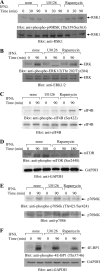
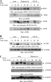
FIGURE 1.
FIGURE 1.
IFNλ-dependent activation of ERK/RSK1 and mTOR signaling cascades in HT-29 cells. A–F, serum-starved HT-29 cells were pretreated for 60 min with U0126 or rapamycin and were either left untreated or treated with IFNλ, in the continuous presence or absence of rapamycin or U0126, as indicated. The cells were lysed, and total cell lysates were resolved by SDS-PAGE and immunoblotted with antibodies against the phosphorylated form of RSK1 on Thr359/Ser363 or against RSK1 (A); with antibodies against the phosphorylated forms of ERK on Thr202/Tyr204 or against ERK (B); with antibodies against the phosphorylated form of eIF4B on Ser422 or against eIF4B (C); with antibodies against the phosphorylated form of mTOR on Ser2448 or against GAPDH (D); with antibodies against the phosphorylated form of p70S6K on Thr421/Ser424 or against p70S6K (E); or with antibodies against the phosphorylated forms of 4E-BP1 on Thr37/46 or against GAPDH (F), as indicated.
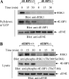
FIGURE 2.
FIGURE 2.
IFNλ-dependent activation of RSK1 and mTOR pathways in ARPE-19 cells. A–C, serum-starved ARPE-19 cells were pretreated with rapamycin or U0126 as indicated and then treated with IFNλ for the indicated times. Total cell lysates were resolved by SDS-PAGE and immunoblotted with anti-phospho-Thr359/Ser363 RSK1, anti-RSK1, anti-phospho-Ser422-eIF4B, or anti-eIF4B antibodies (A); with anti-phospho-Thr37/46-4E-BP1 or anti-GAPDH antibodies (B); or with antibodies against the phosphorylated form of p70S6K on Thr421/Ser424 or against p70S6K or anti-GAPDH (C), as indicated.
FIGURE 3.
FIGURE 3.
Binding of 4E-BP1 and RSK1 to the 7-methylguanosine cap complex prevents recruitment of eIF4G, eIF4A, and eIF4E to the cap complex. A, serum-starved HT-29 cells were pretreated for 60 min with U0126 or rapamycin and were either left untreated or treated with IFNλ, in the continuous presence or absence of rapamycin or U0126, as indicated. Cell lysates were bound to the cap analog m7GTP conjugated to beads, and bound proteins were resolved by SDS-PAGE and immunoblotted with the indicated antibodies. B, HT-29 cells were treated as indicated and assayed as described in A. Bound proteins were immunoblotted with the indicated antibodies. C, serum-starved HT-29 cells were pretreated for 6 h with SL0101-1 and were subsequently treated with IFNλ, as indicated. D, serum-starved HT-29 cells were pretreated for 60 min with U0126 or rapamycin and then treated with IFNλ, in the continuous presence or absence of rapamycin or U0126, as indicated. Equal amounts of cell lysates were immunoprecipitated (IP) with an anti-4E-BP1 antibody, and immune complexes were resolved by SDS-PAGE and immunoblotted with anti-RSK1 or anti-4E-BP1 antibodies, as indicated. E, HT-29 cells were pretreated for 6 h with SL0101-1 and were left untreated or treated with IFNλ, in the continuous presence or absence of inhibitor, as indicated. Equal amounts of cell lysates were immunoprecipitated with anti-4EBP1 antibodies, and immune complexes were resolved by SDS-PAGE for analysis of 4E-BP1 and RSK1, as indicated. F, HT-29 cells were transfected with either control siRNA or siRNA specifically targeting 4E-BP1 and treated with IFNλ, as indicated. Total cell lysates were resolved by SDS-PAGE and immunoblotted with antibodies, against 4E-BP1 or GAPDH, as indicated. G, cells were transfected with either control siRNA or siRNA specifically targeting 4E-BP1 and treated with IFNλ, as indicated. Cell lysates were bound to the cap analog m7GTP conjugated to beads, and bound proteins were resolved by SDS-PAGE and immunoblotted with the indicated antibodies.
FIGURE 4.
FIGURE 4.
Binding of inactive RSK1 to 4E-BP1. A, equal amounts of GST-RSK1 or GST-RSK1 (activated) were annealed with 4E-BP1-His and then bound to the His affinity column. Equal amounts from fractions collected from the affinity column were resolved by SDS-PAGE and immunoblotted with antibodies against GST or 4E-BP1, as indicated. B, input protein levels from the experiment shown in A for GST-RSK1 (sample A) or GST-RSK1 activated (sample B). Immunoblotting with anti-GST or the anti-phospho-Ser221 RSK1 or anti-4E-BP1 (to detect His-4E-BP1) is shown. C, equal amounts of GST-RSK1 were subjected to in vitro kinase assays using active ERK1 and active PDK1, in the presence or absence of the SL0101-1 inhibitor, and then were annealed with 4E-BP1-His and bound to the His affinity column. After extensive washing, proteins were eluted from the column, and equal amounts from each eluted sample were resolved by SDS-PAGE and immunoblotted with antibodies against GST and 4E-BP1, as indicated. D, equal amounts of GST-RSK1 (activated) or GST-RSK1 (activated) that was subjected to an in vitro phosphatase (PP2A) assay were annealed with 4E-BP1-His. After binding to the His affinity column and extensive washing, proteins were eluted from the column, and equal amounts of eluted samples were resolved by SDS-PAGE and immunoblotted with antibodies against GST or 4E-BP1, as indicated. E, input protein levels from the experiment shown in D (panel A) for activated GST-RSK1 (sample A) or activated GST-RSK1 after subjected to in vitro phosphatase (PP2A) assay (sample B) or GST (sample C). Immunoblotting with anti-GST, anti-phospho-Ser221 RSK1 or anti-4E-BP1 (to detect 4E-BP1-His) is shown.
FIGURE 5.
FIGURE 5.
RSK1 activity is required for IFNλ-dependent phosphorylation of 4E-BP1 on Thr37/46. A, serum-starved HT-29 cells were pretreated with SL0101-1 or diluent for 6 h and then treated with IFNλ for the indicated times, in the continuous presence or absence of SL0101-1, as indicated. Total cell lysates were resolved by SDS-PAGE and immunoblotted with the indicated antibodies. Equal cell lysates from the same experiment were analyzed separately by SDS-PAGE and immunoblotted with an anti-phospho-RSK1 (Ser221) and anti-RSK1. B, HT29 cells were transfected with either control siRNA or siRNA targeting RSK1 and treated with IFNλ, as indicated. Total cell lysates were resolved by SDS-PAGE and immunoblotted with the indicated antibodies. C, serum-starved HT-29 cells were pretreated with U0126 for 1 h and then treated with IFNλ for 90 min. The cells were lysed, and equal amounts of protein were immunoprecipitated (IP) with an anti-RSK1 antibody. In vitro kinase assays to detect RSK activity were subsequently carried out on the immunoprecipitates, using a 4E-BP1-His protein as an exogenous substrate. D, serum-starved HT-29 cells were pretreated with SL0101-1 for 6 h or BI-D1870 for 1 h and then treated with IFNλ for the indicated times. The cells were lysed, and equal amounts of protein were immunoprecipitated with an anti-RSK1 antibody. In vitro kinase assays to detect RSK activity were subsequently carried out on the immunoprecipitates, using a GST-4E-BP1 protein as an exogenous substrate.
FIGURE 6.
FIGURE 6.
RSK1 associates with 4E-BP1 in the 7-methylguanosine cap complex. A, serum-starved 4E-BP1+/+ and 4E-BP1−/− cells were treated with mouse IFNα for the indicated times. Equal amounts of cell lysates were incubated with cap analog beads, and after intensive washing, the retained proteins were resolved by SDS-PAGE and immunoblotted with antibodies against RSK1, 4E-BP1, or eIF4E. B, total cell lysates from the same experiment shown in A were resolved by SDS-PAGE and immunoblotted with the indicated antibodies.
FIGURE 7.
FIGURE 7.
Requirement of RSK1 activity in IFNλ-dependent p21WAF1 expression. A, serum-starved HT29 cells were either not pretreated or pretreated with SL0101-1 and then treated with IFNλ for 24 h. Cell lysates were resolved by SDS-PAGE and immunoblotted with antibodies against p21WAF1 or GAPDH, as indicated. B, HT29 cells were either not pretreated or were pretreated with SL0101-1 and then treated with IFNλ for 6 h. Expression of mRNA for the p21 gene was assessed by quantitative real-time RT-PCR. The GAPDH transcript was used for normalization. Data are expressed as -fold increase over IFNλ-untreated samples and represent means ± S.E. (error bars) from three experiments.

Similar articles

Cited by

References

    1. Kotenko S. V., Gallagher G., Baurin V. V., Lewis-Antes A., Shen M., Shah N. K., Langer J. A., Sheikh F., Dickensheets H., Donnelly R. P. (2003) Nat. Immunol. 4, 69–77 - PubMed
    1. Donnelly R. P., Kotenko S. V. (2010) J. Interferon Cytokine Res. 30, 555–564 - PMC - PubMed
    1. Sheppard P., Kindsvogel W., Xu W., Henderson K., Schlutsmeyer S., Whitmore T. E., Kuestner R., Garrigues U., Birks C., Roraback J., Ostrander C., Dong D., Shin J., Presnell S., Fox B., Haldeman B., Cooper E., Taft D., Gilbert T., Grant F. J., Tackett M., Krivan W., McKnight G., Clegg C., Foster D., Klucher K. M. (2003) Nat. Immunol. 4, 63–68 - PubMed
    1. Kotenko S. V., Langer J. A. (2004) Int. Immunopharmacol. 4, 593–608 - PubMed
    1. Lasfar A., Lewis-Antes A., Smirnov S. V., Anantha S., Abushahba W., Tian B., Reuhl K., Dickensheets H., Sheikh F., Donnelly R. P., Raveche E., Kotenko S. V. (2006) Cancer Res. 66, 4468–4477 - PubMed

Publication types

MeSH terms

LinkOut - more resources