

Cloning and characterization of human MUC19 gene
- PMID: 21075863
- PMCID: PMC3175562
- DOI: 10.1165/rcmb.2010-0312OC
Cloning and characterization of human MUC19 gene
Abstract
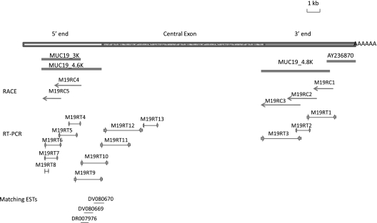
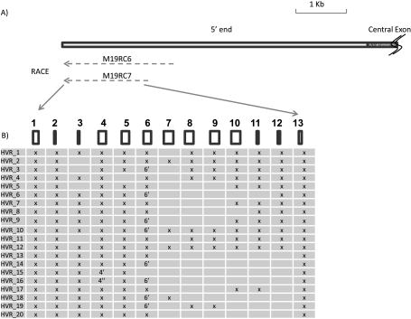
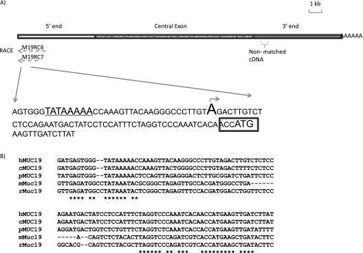
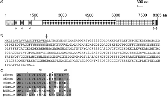
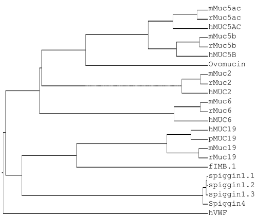
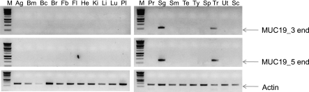
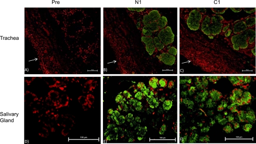
The most recently discovered gel-forming mucin, MUC19, is expressed in both salivary glands and tracheal submucosal glands. We previously cloned the 3'-end partial sequence (AY236870), and here report the complete sequencing of the entire MUC19 cDNA. One highly variable region (HVR) was discovered in the 5' end of MUC19. A total of 20 different splicing variants were detected in HVR, and 18 variants are able to translate into proteins along with the rest of the MUC19 sequence. The longest variant of MUC19 consists of 182 exons, with a transcript of approximately 25 kb. A central exon of approximately 12 kb contains highly repetitive sequences and has no intron interruption. The deduced MUC19 protein has the bona fide gel-forming mucin structure, VWD-VWD-VWD-"threonine/serine-rich repeats"-VWC-CT. An unusual structural feature of MUC19, which is lacking in other gel-forming mucins, is its long amino terminus upstream of the first VWD domain. The long amino terminus is mostly translated from the sequences in HVR, and contains serine-rich repetitive sequences. To validate the integrity of the MUC19 sequence, primers from both the 3' and 5' end were used to demonstrate a similar tissue expression pattern of MUC19 in trachea and salivary glands. In addition, antibodies were developed against either the amino (N) or carboxy (C) terminus of MUC19, and similar antibody staining patterns were observed in both salivary and tracheal submucosal glands. In conclusion, we have cloned and elucidated the entire MUC19 gene, which will facilitate understanding of the function and regulation of this important, yet understudied, mucin gene in airway diseases.
Figures
Similar articles
-
Genome-wide search and identification of a novel gel-forming mucin MUC19/Muc19 in glandular tissues.Am J Respir Cell Mol Biol. 2004 Feb;30(2):155-65. doi: 10.1165/rcmb.2003-0103OC. Epub 2003 Jul 25. Am J Respir Cell Mol Biol. 2004. PMID: 12882755
-
The gene encoding mouse Muc19: cDNA, genomic organization and relationship to Smgc.Physiol Genomics. 2004 Nov 17;19(3):303-18. doi: 10.1152/physiolgenomics.00161.2004. Epub 2004 Aug 31. Physiol Genomics. 2004. PMID: 15340121
-
Tissue distibution of murine Muc19/smgc gene products.J Histochem Cytochem. 2010 Feb;58(2):141-56. doi: 10.1369/jhc.2009.954891. J Histochem Cytochem. 2010. PMID: 19826070 Free PMC article.
-
Proteomic analysis of polymeric salivary mucins: no evidence for MUC19 in human saliva.Biochem J. 2008 Aug 1;413(3):545-52. doi: 10.1042/BJ20080260. Biochem J. 2008. PMID: 18426393
-
Cloning, chromosomal localization and characterization of the murine mucin gene orthologous to human MUC4.Eur J Biochem. 2002 Jul;269(13):3150-9. doi: 10.1046/j.1432-1033.2002.02988.x. Eur J Biochem. 2002. PMID: 12084055
Cited by
-
The Saliva Proteome of Dogs: Variations Within and Between Breeds and Between Species.Proteomics. 2018 Feb;18(3-4):1700293. doi: 10.1002/pmic.201700293. Proteomics. 2018. PMID: 29327448 Free PMC article.
-
Strategies for measuring airway mucus and mucins.Respir Res. 2019 Nov 21;20(1):261. doi: 10.1186/s12931-019-1239-z. Respir Res. 2019. PMID: 31752894 Free PMC article. Review.
-
Risk Factors and Genetic Insights into Coronary Artery Disease-Related Sudden Cardiac Death: A Molecular Analysis of Forensic Investigation.Int J Mol Sci. 2025 Apr 8;26(8):3470. doi: 10.3390/ijms26083470. Int J Mol Sci. 2025. PMID: 40331954 Free PMC article.
-
Mucin deficiency causes functional and structural changes of the ocular surface.PLoS One. 2012;7(12):e50704. doi: 10.1371/journal.pone.0050704. Epub 2012 Dec 18. PLoS One. 2012. PMID: 23272068 Free PMC article.
-
Salivary mucin 19 glycoproteins: innate immune functions in Streptococcus mutans-induced caries in mice and evidence for expression in human saliva.J Biol Chem. 2015 Jan 30;290(5):2993-3008. doi: 10.1074/jbc.M114.597906. Epub 2014 Dec 15. J Biol Chem. 2015. PMID: 25512380 Free PMC article.
References
-
- Rose MC, Voynow JA. Respiratory tract mucin genes and mucin glycoproteins in health and disease. Physiol Rev 2006;86:245–278. - PubMed
-
- Rubin BK. c. Otolaryngol Clin North Am 2010;43:27–34. (vii–viii.). - PubMed
-
- Desseyn JL, Aubert JP, Porchet N, Laine A. Evolution of the large secreted gel-forming mucins. Mol Biol Evol 2000;17:1175–1184. - PubMed
-
- Chen Y, Zhao YH, Kalaslavadi TB, Hamati E, Nehrke K, Le AD, Ann DK, Wu R. Genome-wide search and identification of a novel gel-forming mucin MUC19/MUC19 in glandular tissues. Am J Respir Cell Mol Biol 2004;30:155–165. - PubMed
-
- Offner GD, Troxler RF. Heterogeneity of high-molecular-weight human salivary mucins. Adv Dent Res 2000;14:69–75. - PubMed
Publication types
MeSH terms
Substances
Grants and funding
LinkOut - more resources
Full Text Sources
Molecular Biology Databases
