Review
. 2011 Aug;17(4):351-67.
doi: 10.1177/1073858410381531. Epub 2010 Nov 12.

The essential role of Epstein-Barr virus in the pathogenesis of multiple sclerosis

Affiliations
Free PMC article
Review

The essential role of Epstein-Barr virus in the pathogenesis of multiple sclerosis

Michael P Pender. Neuroscientist. 2011 Aug.
Free PMC article

Abstract

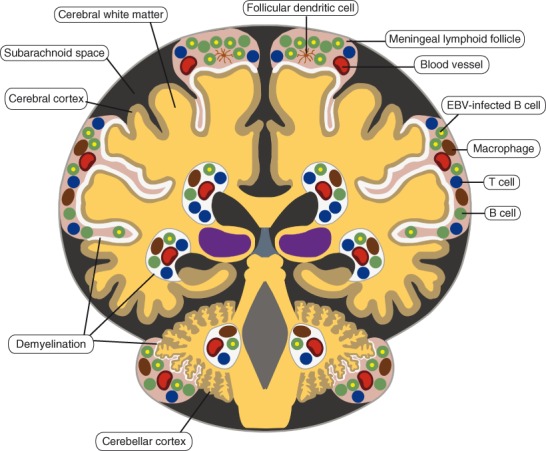
There is increasing evidence that infection with the Epstein-Barr virus (EBV) plays a role in the development of multiple sclerosis (MS), a chronic inflammatory demyelinating disease of the CNS. This article provides a four-tier hypothesis proposing (1) EBV infection is essential for the development of MS; (2) EBV causes MS in genetically susceptible individuals by infecting autoreactive B cells, which seed the CNS where they produce pathogenic autoantibodies and provide costimulatory survival signals to autoreactive T cells that would otherwise die in the CNS by apoptosis; (3) the susceptibility to develop MS after EBV infection is dependent on a genetically determined quantitative deficiency of the cytotoxic CD8+ T cells that normally keep EBV infection under tight control; and (4) sunlight and vitamin D protect against MS by increasing the number of CD8+ T cells available to control EBV infection. The hypothesis makes predictions that can be tested, including the prevention and successful treatment of MS by controlling EBV infection.


Conflict of interest statement

The author(s) declared no potential conflicts of interest with respect to the authorship and/or publication of this article.

Figures

Figure 1.
Figure 1.
Normal sequence of events during infection of the tonsil by Epstein-Barr virus (EBV). During primary infection, EBV entering the tonsil from the saliva infects naive B cells, driving them out of the resting state to become activated B blasts, which then enter germinal centers where they proliferate intensely and differentiate into latently infected memory B cells, which then exit from the tonsil and circulate in the blood. The infected memory B cells do not express any viral proteins except during cell division when they express Epstein-Barr nuclear antigen 1 (EBNA1). Latently infected memory B cells returning to the tonsil can differentiate into plasma cells, which initiates the lytic phase of infection with the production of free virus particles (virions). The virions infect tonsil epithelial cells, where the virus replicates at a high rate and is shed into saliva for transmission to new hosts. Newly formed virus can also infect additional naive B cells in the same host. During primary infection, this cycle initially proceeds unchecked by the immune system. However, the infected host soon mounts an immune response against the virus. EBV-specific cytotoxic CD8+ T cells kill infected B cells expressing viral proteins, and anti-EBV antibodies neutralize viral infectivity by binding to free virus. Red lines with perpendicular bars indicate inhibition. This model is based on work published by Thorley-Lawson and colleagues (Thorley-Lawson and Gross 2004; Laichalk and Thorley-Lawson 2005; Hadinoto and others 2009; Roughan and others 2010).
Figure 2.
Figure 2.
Epstein-Barr virus (EBV)–infected B cells in the brain in secondary progressive multiple sclerosis (MS). EBV-infected B cells are localized in perivascular inflammatory infiltrates in the white matter and in meningeal inflammatory infiltrates overlying the cortex, with the highest concentration of EBV-infected B cells in meningeal infiltrates that resemble lymphoid follicles with germinal centers. The white areas in the cortex and white matter represent demyelinated regions. This distribution of EBV-infected B cells in the MS brain is based on the study of Serafini and others (2007).
Figure 3.
Figure 3.
Proposed role of Epstein-Barr virus (EBV) infection in the development of multiple sclerosis (MS). During primary infection, EBV infects autoreactive naive B cells in the tonsil, driving them to enter germinal centers, where they proliferate intensely and differentiate into latently infected autoreactive memory B cells (step 1), which then exit from the tonsil and circulate in the blood (step 2). The number of EBV-infected B cells is normally controlled by EBV-specific cytotoxic CD8+ T cells, which kill the infected B cells, but not if there is a defect in this defense mechanism. Surviving EBV-infected autoreactive memory B cells enter the CNS, where they take up residence and produce oligoclonal IgG and pathogenic autoantibodies, which attack myelin and other components of the CNS (step 3). Autoreactive T cells that have been activated in peripheral lymphoid organs by cross-reacting infectious antigens circulate in the blood and enter the CNS, where they are reactivated by EBV-infected autoreactive B cells presenting CNS peptides (Cp) bound to major histocompatibility complex (MHC) molecules (step 4). These EBV-infected B cells provide costimulatory survival signals (B7) to the CD28 receptor on the autoreactive T cells and thereby inhibit the activation-induced T cell apoptosis that normally occurs when autoreactive T cells enter the CNS and interact with nonprofessional antigen-presenting cells such as astrocytes and microglia, which do not express B7 costimulatory molecules (Tabi and others 1994; Pender 1998) (step 6). After the autoreactive T cells have been reactivated by EBV-infected autoreactive B cells, they produce cytokines such as interleukin 2 (IL2), interferon γ (IFNγ) and tumor necrosis factor β (TNFβ) and orchestrate an autoimmune attack on the CNS with resultant oligodendrocyte and myelin destruction (step 5). TCR, T cell receptor.
Figure 4.
Figure 4.
Proposed sequence of events leading to the development of multiple sclerosis (MS) and other human autoimmune diseases, for example, autoimmune thyroid disease. In individuals with a genetic deficiency of CD8+ T cells (carried by “autoimmune genes”) and with HLA class II genes predisposing to MS (such as HLA-DR15), late Epstein-Barr virus (EBV) infection leads to the infection of autoreactive B cells, which accumulate in the CNS, where they reactivate autoreactive T cells that orchestrate an autoimmune attack on the CNS leading to the development of MS (upper panel). In individuals with a genetic deficiency of CD8+ T cells and with HLA class II genes predisposing to autoimmune thyroid disease (such as HLA-DR3), late EBV infection leads to the infection of autoreactive B cells, which accumulate in the thyroid gland, where they reactivate autoreactive T cells that orchestrate an autoimmune attack on the thyroid gland, leading to the development of autoimmune thyroid disease (lower panel).
Figure 5.
Figure 5.
Proposed genetic deficiency of CD8+ T cells underlying the development of multiple sclerosis (MS). The upper green panel on the graph represents health, the middle orange panel the development of relapsing MS, and the lower red panel the development of progressive MS. In healthy individuals (Health), the number of CD8+ T cells declines with increasing age but still remains sufficient to control Epstein-Barr virus (EBV) infection. In individuals with a mild genetic deficiency of CD8+ T cells, the deficiency of CD8+ T cells is aggravated by increasing age, eventually leading to insufficient CD8+ T cells to control EBV infection. In individuals carrying HLA class II genes predisposing to MS, this leads to the accumulation of EBV-infected B cells in the CNS and the development of relapsing MS, which ultimately evolves into progressive MS as the CD8+ T cell count further declines with age and as the EBV load in the CNS subsequently increases. In individuals with a severe genetic deficiency of CD8+ T cells, MS develops at a younger age and progresses more rapidly. Lower exposure to sunlight at higher latitudes aggravates the genetic CD8+ T cell deficiency and increases the incidence and progression of MS.

Similar articles

Cited by

References

    1. Acheson ED, Bachrach CA, Wright FM. 1960. Some comments on the relationship of the distribution of multiple sclerosis to latitude, solar radiation, and other variables. Acta Psychiatr Scand Suppl 35:132–47 - PubMed
    1. Amadori A, Zamarchi R, De Silvestro G, Forza G, Cavatton G, Danieli GA. and others. 1995. Genetic control of the CD4/CD8 T-cell ratio in humans. Nat Med 1:1279–83 - PubMed
    1. Antel JP, Peeples DM, Reder AT, Arnason BGW. 1984. Analysis of T regulator cell surface markers and functional properties in multiple sclerosis. J Neuroimmunol 6:93–103 - PubMed
    1. Armengol MP, Juan M, Lucas-Martín A, Fernández-Figueras MT, Jaraquemada D, Gallart T. and others. 2001. Thyroid autoimmune disease: demonstration of thyroid antigen-specific B cells and recombination-activating gene expression in chemokine-containing active intrathyroidal germinal centers. Am J Pathol 159:861–73 - PMC - PubMed
    1. Ascherio A, Munger KL. 2007. Environmental risk factors for multiple sclerosis: Part I. The role of infection. Ann Neurol 61:288–99 - PubMed

Publication types

MeSH terms