. 2011 Jan;300(1):H118-24.
doi: 10.1152/ajpheart.00932.2010. Epub 2010 Nov 12.

Increased propensity for cell death in diabetic human heart is mediated by mitochondrial-dependent pathways

Affiliations

Increased propensity for cell death in diabetic human heart is mediated by mitochondrial-dependent pathways

Ethan J Anderson et al. Am J Physiol Heart Circ Physiol. 2011 Jan.

Abstract

Progressive energy deficiency and loss of cardiomyocyte numbers are two prominent factors that lead to heart failure in experimental models. Signals that mediate cardiomyocyte cell death have been suggested to come from both extrinsic (e.g., cytokines) and intrinsic (e.g., mitochondria) sources, but the evidence supporting these mechanisms remains unclear, and virtually nonexistent in humans. In this study, we investigated the sensitivity of the mitochondrial permeability transition pore (mPTP) to calcium (Ca(2+)) using permeabilized myofibers of right atrium obtained from diabetic (n = 9) and nondiabetic (n = 12) patients with coronary artery disease undergoing nonemergent coronary revascularization surgery. Under conditions that mimic the energetic state of the heart in vivo (pyruvate, glutamate, malate, and 100 μM ADP), cardiac mitochondria from diabetic patients show an increased sensitivity to Ca(2+)-induced mPTP opening compared with nondiabetic patients. This increased mPTP Ca(2+) sensitivity in diabetic heart mitochondria is accompanied by a substantially greater rate of mitochondrial H(2)O(2) emission under identical conditions, despite no differences in respiratory capacity under these conditions or mitochondrial enzyme content. Activity of the intrinsic apoptosis pathway mediator caspase-9 was greater in diabetic atrial tissue, whereas activity of the extrinsic pathway mediator caspase-8 was unchanged between groups. Furthermore, caspase-3 activity was not significantly increased in diabetic atrial tissue. These data collectively suggest that the myocardium in diabetic patients has a greater overall propensity for mitochondrial-dependent cell death, possibly as a result of metabolic stress-imposed changes that have occurred within the mitochondria, rendering them more susceptible to insults such as Ca(2+) overload. In addition, they lend further support to the notion that mitochondria represent a viable target for future therapies directed at ameliorating heart failure and other comorbidities that come with diabetes.


Figures
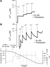
Fig. 1.
Fig. 1.
Use of permeabilized myofibers prepared from human atrial appendage biopsy as an experimental model of mitochondrial Ca2+ (mCa2+) uptake and retention capacity. A: representative experimental trace showing increases in Calcium Green 5-N fluorescence in response to 4-nmol pulses of exogenous Ca2+ in a cuvette containing permeabilized human heart fibers maintained in a submaximal phosphorylating state as outlined in methods in the presence of 1 μM thapsigargin [sarco(endo)plasmic reticulum Ca2+-ATPase inhibitor] and 1 μM Ru-360, an inhibitor of mCa2+ uptake. B: permeabilized heart fibers were incubated under identical conditions as in A, without Ru-360. Here, a tandem oximeter/spectrofluorimeter system was used to simultaneously measure O2 consumption and Ca2+ uptake, and the traces shown were acquired simultaneously from the same preparation of permeabilized fibers. At the start of the experiment, a permeabilized fiber bundle (PmFB) + 100 μM ADP was added to the respiratory medium, followed by the substrates and Ca2+ where indicated. The trace on top shows fluorescence of Calcium Green 5-N in response to Ca2+ pulses, with a corresponding decrease in fluorescence after each pulse indicating mitochondrial uptake of Ca2+. The trace on bottom shows O2 tension in respiratory medium (dark gray trace, y-axis on left) and the rate of O2 consumption (light gray trace, y-axis on right) as each substrate and Ca2+ pulse (vertical lines) is added to the chamber. Jo2, O2 flux/O2 consumption.
Fig. 2.
Fig. 2.
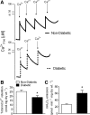
Mitochondria in cardiac tissue from diabetic patients have increased sensitivity to exogenous Ca2+ and increased H2O2 emission. Shown in A are representative experimental traces of mCa2+ uptake in permeabilized myofibers prepared from atrial appendage biopsies in nondiabetic (top) and diabetic (bottom) patients. B and C: the quantified amount of total mitochondrial Ca2+ retained before opening of mitochondrial permeability transition pore (mPTP) (B) and quantified rates of mitochondrial H2O2 emission in paired experiments (C). Data shown are means ± SE, representative of n = 9–12, *P < 0.05.
Fig. 3.
Fig. 3.
Mitochondrial enzyme content and rates of O2 consumption under study conditions are unchanged in cardiac tissue from diabetic patients. A: quantified rates of submaximal state 3 O2 consumption (100 μM ADP) in permeabilized cardiac fibers energized with pyruvate, glutamate, and malate (PGM) and PGM + succinate (PGMS) from nondiabetic and diabetic patients. B: rates of citrate synthase activity in tissue homogenates prepared from both groups. C: representative immunoblot of cardiac tissue protein from 6 diabetic and 6 nondiabetic patients using a primary antibody cocktail that recognizes polypeptides from complex I-IV and the F1F0-ATPase in the electron transport system (green bands), with glyceraldehyde-3-phosphate dehydrogenase as loading control (red band). Protein-ladder standards are shown in lane 1 and lanes 14 and 15. D: densitometry analysis of OxPhos protein from each patient group. Quantified data shown are means ± SE, representative of n = 8–10. AU, arbitrary units; OxPhos, oxidative phosphorylation.
Fig. 4.
Fig. 4.
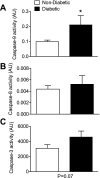
Activity of caspase-9 is increased in cardiac tissue from diabetic patients. Quantified rates of caspase-9 (A), caspase-8 (B), and caspase-3 (C) in cardiac tissue from nondiabetic and diabetic patients are given. Data shown are means ± SE, representative of n = 9–12, *P < 0.05.

References

    1. Anderson EJ, Kypson AP, Rodriguez E, Anderson CA, Lehr EJ, Neufer PD. Substrate-specific derangements in mitochondrial metabolism and redox balance in the atrium of the type 2 diabetic human heart. J Am Coll Cardiol 54: 1891–1898, 2009 - PMC - PubMed
    1. Anderson EJ, Lustig ME, Boyle KE, Woodlief TL, Kane DA, Lin CT, Price JW, 3rd, Kang L, Rabinovitch PS, Szeto HH, Houmard JA, Cortright RN, Wasserman DH, Neufer PD. Mitochondrial H2O2 emission and cellular redox state link excess fat intake to insulin resistance in both rodents and humans. J Clin Invest 119: 573–581, 2009 - PMC - PubMed
    1. Anderson EJ, Yamazaki H, Neufer PD. Induction of endogenous uncoupling protein 3 suppresses mitochondrial oxidant emission during fatty acid-supported respiration. J Biol Chem 282: 31257–31266, 2007 - PubMed
    1. Ares-Carrasco S, Picatoste B, Benito-Martin A, Zubiri I, Sanz AB, Sanchez-Nino MD, Ortiz A, Egido J, Tunon J, Lorenzo O. Myocardial fibrosis and apoptosis, but not inflammation, are present in long-term experimental diabetes. Am J Physiol Heart Circ Physiol 297: H2109–H2119, 2009 - PubMed
    1. Baines CP. The cardiac mitochondrion: nexus of stress. Annu Rev Physiol 72: 61–80, 2010 - PubMed

Publication types

MeSH terms

Substances