. 2011 Mar;84(3):526-36.
doi: 10.1095/biolreprod.110.086488. Epub 2010 Nov 10.

Zinc requirement during meiosis I-meiosis II transition in mouse oocytes is independent of the MOS-MAPK pathway

Affiliations

Zinc requirement during meiosis I-meiosis II transition in mouse oocytes is independent of the MOS-MAPK pathway

Miranda L Bernhardt et al. Biol Reprod. 2011 Mar.

Abstract

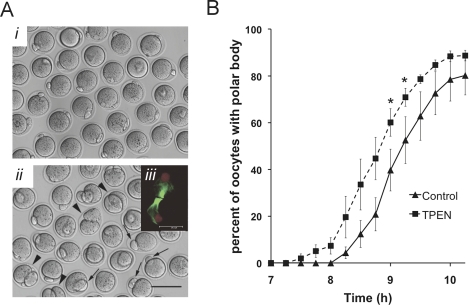
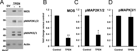
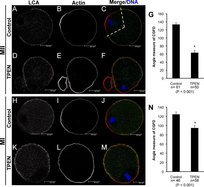
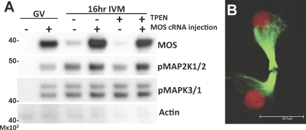
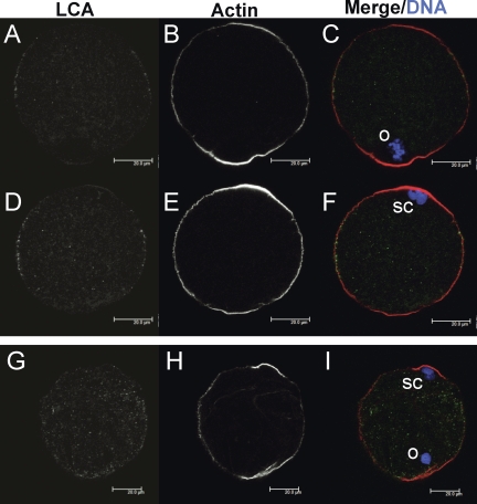
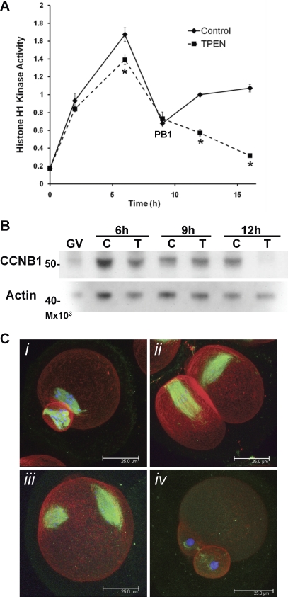
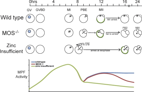
Zinc is essential for many biological processes, including proper functioning of gametes. We recently reported that zinc levels rise by over 50% during oocyte maturation and that attenuation of zinc availability during this period could be achieved using the membrane-permeable heavy metal chelator N,N,N',N'-tetrakis(2-pyridylmethyl)ethylenediamine (TPEN). This zinc insufficiency resulted in formation of large polar bodies, failure to establish metaphase II arrest, and impaired establishment of cortical polarity. As these phenotypes resemble those of MOS null oocytes, we examined the impact of zinc insufficiency on the MOS-MAPK pathway. Reduced levels of both MOS protein and phosphorylation of MAP2K1/2 are observed in zinc-insufficient oocytes; however, these differences appear only after completion of the first meiotic division. In addition, activation of the downstream effector of the MOS pathway, MAPK3/1, is not affected by zinc insufficiency, and reduced MOS levels are observed only with the presence of TPEN after the first polar body extrusion. These data are inconsistent with the hypothesis that reduced MOS mediates the observed phenotype. Finally, MOS overexpression does not rescue the phenotype of zinc-insufficient oocytes, confirming that the observed disruption of asymmetric division and spindle abnormalities cannot be attributed to impaired MOS signaling. Zinc-insufficient oocytes do not increase maturation promoting factor (MPF) activity following the first meiotic division, and increasing MPF activity through expression of nondegradable cyclin B1 partially rescues the ability of zinc-insufficient oocytes to enter metaphase II. Although we have shown that zinc has a novel role in the meiotic cell cycle, it is not mediated through the MOS-MAPK pathway.


Figures

FIG. 1.
FIG. 1.
The first meiotic division in zinc-insufficient oocytes is accelerated and produces a large first polar body. A) Denuded oocytes are shown from COCs matured in vitro in control medium (i) or medium containing 10 μM TPEN (ii). Bar = 100 μm. A, iii) inset shows a Z stack projection of confocal images of the spindle from an oocyte matured in the presence of TPEN; microtubules are shown in green and chromatin in red; bar = 20μm. Arrows (ii) mark oocytes with large polar bodies; arrowheads show examples of symmetric division. B) Timing of first polar body extrusion for oocytes IVM in control (solid line) or TPEN-containing medium (dashed line) based on the percentage of oocytes displaying a polar body. Data are average percentages over five separate experiments ± SEM; a total of 207 control and 240 TPEN-treated oocytes were scored. Asterisks indicate statistical significance according to Student t-test (P < 0.05).
FIG. 2.
FIG. 2.
Oocytes matured in the presence of TPEN have reduced levels of MOS and phospho-MAP2K1/2, while phospho-MAPK3/1 levels remain unchanged. A) Western blot analysis of freshly isolated intact (GV) oocytes and oocytes in vitro matured in the absence (−) or presence (+) of 10 μM TPEN for 16 to 18 h. BD) Graphs present densitometric analyses of at least three experiments normalized to actin, with 25 to 40 oocytes per well ± SEM. Single asterisks indicate statistical significance according to Student t-test (P < 0.05; double asterisks indicate P < 0.005).
FIG. 3.
FIG. 3.
CG distribution is disrupted in zinc-insufficient oocytes. Confocal images of LCA-labeled CGs are shown in panels A, D, H, and K; CG staining appears in green in the merged images (C, F, J, M). Oocytes were also stained for actin (B, E, I, L; red in merged images), and DAPI-stained chromatin is shown in blue (C, F, J, M). Proper CG redistribution and actin cap formation in a control IVM MII egg are shown in panels AC. Reduced formation of the CGFD is observed in oocytes matured in the presence of TPEN for 15 h (D and F); actin cap formation is shown in panels E and F. Cortical reorganization in MI oocytes matured for 7.5 h in control (HJ) or TPEN-containing medium (KM) are shown. Representative images of at least 20 oocytes analyzed per group are shown. Measurement of the angle occupied by the CGFD for control and TPEN-treated oocytes after 15–16 h and after 7.5 h of IVM are shown in panels G and N, respectively; the region measured is demonstrated by the dotted yellow line in panel C. Error bars show SEM; asterisk indicates statistical significance according to Student t-test (P values and number of oocytes measured are indicated below the graphs).
FIG. 4.
FIG. 4.
The critical window for zinc action in maintaining MOS levels is during the later portion of meiotic maturation. Western blot analysis shows changes in relative levels of MOS, phoshpo-MAP2K1/2, and phospho-MAPK3/1 in oocytes in vitro matured in the presence of TPEN versus levels in control oocytes at time points from 4 to 16 h, as indicated. Graphs present densitometric analysis to show protein levels relative to that of controls for each time point ± SEM, with normalization to actin (AC). DI) Western blot analysis of oocytes matured for 18 h with 10 μM TPEN present for a portion of the culture period, as designated graphically in DF. Designation C14 indicates that oocytes were cultured in control medium for the first 14 h of culture, then transferred to TPEN-containing medium for the remaining 4 h of the 18-h culture period; T4 indicates culture in the presence of TPEN for the first 4 h followed by 14 h in control medium; and so on. Protein levels were normalized to that of actin, and error bars show SEM. Graphs present combined data from two to five experiments for each condition with 20 to 50 oocytes per well; asterisks indicate statistical significance according to Student t-test (P < .05).
FIG. 5.
FIG. 5.
Overexpression of MOS in zinc-insufficient oocytes does not rescue transition to meiosis II. A) Western blot analysis shows overexpression of MOS protein and phosphorylation of MAP2K1/2 and MAPK3/1 in oocytes injected with MOS cRNA (+) or buffer only (−). Actin is shown as a loading control. Oocytes with intact GV were cultured in IBMX-containing medium for 6 h to allow protein expression, followed by 16 h of culture in control or TPEN-containing medium. B) Three-dimensional projection of a confocal Z stacks through the oocyte spindle structure in MOS-overexpressing oocytes cultured in TPEN-containing medium for 16 h shows a telophase arrest-like spindle with decondensing chromatin. A total of 33 MOS-overexpressing oocytes were analyzed; a representative image is shown. Microtubules appear in green, chromatin in red. Bar = 20 μm.
FIG. 6.
FIG. 6.
Exogenous chromatin induces actin cap formation but not CG redistribution in zinc-insufficient oocytes. Confocal images from two different planes of the same control IVM MII egg are shown in panels AF. AC) Images are in the plane of the egg's MII spindle (o), while images in panels DF are in the plane of the exogenously introduced heat-inactivated sperm chromatin (sc). The egg shown was injected with three sperm nuclei. GI) Images from a single plane of a TPEN IVM oocyte injected with sperm chromatin, with both the oocyte's endogenous chromatin (o) and microinjected sperm chromatin (sc) in the same plane. LCA-labeled CGs are shown in panels A, D, and G and appear in green in merged images (C, F, I). Oocytes were also stained for actin (B, E, H; red in merged images), and DAPI-stained chromatin is shown in blue (C, F, I). At least 20 control and TPEN-treated IVM oocytes injected with sperm chromatin were analyzed over three separate experiments; representative images are shown. Bars = 20 μm.
FIG. 7.
FIG. 7.
Zinc-insufficient oocytes fail to increase MPF activity following the first meiotic division. A) Kinetics of histone H1 kinase activity over the time course of IVM are shown. Values are in arbitrary units and represent densitometric analysis normalized to control oocytes matured for 12h ± SEM. Four to eleven oocytes were assayed for each data point. Asterisks indicate statistical significance according to Student t-test (P < 0.01). B) Western blot analysis for CCNB1 in oocytes matured in control (C) or TPEN (T)-containing medium for 6, 9, or 12 h. The experiment was repeated three times; a representative blot is shown. C) Three-dimensional projected confocal Z stacks of eggs stained for tubulin (green), actin (red), and DNA (blue) are shown. Following injection with CCNB1(Δ90)-EGFP cRNA during IVM in TPEN-containing medium, 26% of the injected oocytes had MII spindle-like structures (C, i and ii), while 32% did not produce polar bodies and had two spindle-like structures (C, iii), and 38% remained in MI (not shown). Uninjected oocytes matured in the presence of TPEN displayed telophase I-arrested spindles (C, iv). A total of 156 injected oocytes were analyzed. Bar = 25 μm.
FIG. 8.
FIG. 8.
Proposed model of the relationship between zinc-insufficient and MOS null oocytes. Timeline shows number of h after initiation of IVM. GVBD and formation of the MI spindle proceed normally in both MOS null and zinc-insufficient oocytes. The MI spindle in MOS null oocytes does not migrate to the oocyte cortex, and the first meiotic division produces a large polar body [19]. Spindle migration appears to occur normally in zinc-insufficient oocytes; however, the first meiotic division is slightly accelerated and produces a large polar body. MOS null oocytes fail to arrest in MII, and a majority expel a second polar body and transition into a third M phase state with monopolar spindles [28]. Zinc-insufficient oocytes do not form an MII spindle and instead arrest with telophase-like spindles. CG reorganization (green outlines within cells) is disrupted in both MOS null eggs and zinc-insufficient oocytes. A schematic of MPF activity is shown below. MPF activity increases after GVBD in wild-type, MOS null, and zinc-insufficient oocytes and then declines just before polar body extrusion (PBE), allowing anaphase to take place. In wild type and MOS null oocytes, MPF activity rises once again as the MII spindle is established and is maintained at high levels in MII arrested eggs, while in MOS null eggs, MPF activity once again drops as oocytes expel a second polar body [28]. After the decline with the first meiotic division, MPF activity in zinc-insufficient oocytes remains low, and these cells fail to establish an MII spindle. Although MOS absence and zinc insufficiency produce visibly similar phenotypes, different mechanisms are involved.

References

    1. Vallee BL, Falchuk KH. The biochemical basis of zinc physiology. Physiol Rev 1993; 73: 79 118 - PubMed
    1. Kambe T, Yamaguchi-Iwai Y, Sasaki R, Nagao M. Overview of mammalian zinc transporters. Cell Mol Life Sci 2004; 61: 49 68 - PMC - PubMed
    1. Yamasaki S, Sakata-Sogawa K, Hasegawa A, Suzuki T, Kabu K, Sato E, Kurosaki T, Yamashita S, Tokunaga M, Nishida K, Hirano T. Zinc is a novel intracellular second messenger. J Cell Biol 2007; 177: 637 645 - PMC - PubMed
    1. Andrews JC, Nolan JP, Hammerstedt RH, Bavister BD. Role of zinc during hamster sperm capacitation. Biol Reprod 1994; 51: 1238 1247 - PubMed
    1. de Lamirande E, Lamothe G, Villemure M. Control of superoxide and nitric oxide formation during human sperm capacitation. Free Radic Biol Med 2009; 46: 1420 1427 - PubMed

Publication types

MeSH terms