. 2011 Mar;39(6):2221-33.
doi: 10.1093/nar/gkq898. Epub 2010 Nov 13.

The highly conserved eukaryotic DRG factors are required for efficient translation in a manner redundant with the putative RNA helicase Slh1

Affiliations

The highly conserved eukaryotic DRG factors are required for efficient translation in a manner redundant with the putative RNA helicase Slh1

Marie-Claire Daugeron et al. Nucleic Acids Res. 2011 Mar.

Abstract

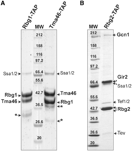
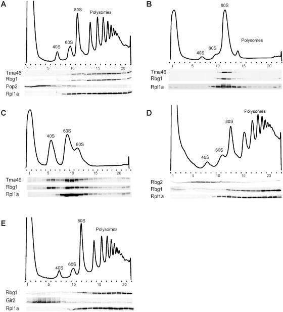
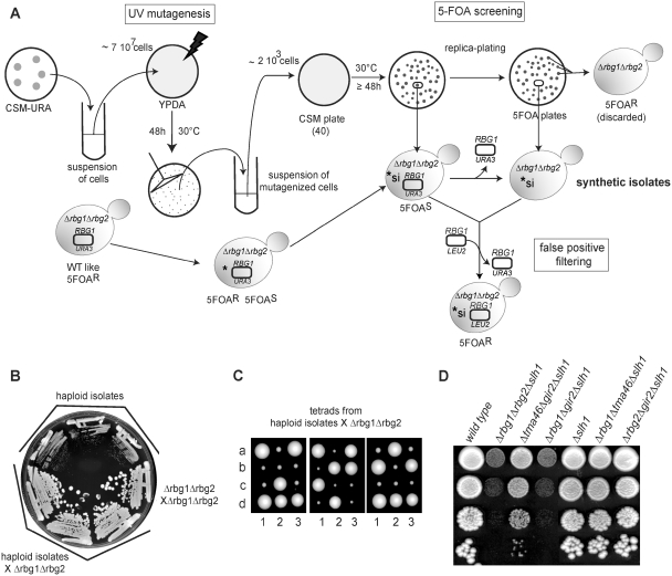
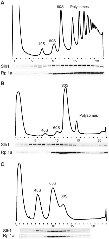
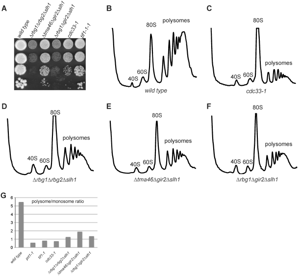
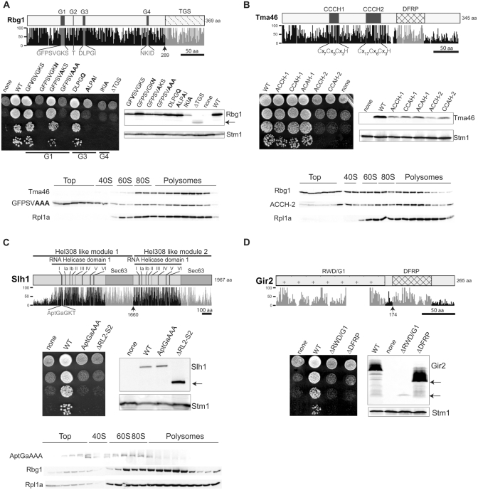
Eukaryotic and archaeal DRG factors are highly conserved proteins with characteristic GTPase motifs. This suggests their implication in a central biological process, which has so far escaped detection. We show here that the two Saccharomyces cerevisiae DRGs form distinct complexes, RBG1 and RBG2, and that the former co-fractionate with translating ribosomes. A genetic screen for triple synthetic interaction demonstrates that yeast DRGs have redundant function with Slh1, a putative RNA helicase also associating with translating ribosomes. Translation and cell growth are severely impaired in a triple mutant lacking both yeast DRGs and Slh1, but not in double mutants. This new genetic assay allowed us to characterize the roles of conserved motifs present in these proteins for efficient translation and/or association with ribosomes. Altogether, our results demonstrate for the first time a direct role of the highly conserved DRG factors in translation and indicate that this function is redundantly shared by three factors. Furthermore, our data suggest that important cellular processes are highly buffered against external perturbation and, consequently, that redundantly acting factors may escape detection in current high-throughput binary genetic interaction screens.


Figures

Figure 1.
Figure 1.
Rbg1 and Rbg2 are not associated with the same protein partners in vivo. Protein profiles observed after TAP purification of the Rbg1-TAP or Tma46-TAP fusions (A) or Rbg2-TAP fusion (B). TAP purified proteins were fractionated on a 5–20% gradient SDS-PAGE gel and stained with Coomassie blue. Molecular weight markers are indicated (MW). Proteins identified by mass-spectrometry are indicated. Gir2 is a highly acidic 31-kDa protein and has an anomalous electrophoretic behavior (38). The protein chaperones, Ssa1 and Ssa2 also observed in other TAP purified purifications, are likely to be nonspecific interactants. Some remaining TEV protease was detected in the gel shown in (B). An asterisk indicates bands identified as degradation products of Rbg1 or Tma46 proteins.
Figure 2.
Figure 2.
Factors Rbg1 and Tma46, but not Rbg2 and Gir2, co-fractionate with translating ribosomes. Polysomes extracts were prepared from cells expressing several epitope-tagged proteins. Tagged proteins were expressed from their genomic loci to avoid overproduction, except when otherwise stated. Polysomes were resolved by density sedimentation in 10–50% sucrose gradient. The UV absorbance trace (254 nm) obtained by continuous monitoring during fractionation is shown with the position of the 40S, 60S, 80S and polysomes peaks indicated. Fractions (numbered) were analyzed by western blotting to detect the TAP, HA and/or VSV tags. The 60S ribosomal protein Rpl1a was used as a positive control for polysome/ribosome/60S subunit association and detected with specific rabbit polyclonal antibodies. The Pop2 deadenylase fused to the VSV tag was used as a marker for nonpolysomal association. To demonstrate the specificity of the association of factors to polysomes or ribosomes, those were dissociated by treating extracts with micrococcal nuclease or micrococcal nuclease and EDTA prior to fractionation on the sucrose gradient. (A) Distribution of Tma46-TAP, Rbg1-HA, Pop2-VSV and RPL1A. (B) Distribution of Tma46-TAP, Rbg1-HA and Rpl1a after micrococcal nuclease treatment of the extract. (C) Distribution of Tma46-TAP, Rbg1-HA and Rpl1a after micrococcal nuclease treatment of the extract followed by EDTA addition. (D) Distribution of Rbg1-HA, Rbg2-TAP and Rpl1a. (E) Distribution of Rbg1-TAP, Gir2-HA and Rpl1a. For this experiment the Gir2-HA protein was encoded by a centromeric (LEU2) plasmid (pMCD-G2) and expressed in a Δgir2 strain that also carried a genomic RBG1-TAP fusion.
Figure 3.
Figure 3.
Identification of Slh1 as a genetic partner of the RBG complexes. (A) Description of the screen for gene having a negative synthetic effect in a Δrbg1Δrbg2 strain. A Δrbg1 Δrbg2 ura3-52 yeast strain containing an URA3-plasmid carrying a RBG1 allele was grown on a CSM-URA plate to stationary phase. Colonies from the plate were resuspended in water, plated on YPDA solid medium (7 × 107 cells /plate) and UV-irradiated. After incubation at 30°C for 48 h, colonies were scraped from the irradiated YPDA plates, resuspended in water and plated on CMS plates (2 × 103 cells/plate; 40 plates). After incubation at 30°C for 48 h, CSM plates were replica-plated on 5FOA plates. The original strain grows well upon loss of the URA3-plasmid [WT like] and is thus resistant to 5-FOA [5-FOAR]. Mutants unable to grow (or growing poorly) on 5-FOA plates [5-FOAS] were retained as putative candidates containing a mutation with a negative synthetic interaction (*si) with the double deletion Δrbg1Δrbg2. To eliminate false positive, 5-FOAS candidates were transformed with a LEU2-plasmid carrying a RBG1 allele. Those were plated on 5-FOA plates and only 5-FOAR transformants were kept for further analysis. (B) The three synthetic slow growth strains isolated in the screen carry a recessive mutation. The original isolates and diploids resulting from the cross of these isolates with an isogenic Δrbg1Δrbg2 strain were streaked in parallel with a Δrbg1/Δrbg1; Δrbg2/Δrbg2 isogenic diploid on YPDA plates and incubated 3 days at 30°C to monitor growth. (C) Meiotic segregation analysis indicates that a single gene is responsible for the synthetic slow growth. Diploid obtained by crossing the three synthetic slow growth strains isolated in the screen with a Δrbg1Δrbg2 strain were sporulated and tetrads were dissected on YPDA plates. Pictures were taken after 4 days at 30°C. Tetrads are numbered 1, 2 and 3 and spores are labeled a, b, c and d. (D) Growth phenotype of yeast strains carrying combinations of deletions of RBG1, TMA46, RBG2 and GIR2 and SLH1 genes. Serial dilutions of exponential liquid cultures were spotted on YPDA plates and incubated 3 days at 30°C.
Figure 4.
Figure 4.
Slh1 protein is associated with translating ribosomes. (A) Whole cell extracts were fractionated on 10–50% sucrose gradient and analyzed as described in Figure 3. Extracts were prepared from cells expressing the Slh1–TAP fusion protein expressed from its natural genomic locus to avoid over expression. In (B), the extract was treated with micrococcal nuclease before loading onto the gradient. In (C), micrococcal nuclease treatment of the extract was followed by EDTA addition before loading onto the gradient.
Figure 5.
Figure 5.
Mutant strains lacking the Rbg1, Rbg2 and Slh1 proteins exhibit polysome profiles indicative of translation initiation defects. (A) Growth behavior of yeast strains carrying combinations of deletions of RBG1, TMA46, RBG2 and GIR2 and SLH1 genes is compared to the growth behavior of cells carrying conditional mutations of translation initiation factors. These phenotypes can be correlated with the polysome profiles of the same strains (B–F). Serial dilutions of exponential liquid cultures were spotted on YPDA plates and incubated 3 days at 30°C. Polysomes extracts from wild type (B), cdc33-1 (eIF4E) (C), Δrbg1Δrbg2Δslh1 (D), Δtma46Δgir2Δslh1 (E) and Δrbg1Δgir2Δslh1 (F) strains were resolved by sedimentation in 10–50% sucrose gradient. The UV absorbance trace (254 nm) is drawn and the position of the 40S, 60S, 80S and polysomes peaks is indicated. (G) Histograms of polysomes/monosomes ratios derived from the polysomes profiles of the indicated strains.
Figure 6.
Figure 6.
Structurally conserved motifs of Rbg1, Tma46, Slh1 and Gir2 are critical for function. Schematic representations of the Rbg1 (A), Tma46 (B), Slh1 (C) and Gir2 (D) protein sequences with a plot of the percentage of conservation to the human, mouse, chicken, zebrafish, nematode, drosophila and arabidopsis orthologs. [Percentage conservation was derived from ClustalX alignments (38).] Boxes depict some characteristic domains of each protein and the corresponding plots are in light gray. The characteristic residues displaying 100% of conservation among eukaryote species are plotted in dark gray. Black lines indicate amino acid scales. Arrows indicate the position of truncations used in the structure/function analysis. To monitor cellular growth, serial dilutions of mutant strains (Δrbg1Δrbg2Δslh1 for (A) and Δtma46Δgir2Δslh1 for (B–D) expressing none, wild type (WT) or mutant proteins, were spotted on YPDA plates and incubated at 30°C for 2–3 days. Amounts of the WT or mutant protein were determined by western blotting. The ribosome associated Stm1 protein was used as a loading control. Distributions of mutant proteins in polysomes sucrose gradient were analyzed by western blotting. Polysomes extracts were prepared from: (A) Δrbg1 cells expressing a Tma46-TAP fusion encoded in the genome and the HA-Rbg1 (GFPSVAAA) mutant encoded by a plasmid. (B) Δtma46 cells expressing a Rbg1-TAP fusion encoded in the genome and HA-Tma46 (ACCH-2) mutant encoded by a plasmid. (C) Δslh1 cells expressing a Rbg1-TAP fusion encoded in the genome and Slh1-ProtA (AptGaAAA) mutant encoded by a plasmid. Positions of the 40S, 60S, 80S monosomes and the polysomes are indicated.

Similar articles

Cited by

References

    1. Leipe DD, Wolf YI, Koonin EV, Aravind L. Classification and evolution of P-loop GTPases and related ATPases. J. Mol. Biol. 2002;317:41–72. - PubMed
    1. Caldon CE, March PE. Function of the universally conserved bacterial GTPases. Curr. Opin. Microbiol. 2003;6:135–139. - PubMed
    1. Li B, Trueb B. DRG represents a family of two closely related GTP-binding proteins. Biochim. Biophys. Acta. 2000;1491:196–204. - PubMed
    1. O'Connell A, Robin G, Kobe B, Botella JR. Biochemical characterization of Arabidopsis developmentally regulated G-proteins (DRGs) Protein Expr. Purif. 2009;67:88–95. - PubMed
    1. Sazuka T, Tomooka Y, Ikawa Y, Noda M, Kumar S. DRG: a novel developmentally regulated GTP-binding protein. Biochem. Biophys. Res. Commun. 1992;189:363–370. - PubMed

Publication types

MeSH terms

Substances