. 2010 Dec;27(6):418-24.
doi: 10.1097/WNP.0b013e3181fe0709.

Interictal spikes precede ictal discharges in an organotypic hippocampal slice culture model of epileptogenesis

Affiliations

Interictal spikes precede ictal discharges in an organotypic hippocampal slice culture model of epileptogenesis

J Dyhrfjeld-Johnsen et al. J Clin Neurophysiol. 2010 Dec.

Abstract

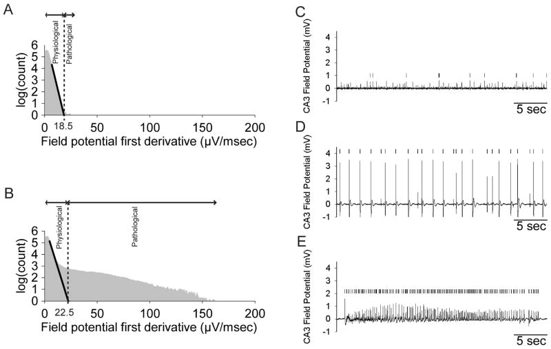
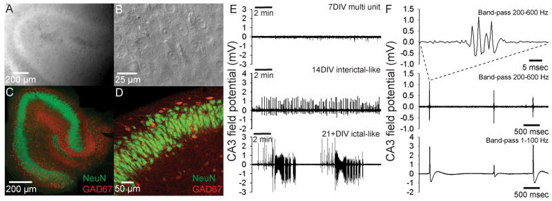
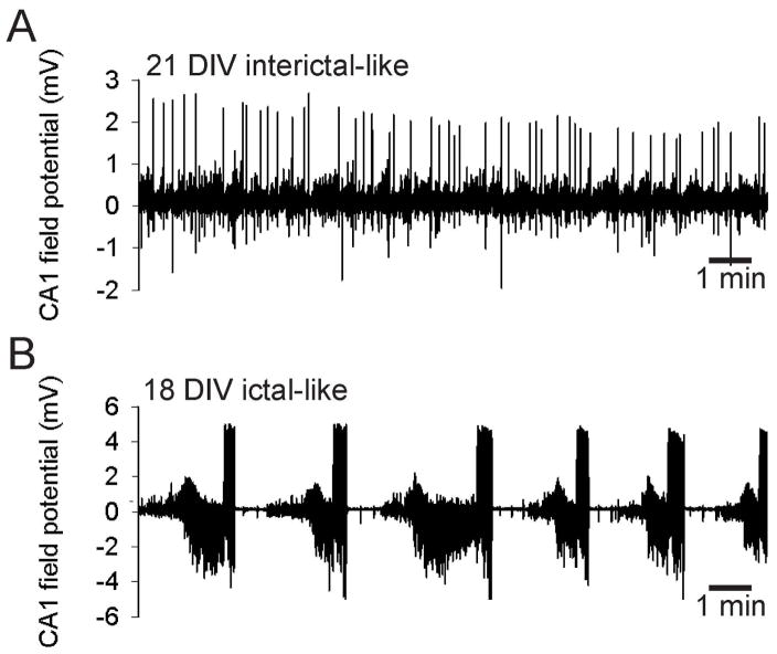
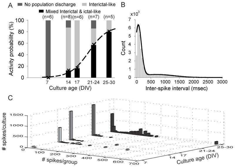
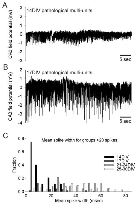
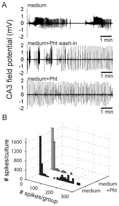
In organotypic hippocampal slice cultures, principal neurons form aberrant excitatory connections with other principal cells in response to slicing induced deafferentation, similar to mechanisms underlying epileptogenesis in posttraumatic epilepsy. To investigate the consequences of this synaptogenesis, the authors recorded field-potential activity from area CA3 during perfusion with the complete growth medium used during incubation. At 7 days in vitro, slice cultures only displayed multiunit activity. At 14 days in vitro, the majority displayed population bursts reminiscent of interictal-like spikes, but sustained synchronous activity was rare. Band-pass filtering of interictal discharges revealed fast ripple-like complexes, similar to in vivo recordings. Spontaneous ictal-like activity became progressively more prevalent with age: at 21 days in vitro, 50% of organotypic hippocampal slice cultures displayed long-lasting, ictal-like discharges that could be suppressed by phenytoin, whereas interictal activity was not suppressed. The fraction of cultures displaying ictal events continually increased with incubation time. Quantification of population spike activity throughout epileptogenesis using automatic detection and clustering algorithms confirmed the appearance of interictal-like activity before ictal-like discharges and also revealed high-frequency pathologic multiunit activity in slice cultures at 14 to 17 days in vitro. These experiments indicate that interictal-like spikes precede the appearance of ictal-like activity in a reduced in vitro preparation. Epileptiform activity in cultures resembled in vivo epilepsy, including sensitivity to anticonvulsants and steadily increasing seizure incidence over time, although seizure frequency and rate of epileptogenesis were higher in vitro. Organotypic hippocampal slice cultures comprise a useful model system for investigating mechanisms of epileptogenesis as well as developing antiepileptic and antiepileptogenic drugs.


Figures

Figure 1
Figure 1. Automated detection of pathological spike activity
A,B. Pathological spikes were detected in CA3 field potential traces using a previously published method (White et al., 2006). Briefly, a powerlaw (black line) is fitted to the steepest slope of the initial part of the first-derivative distribution of the signal (in grey, A. a 7 DIV slice culture with some pathological multi-unit activity, B. a 14 DIV slice culture with interictal-like discharges), yielding the cutoff between physiological and pathological activity as the intercept with the x-axis. This method reliably detected individual spikes (marked with vertical ticks | ) from C. pathological multi-unit activity, D. interictal-like activity and E. ictal-like discharges.
Figure 2
Figure 2. Organotypic hippocampal slice cultures spontaneously develop epileptiform discharges in the growth medium
A. Low power IR-DIC image of hippocampal slice culture in the recording chamber shows preserved slice integrity and organotypic organization. B. High power IR-DIC image showing healthy pyramidal neurons in CA3. C. Immunohistochemical staining of a 30DIV slice with antibodies against NeuN (green) and GAD67 (red), respectively labeling neuronal nuclei and GABAergic cell bodies and processes. D. Higher power image showing CA3 pyramidal neurons labeled in green, GABAergic processes in red and double-labeled GABAergic interneuron cell-bodies in orange. E. Field potential recordings of spontaneous activity from CA3 of organotypic slice cultures. At 7 DIV only physiological multi-unit activity was recorded, but after 14 DIV the majority of slices displayed epileptiform, interictal-like spikes. This spontaneous epileptiform activity developed into a mix of interictal- and ictal-like discharges in slice cultures older than 21 DIV. F. Band-pass filtered (8-pole Bessel filter at 200 Hz, Gaussian filter at 600 Hz) field potential recording from area CA3 of a 14 DIV organotypic slice culture reveals fast ripple-like complexes (top trace). These fast ripple-like complexes occured at intervals larger than 1 sec (middle trace). Fast ripple-like complexes were associated with interictal-like spikes (bottom trace filtered w. 8 pole Bessel filter at 1 Hz, Gaussian filter at 100 Hz).
Figure 3
Figure 3. Spontaneous epileptiform activity in interface-type cultures
A. Interictal-like acticity recorded from area CA1 in an interface-type culture after 21 DIV. B. Ictal-like activity recorded from area CA1 in an interface-type culture after 18 DIV.
Figure 4
Figure 4. Spontaneous ictogenesis in organotypic hippocampal slice cultures mirrors in vivo developmental profile
A. Spontaneous activity recorded in growth medium from CA3 of 32 organotypic slice cultures aged 7–30 DIV categorized as “No population discharge” (multi-unit activity only), “Interictal-like” (units and interictal-like spikes & bursts) and “Mixed interictal- and ictal-like” (units, interictal-like spikes & bursts and ictal-like discharges). The probability P of ictal-like discharges as function of age was fitted with a sigmoidal function (dashed line) with P50 = 21 DIV (r2 = 0.99). B. Pooled inter-spike-interval (ISI) distribution for all spikes detected (Fig. 1) in 10 cultures 14–30 DIV displaying mixed interictal- and ictal-like discharges. The distribution was fitted with the sum of two Gaussian distributions (black line), reflecting a population of low ISI (60±110 msec, high frequency, ictal-like activity) and a population of high ISI (700±600 msec, low frequency, interictal-like activity). C. The development of spike activity in groups of discharges with ISI ≤500 msec (frequency ≥2 Hz) as a function of organotypic slice culture age. The first bin of up to 10 consecutive spikes at ≥2 Hz discharge frequency includes single spikes with ISI ≥500 msec. Prolonged periods of pathological multi-unit activity with mean spike width <20 msec (Fig. 5) are excluded.
Figure 5
Figure 5. Long lasting ≥2 Hz activity at 14
17 DIV. A. High frequency pathological multi-unit activity at 14 DIV. B. High frequency pathological multi-unit activity at 14 DIV. C. Mean spike width distributions for groups of consecutive high frequency spikes at 14–30 DIV. Note that mean spike widths at 14–17 DIV predominantly fall below 20 msec, consistent with multi-unit discharges.
Figure 6
Figure 6. Spontaneous epileptiform discharges are the result of intrinsic alterations in organotypic slices but requires growth medium
A. Robust ictal-like discharges in growth medium disappear within ~5 min of washing in standard ACSF. B. 100 min field potential recording from a 7 DIV hippocampal slice culture without GlutaMAX in the growth medium. C. 100 min field potential recording from a 7 DIV hippocampal slice culture with GlutaMAX in the growth medium. D. The threshold between pathological activity and physiological activity (Fig. 1) calculated in 5 min epochs of 100 min recordings with and without GlutaMAX for 7–8 DIV cultures. E. Without the glutamine supplement GlutaMAX in the recording medium, epileptiform activity in 21+ DIV cultures is delayed beyond the standard 60 min recording period.
Figure 7
Figure 7. The anti-epileptic drug Phenytoin converts spontaneous ictal-like discharges to interictal-like activity
A. The duration of robust ictal-like discharges recorded in the growth medium of 3 hippocampal slice cultures (upper panel) was gradually shortened during wash-in of medium containing 100 μM phenytoin (middle panel). After 20 min wash-in, only interictal-like spikes and short burst persisted in the medium containing 100 μM phenytoin (lower panel). B. The transition from ictal-like activity in growth medium (black bars) to inter-ictal like activity following 20 min wash-in of medium containing 100 μM phenytoin (grey bars) is reflected in the decrease of ≥2 Hz spike activity spanning more than 20 consecutive spikes.

References

    1. Abegg MH, Savic N, Ehrengruber MU, McKinney RA, Gahwiler BH. Epileptiform activity in rat hippocampus strengthens excitatory synapses. J Physiol. 2004;554:439–448. - PMC - PubMed
    1. Bains JS, Longacher JM, Staley KJ. Reciprocal interactions between CA3 network activity and strength of recurrent collateral synapses. Nat Neurosci. 1999;2:720–726. - PubMed
    1. Bausch SB, McNamara JO. Contributions of mossy fiber and CA1 pyramidal cell sprouting to dentate granule cell hyperexcitability in kainic acid-treated hippocampal slice cultures. J Neurophysiol. 2004;92:3582–3595. - PubMed
    1. Behrens CJ, van den Boom LP, de HL, Friedman A, Heinemann U. Induction of sharp wave-ripple complexes in vitro and reorganization of hippocampal networks. Nat Neurosci. 2005;8:1560–1567. - PubMed
    1. Berdichevsky Y, Sabolek H, Levine JB, Staley KJ, Yarmush ML. Microfluidics and multielectrode array-compatible organotypic slice culture method. J Neurosci Methods. 2009;178:59–64. - PMC - PubMed

Publication types