. 2010 Dec 15;29(24):4106-17.
doi: 10.1038/emboj.2010.280. Epub 2010 Nov 12.

The tumour suppressor C/EBPδ inhibits FBXW7 expression and promotes mammary tumour metastasis

Affiliations

The tumour suppressor C/EBPδ inhibits FBXW7 expression and promotes mammary tumour metastasis

Kuppusamy Balamurugan et al. EMBO J. .

Abstract

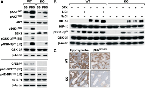
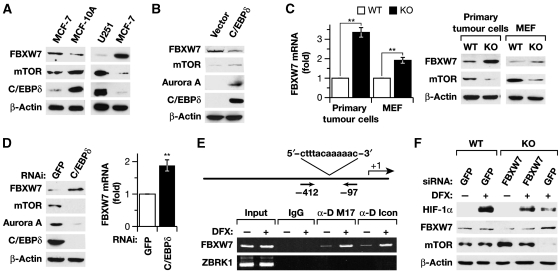
Inflammation and hypoxia are known to promote the metastatic progression of tumours. The CCAAT/enhancer-binding protein-δ (C/EBPδ, CEBPD) is an inflammatory response gene and candidate tumour suppressor, but its physiological role in tumourigenesis in vivo is unknown. Here, we demonstrate a tumour suppressor function of C/EBPδ using transgenic mice overexpressing the Neu/Her2/ERBB2 proto-oncogene in the mammary gland. Unexpectedly, this study also revealed that C/EBPδ is necessary for efficient tumour metastasis. We show that C/EBPδ is induced by hypoxia in tumours in vivo and in breast tumour cells in vitro, and that C/EBPδ-deficient cells exhibit reduced glycolytic metabolism and cell viability under hypoxia. C/EBPδ supports CXCR4 expression. On the other hand, C/EBPδ directly inhibits expression of the tumour suppressor F-box and WD repeat-domain containing 7 gene (FBXW7, FBW7, AGO, Cdc4), encoding an F-box protein that promotes degradation of the mammalian target of rapamycin (mTOR). Consequently, C/EBPδ enhances mTOR/AKT/S6K1 signalling and augments translation and activity of hypoxia-inducible factor-1α (HIF-1α), which is necessary for hypoxia adaptation. This work provides new insight into the mechanisms by which metastasis-promoting signals are induced specifically under hypoxia.


Conflict of interest statement

The authors declare that they have no conflict of interest.

Figures

Figure 1
Figure 1
Analysis of the role of C/EBPδ in mammary tumourigenesis and tumour cells. (A) C/EBPδ is a mammary tumour suppressor. Average number of tumours (mean±s.e.m.) per mouse at the study end point, which was determined by the size of the primary tumour (+/+ WT, n=22; +/− Het, n=15; −/− KO, n=17). *P=0.043 and **P=0.0065 by Poisson regression analysis compared with tumour frequency in KO mice. (B) C/EBPδ promotes lung metastasis. Incidence of lung metastasis in the tumour-bearing mice shown in A. Depicted is the percentage of mice with metastasis regardless of the number or size of metastases per lung (see text for analysis of metastatic load). *P=0.028 by odds ratio analysis and P=0.018 by Fisher exact test. (C) C/EBPδ is induced by hypoxia in vivo. Immunohistochemistry of parallel sections of an MMTV-Neu mammary tumour stained with anti-hypoxyprobe and anti-C/EBPδ antibodies. Original magnification: × 400. (D) Loss of C/EBPδ reduces HIF-1α and CXCR4 expression. Primary MMTV-Neu tumour cells isolated from C/EBPδ WT or KO mice were cultured at 1% or at 20% O2 in the presence of 200 μM DFX for 8 h followed by western analysis of whole cell extracts with antibodies against the indicated proteins. C/EBPδ expression was analysed in nuclear extracts (NE), because it is not detectable in whole cell extracts of these cells. (E) C/EBPδ is necessary for glycolytic adaptation. Analysis of glucose and lactate levels in culture medium of primary tumour cells after 24 h at 20 or 1% O2. (F) C/EBPδ promotes cell survival under hypoxia. Cell viability of primary tumour cells was analysed by MTT assay after 48 h at 1% O2 relative to cells cultured at 20% O2. (E, F) Results are mean±s.e.m. of three experiments with independent preparations of primary tumour cells. *P<0.05, **P<0.001; NS, not significant.
Figure 2
Figure 2
C/EBPδ is required for HIF-1α expression in human tumour cells. (A) MCF-7 breast tumour cells were transfected with shRNA against C/EBPδ or GFP as negative control and, 24 h later, was exposed to 1% O2, 100 μM DFX or 100 μM CoCl2 for 16 h. Nuclear extracts were analysed by western analysis, as indicated. (B) U251 glioblastoma cells harbouring an HRE–luciferase reporter were transfected as in A. After 2 days, cells were treated with 100 μM DFX for 16 h followed by western analysis of the indicated proteins. (C) U251 cells were treated as in B and whole cell lysates were analysed for luciferase reporter activity. Reporter activity (mean±s.e.m. of three independent experiments) is shown relative to untreated control samples. *P<0.05.
Figure 3
Figure 3
C/EBPδ-deficient cells have reduced AKT and increased GSK-3β activity. (A) C/EBPδ WT and KO MEFs were serum-starved (SS) for 16 h and then stimulated with 10% FBS (FBS) for 1 h. Whole cell extracts were prepared and immunoblotting was performed with the indicated antibodies on two membranes, as shown by the separating line. The WT and KO samples were run on the same gel, with intermediate lanes deleted. The AKT antibody used here recognizes all three isoforms of AKTs. (SE, short exposure; LE, long exposure). (B) MEFs were cultured at 20% O2 in the presence of 100 μM DFX and/or 25 mM LiCl or NaCl for 16 h, followed by western analysis of whole cell extracts with the indicated antibodies. (C) Representative images of immunohistochemistry on parallel sections from MMTV-Neu mammary tumours of C/EBPδ WT and KO mice stained for anti-hypoxyprobe and pS6Ser235/236. Original magnification: × 200.
Figure 4
Figure 4
C/EBPδ promotes HIF-1α protein expression through mTOR signalling. (A) Western analysis of whole cell extracts with the indicated antibodies from C/EBPδ KO MEFs 2 days after transfection with expression vectors for C/EBPδ or mTOR followed by 100 μM DFX treatment for 16 h. The endogenous C/EBPδ protein of WT cells is not detected because exposure time was optimized for the overexpressed protein. Relative quantification of mTOR expression levels normalized to actin are shown at the bottom. (B) MEFs were pretreated with 200 nM rapamycin for 30 min followed by 100 μM DFX for 16 h. Western analysis was performed with whole cell extracts and the indicated antibodies. (C) Western analysis of whole cell lysates from primary WT and C/EBPδ KO mammary tumour cells, or from MCF-10A cells 48 h after transfection with shRNA against C/EBPδ or GFP as control. (D) C/EBPδ WT and KO primary tumour cells or immortalized MEFs were treated with 25 μM MG132 or DMSO control for 6 h. Whole cell extracts were analysed for expression of mTOR and β-actin. (E) C/EBPδ WT and KO MEFs were transfected with HA-ubiquitin expression vector. The next day, whole cell lysates were prepared and subjected to immunoprecipitation of mTOR followed by western analysis with antibodies for HA-tag, mTOR or FBXW7·IgG was used as negative control. (F) Western analysis of whole cell extracts from MEFs treated with 100 μM CHX for the indicated time periods. Quantification of the mTOR/actin signal from three independent experiments is shown on the right. *P<0.05.
Figure 5
Figure 5
C/EBPδ augments mTOR and HIF-1α expression by direct inhibition of FBXW7 expression. (A) Inverse correlation of C/EBPδ and FBXW7. Western analysis of whole cell extracts from MCF-7, MCF-10A and U251 cells with the indicated antibodies. (B) Ectopic C/EBPδ downregulates FBXW7 protein levels. Western analysis of whole cell extracts from MCF-7 cells 48 h after transfection with vector or C/EBPδ expression construct. (C) FBXW7 mRNA and protein analysis in C/EBPδ WT and KO MEFs and in primary tumour cells. Left: qRT-PCR analysis of FBXW7 mRNA levels (mean±s.e.m., normalized to actin) of three independent experiments. WT levels were set at 1. **P<0.001. Right: Western analysis of whole cell lysates. (D) C/EBPδ depletion increases FBXW7 expression. MCF-10A cells were nucleofected with shRNA constructs against GFP or C/EBPδ, harvested 2 days later and analysed as in C. **P<0.001. (E) C/EBPδ targets the FBXW7α promoter. Schematic of the FBXW7α promoter with the sequence of the C/EBP-binding site and the position of the primers for ChIP analysis. MCF-7 cells were treated with 100 μM DFX for 2 h. The chromatin was immunoprecipitated with two different C/EBPδ antibodies (M17, Icon) or IgG as control, and amplified with primers for the FBXW7α or ZBRK1 promoter (Lin et al, 2010) as negative control. (F) FBXW7 depletion increases HIF-1α expression. C/EBPδ KO MEFs were nucelofected with siRNA against FBXW7 or GFP as control. After 24 h 100 μM DFX was added for 16 h. Western analysis was carried out with whole cell extracts as indicated.
Figure 6
Figure 6
Model showing how C/EBPδ may integrate and amplify hypoxia and inflammatory signals to promote HIF-1α protein expression, hypoxic adaptation and tumour metastasis. Colours indicate effects mediated by regulation of mRNA expression (blue), protein translation (green), protein stability (red) or protein activity (black). The mTOR protein is part of both TORC1 and TORC2 complexes. The dashed line indicates that the mechanism by which hypoxia induces C/EBPδ expression remains to be determined.

Comment in

References

    1. Abraham RT (2004) mTOR as a positive regulator of tumor cell responses to hypoxia. Curr Top Microbiol Immunol 279: 299–319 - PubMed
    1. Agrawal S, Hofmann WK, Tidow N, Ehrich M, van den Boom D, Koschmieder S, Berdel WE, Serve H, Muller-Tidow C (2007) The C/EBPdelta tumor suppressor is silenced by hypermethylation in acute myeloid leukemia. Blood 109: 3895–3905 - PubMed
    1. Barresi V, Vitarelli E, Cerasoli S, Barresi G (2009) The cell growth inhibitory transcription factor C/EBPdelta is expressed in human meningiomas in association with low histological grade and proliferation index. J Neurooncol 97: 233–240 - PubMed
    1. Blouw B, Song H, Tihan T, Bosze J, Ferrara N, Gerber HP, Johnson RS, Bergers G (2003) The hypoxic response of tumors is dependent on their microenvironment. Cancer Cell 4: 133–146 - PubMed
    1. Carmeliet P, Dor Y, Herbert JM, Fukumura D, Brusselmans K, Dewerchin M, Neeman M, Bono F, Abramovitch R, Maxwell P, Koch CJ, Ratcliffe P, Moons L, Jain RK, Collen D, Keshert E (1998) Role of HIF-1alpha in hypoxia-mediated apoptosis, cell proliferation and tumour angiogenesis. Nature 394: 485–490 - PubMed

Publication types

Substances